SNAPSHOT
Created at: 2018-12-03 20:36
AOP ID and Title:
Graphical Representation

Status
| Author status | OECD status | OECD project | SAAOP status |
|---|---|---|---|
| Open for comment. Do not cite | EAGMST Under Review | 1.7 | Included in OECD Work Plan |
Abstract
Interference with endogenous developmental processes that are regulated by the aryl hydrocarbon receptor (AHR), through sustained exogenous activation, causes molecular, structural, and functional cardiac abnormalities in avian, mammalian and piscine embryos; this cardiotoxicity ultimately leads to severe edema and embryo death in birds and fish and some strains of rat (Carney et al. 2006; Huuskonen et al. 1994; Kopf and Walker 2009). There have been numerous proposed mechanisms of action for this toxicity profile, many of which include the dysregulation of vascular endothelial growth factor (VEGF) as a key event, as it is essential for normal vasculogenesis and therefore cardiogenesis (Ivnitski-Steele and Walker 2005). This AOP describes the indirect suppression of VEGF expression through the sequestration of the aryl hydrocarbon receptor nuclear translocator (ARNT) by AHR. ARNT is common dimerization partner for both AHR and hypoxia inducible factor alpha (HIF-1α), which stimulates angiogenesis through the transcriptional regulation of VEGF (Ivnitski-Steele and Walker 2005). There is considerable cross talk between these two signaling pathways (AHR and HIF-1α), leading to the hypothesis that AHR activation leads to sustained AHR/ARNT dimerization and reduced HIF-1α/ARNT dimerization, preventing the adequate transcription of essential angiogenic factors, such as VEGF. The suppression of VEGF thereby reduces cardiomyocyte and endothelial cell proliferation, altering cardiovascular morphology and reducing cardiac output, which ultimately leads to congestive heart failure and death (Lanham et al. 2014).
The biological plausibility of this AOP is strong, and there is significant evidence in the literature to support it; however, there exist some contradictory data regarding the effect of AHR on VEGF, which seem highly dependent on tissue type and life stage. There are also multiple targets of AHR activation, such as the COX-2 signaling pathway, that could potentially interact. These contradictions and alternate pathways are discussed below. The quantitative understanding of individual key even relationships (KERs) in this AOP is weak; however, there is a strong correlation between the molecular initiating event (MIE: AHR activation) and adverse outcome (AO: embryolethality), and a quantitative relationship is described for birds.
Background
In 1957, millions of broiler chickens died due to a mysterious chick edema disease characterized by pericardial, subcutaneous and peritoneal edema (SCHMITTLE et al. 1958). This disease was later ascribed to the ingestion of feed contaminated with halogenated aromatic hydrocarbons (HAHs), including 2,3,7,8-tetrachlorodibenzo-p-dioxin (TCDD) (Higginbotham et al. 1968; Metcalfe 1972). It has since become evident that TCDD is a prototypical agonist of the AHR: a transcription factor that modulates the expression of a vast array of genes involved in endogenous development and physiological responses to exogenous chemicals (Denison et al. 2011). A general study in the 1980’s found that mothers exposed to herbicides during pregnancy had a 2.8-fold increase in risk of having a baby with congenital cardiovascular malformations (Loffredo et al. 2001). Epidemiological studies have correlated long-term TCDD exposure with ischemic heart disease (Bertazzi et al. 1998; Flesch-Janys et al. 1995); interestingly, and consistent with this AOP, sectioned and stained heart samples from patients with this disease lack epicardial cells (Di et al. 2010). Mammalian studies have confirmed that in utero exposure to TCDD increases susceptibility to cardiovascular dysfunction in adulthood (Aragon et al. 2008; Thackaberry et al. 2005b). The developing heart is highly dependent on oxygen saturation levels; somewhat counterintuitively, a state of hypoxia (relative to adult oxygen tension) drives normal formation and maturation. Deviation from this optimal oxygen level, either above or below normal, hinders myocardial and endothelial development, altering coronary artery connections, ventricle wall thickness and chamber formation (Patterson and Zhang 2010; Wikenheiser et al. 2009). Interestingly, AHR activation (by TCDD), inhibition, and knockdown significantly inhibited the formation of contractile cardiomyocyte nodes during spontaneous differentiation of embryonic stem cells into cardiomyocytes (in vitro) (Wang et al. 2013), indicating that AHR also has an optimal window of expression for normal cardiogenesis. TCDD significantly reduces the degree of myocardial hypoxia that normally occurs during myocyte proliferation and ventricular wall thickening in the developing embryo (Ivnitski-Steele et al. 2004; Lee et al. 2001). This reduction in hypoxia is associated with reduced expression of both HIF-1and the VEGF splice variant, VEGF166 mRNA, which is one of the primary VEGF variants required to mediate coronary vascularization (Ivnitski-Steele et al. 2004). Therefore, it is biologically plausible that sustained AHR activation sequesters ARNT from HIF-1α impairing hypoxia stimulated coronary angiogenesis.
Summary of the AOP
Events
Molecular Initiating Events (MIE), Key Events (KE), Adverse Outcomes (AO)
| Sequence | Type | Event ID | Title | Short name |
|---|---|---|---|---|
| 1 | MIE | 18 | Activation, AhR | Activation, AhR |
| 2 | KE | 944 | dimerization, AHR/ARNT | dimerization, AHR/ARNT |
| 3 | KE | 945 | reduced dimerization, ARNT/HIF1-alpha | reduced dimerization, ARNT/HIF1-alpha |
| 4 | KE | 948 | reduced production, VEGF | reduced production, VEGF |
| 5 | KE | 110 | Impairment, Endothelial network | Impairment, Endothelial network |
| 6 | KE | 317 | Altered, Cardiovascular development/function | Altered, Cardiovascular development/function |
| 7 | AO | 947 | Increase, Early Life Stage Mortality | Increase, Early Life Stage Mortality |
Key Event Relationships
| Upstream Event | Relationship Type | Downstream Event | Evidence | Quantitative Understanding |
|---|---|---|---|---|
| Activation, AhR | adjacent | dimerization, AHR/ARNT | High | Moderate |
| dimerization, AHR/ARNT | adjacent | reduced dimerization, ARNT/HIF1-alpha | Moderate | Low |
| reduced dimerization, ARNT/HIF1-alpha | adjacent | reduced production, VEGF | Moderate | Moderate |
| reduced production, VEGF | adjacent | Impairment, Endothelial network | High | Low |
| Impairment, Endothelial network | adjacent | Altered, Cardiovascular development/function | Moderate | Low |
| Altered, Cardiovascular development/function | adjacent | Increase, Early Life Stage Mortality | High | Low |
| Activation, AhR | non-adjacent | Increase, Early Life Stage Mortality | High | Moderate |
Stressors
| Name | Evidence |
|---|---|
| Polychlorinated biphenyl | High |
| Polychlorinated dibenzodioxins | High |
| Polychlorinated dibenzofurans | High |
Polychlorinated biphenyl
Certain polychlorinated biphenyl (PCB) congeners are potent AHR agonists, and lead to dioxin-like cardiotoxicity in birds (Carro et al. 2013a; Carro et al. 2013b; Brunstrom, B. 1989; Rifkind et al. 1984) and fish (Clark et al. 2010; Olufsen and Arukwe 2011; Grimes et al. 2008). Furthermore, PCB contamination in wild avian species has been correlated with altered heart size and morphology (DeWitt et al. 2006; Henshel and Sparks 2006).
Of the dioxin-like PCBs, non-ortho congeners are the most toxicologically active, while mono-ortho PCBs are generally less potent (McFarland and Clarke 1989; Safe 1994). Chlorine substitution at ortho positions increases the energetic costs of assuming the coplanar conformation required for binding to the AHR (McFarland and Clarke 1989). Thus, a smaller proportion of mono-ortho PCB molecules are able to bind to the AHR and elicit toxic effects, resulting in reduced potency of these congeners. Other PCB congeners, such as di-ortho substituted PCBs, are very weak AHR agonists and do not likely contribute to dioxin-like effects (Safe 1994).
References
Carro, T., Dean, K., and Ottinger, M. A. (2013a). Effects of an environmentally relevant polychlorinated biphenyl (PCB) mixture on embryonic survival and cardiac development in the domestic chicken. Environ.Toxicol.Chem.
Carro, T., Taneyhill, L. A., and Ottinger, M. A. (2013b). The effects of an environmentally relevant 58 congener polychlorinated biphenyl (PCB) mixture on cardiac development in the chick embryo. Environ.Toxicol.Chem.
Brunstrom, B. (1989). Toxicity of coplanar polychlorinated biphenyls in avian embryos. Chemosphere 19, 765-768.
Rifkind, A. B., Hattori, Y., Levi, R., Hughes, M. J., Quilley, C., and Alonso, D. R. (1984). The chick embryo as a model for PCB and dioxin toxicity: Evidence of cardiotoxicity and increased prostaglandin synthesis. In Biological Mechanisms of Dioxin Action (A. Poland and R. D. Kimbrough, Eds.), pp. 255–266. Cold Spring Harbor Press, Cold Spring Harbor, NY.
Clark, B.W.; Matson, C.W.; Jung, D.; Di Giulio, R.T. 2010. AHR2 mediates cardiac teratogenesis of polycyclic aromatic hydrocarbons and PCB-126 in Atlantic killifish (Fundulus heteroclitus). Aquat. Toxicol. 99, 232-240.
Olufsen, M. and Arukwe, A. (2011) Developmental effects related to angiogenesis and osteogenic differentiation in Salmon larvae continuously exposed to dioxin-like 3,3’,4,4’-tetrachlorobiphenyl (congener 77). Aquatic Toxicology 105: 669– 680
A.C. Grimes, K.N. Erwin, H.A. Stadt, G.L. Hunter, H.A. Gefroh, H.J. Tsai, M.L. Kirby (2008) PCB126 exposure disrupts zebrafish ventricular and branchial but not early neural crest development Toxicol. Sci., 106, pp. 193-205
DeWitt, J. C., Millsap, D. S., Yeager, R. L., Heise, S. S., Sparks, D. W., and Henshel, D. S. (2006). External heart deformities in passerine birds exposed to environmental mixtures of polychlorinated biphenyls during development. Environ.Toxicol.Chem. 25, 541-551.
Henshel, D. and Sparks, D.W. (2006) Site Specific PCB-Correlated Interspecies Differences in Organ Somatic Indices. Ecotoxicology, 1: 9-18
Safe, S. (1994). Polychlorinated biphenyls (PCBs): Environmental impact, biochemical and toxic responses, and implications for risk assessment. Critical Reviews in Toxicology 24, 87-149.
McFarland, V. A., and Clarke, J. U. (1989). Environmental occurrence, abundance, and potential toxicity of polychlorinated biphenyl congeners: Considerations for a congener-specific analysis. Environ.Health Perspect. 81, 225-239.
Polychlorinated dibenzodioxins
- Polychlorinated dibenzo-p-dioxins (PCDDs), which includes 2,3,7,8-tetrachlorodibenzo-p-dioxin (TCDD), represent some of the most potent AHR ligands (Denison et al. 2011).
- When screened for their ability to induce aryl hydrocarbon hydroxylase activity, an indirect measurement of AHR activation, dioxins with chlorine atoms at a minimum of three out of the four lateral ring positions, and with at least one non-chlorinated ring position are the most active (Poland and Glover 1973).
- Until recently, TCDD was considered to be the most potent dioxin-like compound (DLC) (van den Berg et al. 1998); however, recent reports indicate that 2,3,4,7,8-pentachlorodibenzofuran (PeCDF) is more potent than TCDD in some species of birds (Cohen-Barnhouse et al. 2011; Farmahin et al. 2013; Hervé et al. 2010)
- TCDD induced cardiotoxicity in developing chick (Heid et al. 2001; Walker et al. 1997; Walker and Catron 2000) and zebrafish (Antkiewicz et al. 2005; Belair et al. 2001; Henry et al. 1997; Plavicki et al. 2013) embryos.
- Kopf and Walker (2009) provide a concise overview of DLC induced heart defects in fish, birds and mammals.
References:
Denison, M. S., Soshilov, A. A., He, G., DeGroot, D. E., and Zhao, B. (2011). Exactly the same but different: promiscuity and diversity in the molecular mechanisms of action of the aryl hydrocarbon (dioxin) receptor. Toxicol. Sci. 124(1), 1-22.
Poland, A., and Glover, E. (1973). Studies on the mechanism of toxicity of the chlorinated dibenzo-p-dioxins. Environ. Health Perspect. 5, 245-251.
van den Berg, M., Birnbaum, L. S., Bosveld, A. T., Brunstrom, B., Cook, P., Feeley, M., Giesy, J. P., Hanberg, A., Hasegawa, R., Kennedy, S. W., Kubiak, T. J., Larsen, J. C., Van Leeuwen, F. X. R., Liem, A. K. D., Nolt, C., Peterson, R. E., Poellinger, L., Safe, S., Schrenk, D., Tillitt, D. E., Tysklind, M., Younes, M., Wærn, F., and Zacharewski, T. R. (1998). Toxic equivalency factors (TEFs) for PCBs, PCDDs, PCDFs for humans and wildlife. Environ. Health Perspect. 106(12), 775-792.
Cohen-Barnhouse, A. M., Zwiernik, M. J., Link, J. E., Fitzgerald, S. D., Kennedy, S. W., Hervé, J. C., Giesy, J. P., Wiseman, S. B., Yang, Y., Jones, P. D., Wan, Y., Collins, B., Newsted, J. L., Kay, D. P., and Bursian, S. J. (2011b). Sensitivity of Japanese quail (Coturnix japonica), Common pheasant (Phasianus colchicus), and White Leghorn chicken (Gallus gallus domesticus) embryos to in ovo exposure to TCDD, PeCDF, and TCDF. Toxicol. Sci. 119(1), 93-103.
Farmahin, R., Manning, G. E., Crump, D., Wu, D., Mundy, L. J., Jones, S. P., Hahn, M. E., Karchner, S. I., Giesy, J. P., Bursian, S. J., Zwiernik, M. J., Fredricks, T. B., and Kennedy, S. W. (2013). Amino acid sequence of the ligand binding domain of the aryl hydrocarbon receptor 1 (AHR1) predicts sensitivity of wild birds to effects of dioxin-like compounds. Toxicol. Sci. 131(1), 139-152.
Hervé, J. C., Crump, D. L., McLaren, K. K., Giesy, J. P., Zwiernik, M. J., Bursian, S. J., and Kennedy, S. W. (2010). 2,3,4,7,8-pentachlorodibenzofuran is a more potent cytochrome P4501A inducer than 2,3,7,8-tetrachlorodibenzo-p-dioxin in herring gull hepatocyte cultures. Environ. Toxicol. Chem. 29(9), 2088-2095.
Heid, S. E., Walker, M. K., and Swanson, H. I. (2001). Correlation of cardiotoxicity mediated by halogenated aromatic hydrocarbons to aryl hydrocarbon receptor activation. Toxicol. Sci 61(1), 187-196.
Walker, M. K., and Catron, T. F. (2000). Characterization of cardiotoxicity induced by 2,3,7,8-tetrachlorodibenzo-p-dioxin and related chemicals during early chick embryo development. Toxicol. Appl. Pharmacol. 167(3), 210-221.
Walker, M. K., Pollenz, R. S., and Smith, S. M. (1997). Expression of the aryl hydrocarbon receptor (AhR) and AhR nuclear translocator during chick cardiogenesis is consistent with 2,3,7,8-tetrachlorodibenzo-p-dioxin-induced heart defects. Toxicol. Appl. Pharmacol. 143(2), 407-419.
Antkiewicz, D. S., Burns, C. G., Carney, S. A., Peterson, R. E., and Heideman, W. (2005). Heart malformation is an early response to TCDD in embryonic zebrafish. Toxicol. Sci. 84(2), 368-377.
Belair, C. D., Peterson, R. E., and Heideman, W. (2001). Disruption of erythropoiesis by dioxin in the zebrafish. Dev. Dyn. 222(4), 581-594.
Henry, T. R., Spitsbergen, J. M., Hornung, M. W., Abnet, C. C., and Peterson, R. E. (1997). Early life stage toxicity of 2,3,7,8-tetrachlorodibenzo-p-dioxin in zebrafish (Danio rerio). Toxicol. Appl. Pharmacol. 142(1), 56-68.
Plavicki, J., Hofsteen, P., Peterson, R. E., and Heideman, W. (2013). Dioxin inhibits zebrafish epicardium and proepicardium development. Toxicol. Sci. 131(2), 558-567.
Kopf, P. G., and Walker, M. K. (2009). Overview of developmental heart defects by dioxins, PCBs, and pesticides. J. Environ. Sci. Health C. Environ. Carcinog. Ecotoxicol. Rev. 27(4), 276-285.
Polychlorinated dibenzofurans
Polychlorinated dibenzofurans (PCDFs) are potent AHR ligands (Denison et al. 2011). Recent reports indicate that 2,3,4,7,8-pentachlorodibenzofuran is more potent than TCDD, the prototypical AHR ligand, in some species of birds (Cohen-Barnhouse et al. 2011; Farmahin et al. 2013; Hervé et al. 2010). 2,3,7,8-tetrachlorodibenzofuran and 2,3,4,7,8-pentachlorodibenzofuran have been shown to induce cardiotoxicity in chicken embryos (Heid et al. 2001). Variouse PCDF congener were shown to cause early life-stage mortality in ranbow trout (Walker and Peterson 1995; Walker et al. 1997).
References:
Denison, M. S., Soshilov, A. A., He, G., DeGroot, D. E., and Zhao, B. (2011). Exactly the same but different: promiscuity and diversity in the molecular mechanisms of action of the aryl hydrocarbon (dioxin) receptor. Toxicol. Sci. 124(1), 1-22.
Cohen-Barnhouse, A. M., Zwiernik, M. J., Link, J. E., Fitzgerald, S. D., Kennedy, S. W., Hervé, J. C., Giesy, J. P., Wiseman, S. B., Yang, Y., Jones, P. D., Wan, Y., Collins, B., Newsted, J. L., Kay, D. P., and Bursian, S. J. (2011b). Sensitivity of Japanese quail (Coturnix japonica), Common pheasant (Phasianus colchicus), and White Leghorn chicken (Gallus gallus domesticus) embryos to in ovo exposure to TCDD, PeCDF, and TCDF. Toxicol. Sci. 119(1), 93-103.
Farmahin, R., Manning, G. E., Crump, D., Wu, D., Mundy, L. J., Jones, S. P., Hahn, M. E., Karchner, S. I., Giesy, J. P., Bursian, S. J., Zwiernik, M. J., Fredricks, T. B., and Kennedy, S. W. (2013). Amino acid sequence of the ligand binding domain of the aryl hydrocarbon receptor 1 (AHR1) predicts sensitivity of wild birds to effects of dioxin-like compounds. Toxicol. Sci. 131(1), 139-152.
Hervé, J. C., Crump, D. L., McLaren, K. K., Giesy, J. P., Zwiernik, M. J., Bursian, S. J., and Kennedy, S. W. (2010). 2,3,4,7,8-pentachlorodibenzofuran is a more potent cytochrome P4501A inducer than 2,3,7,8-tetrachlorodibenzo-p-dioxin in herring gull hepatocyte cultures. Environ. Toxicol. Chem. 29(9), 2088-2095.
Walker, M.K.; Cook, P.M.; Butterworth, B.C.; Zabel, E.W. and Peterson, R.E. (1995) Potency of a Complex Mixture of Polychlorinated Dibenzo-p-dioxin, Dibenzofuran, and Biphenyl Congeners Compared to 2,3,7,8-Tetrachlorodibenzo-p-dioxin in Causing Fish Early Life Stage Mortality. Fundamental and Applied Toxicology 30(2): 178-186.
Walker, M.K. and Peterson, R.E. (1991) Potencies of polychlorinated dibenzo-p-dioxin, dibenzofuran, and biphenyl congeners, relative to 2,3,7,8-tetrachlorodibenzo-p-dioxin, for producing early life stage mortality in rainbow trout (Oncorhynchus mykiss) Aquatic Toxicology 21: 219-238.
Overall Assessment of the AOP
Domain of Applicability
Life Stage Applicability| Life Stage | Evidence |
|---|---|
| Embryo | High |
| Term | Scientific Term | Evidence | Links |
|---|---|---|---|
| chicken | Gallus gallus | High | NCBI |
| zebrafish | Danio rerio | High | NCBI |
| mouse | Mus musculus | Low | NCBI |
| Rattus norvegicus | Rattus norvegicus | Low | NCBI |
| Sex | Evidence |
|---|---|
| Male | High |
| Female | High |
Life Stage Applicability, Taxonomic Applicability, Sex Applicability
Elaborate on the domains of applicability listed in the summary section above. Specifically, provide the literature supporting, or excluding, certain domains.
Life Stage Applicability: Exposure must occur early in embryo development in utero (mammals) or in ovo (birds and fish). Mammalian studies often dose between gestational days 14.5 and 17.5 as it represents a developmental window of cardiomyocyte proliferation (Kopf and Walker 2009). Cardiotoxicity has been observed in birds dosed on day zero or day 5 of incubation (Ivnitski-Steele et al. 2005; Walker et al. 1997). Zebrafish seem to have a particular sensitive window of cardio-development between 48 hours-post-fertilization (hpf) and 5 days pf, and become resistant to AHR-mediated cardiotoxicity if exposed after epicardium formation is complete (2 weeks pf) (Plavicki et al. 2013).
Taxonomic Applicability: Early embryonic exposure to AHR-agonists in mice causes cardiotoxicity that persists into adulthood, increasing susceptibility to heart disease (Thackaberry et al. 2005b) and can increased resorptions and late stage fetal death with edema in certain strains of rat (Huuskonen et al. 1994). AHR-agonists also cause cadivascular malformations in birds and fish, and the resulting reduction in cardiac output is fatal (Kopf and Walker 2009).
Therefore, this AOP is most strongly applicable to birds and fish. Although strong AHR agonists cause foetal mortality in mice and rats (Kawakami et al. 2005; Hassoun et al. 1997; Sparschu et al. 1970; Debdas Mukerjee 1998), cardiac malformation is rarely cited as a cause of death. It appears that AHR-mediated effects on cardiaovascular development in mammals more frequently lead to long-term functional deficiencies rather than foetal death.
Sex applicability: Embryonic dysfunction is equally robust in males and females, but adult abnormalities of mice exposed in utero are more prevalent in females (Carreira et al. 2015)
Essentiality of the Key Events
Molecular Initiating Event Summary, Key Event Summary
Provide an overall assessment of the essentiality for the key events in the AOP. Support calls for individual key events can be included in the molecular initiating event, key event, and adverse outcome tables above.
Molecular initiating event: AHR activation (Essentiality = Strong)
- Zebrafish AHR2 morphants (transient knock-out of function) are protected against reduced blood flow, pericardial edema, erythrocyte maturation, and common cardinal vein migration (Bello et al. 2004; Carney et al. 2004; Prasch et al. 2003; Teraoka et al. 2003)
- AHR2-/- zebrafish mutants were protected against TCDD toxicity, including pericardial edema and epicardium development (Goodale et al. 2012; Plavicki et al. 2013)
- AHR activation specifically within cardiomyocytes accounts for heart failure (cardiac malformations, loss of circulation, pericardial edema) induced by TCDD as well as non-cardiac toxicity (swim bladder inflation and craniofacial defects) in zebrafish (Lanham et al. 2014).
- AHR-null mice have impaired angiogenesis in vivo: endothelial cells failed to branch and form tube-like structures (Roman et al. 2009).
- Ischemia-induced angiogenesis was markedly enhanced in AHR-null mice compared with that in wild-type animals (Ichihara et al. 2007)
Key Event 1: AHR/ARNT dimerization (Essentiality = Strong)
- ARNT1 is essential for normal vascular and hematopoietic development (Abbott and Buckalew 2000; Kozak et al. 1997; Maltepe et al. 1997)
- zfarnt2-/- mutation is larval lethal, and the mutants have enlarged heart ventricles and an increased incidence of cardiac arrhythmia (Hill et al. 2009)
- ARNT1 morpholono knock-down protected against pericardial edema and reduced blood flow in zebrafish (Prasch et al. 2006)
- ARNT overexpression rescued cells from the inhibitory effect of hypoxia on AHR-mediated luciferase reporter activity; therefore, the mechanism of interference of the signaling cross-talk between AHR and hypoxia pathways is at least partially dependent on ARNT availability (Vorrink et al. 2014).
Key Event 2: Reduced HIF1α/ARNT dimerization (Essentiality = Moderate)
- Both ARNT–/– and HIF1α–/– mice display embryonic lethality with blocks in developmental angiogenesis and cardiovascular malformations (Iyer et al. 1998; Kozak et al. 1997; Maltepe et al. 1997; Ryan et al. 1998) demonstrating that signaling through the HIF-1 pathway is required for normal development of the cardiovascular system.
- The myocardium exhibits a reduced oxygen status during the later stages of coronary vascular development in chick and mouse embryos (Ivnitski-Steele et al. 2004; Lee et al. 2001)
- Rearing fish embryos in a hypoxic environment can modify cardiac activity, organ perfusion, and blood vessel formation (Pelster 2002)
- TCDD toxicity in fish resembles defects in hypoxia sensing (Prasch et al. 2004)
- Deviation in oxygen levels, below or above normal, during early chick embryogenesis results in abnormal coronary vasculature (Wikenheiser et al. 2009)
- Hypoxia stimulates vasculogenesis and regulates VEGF transcription in vivo and in vitro (Goldberg and Schneider 1994; Levy et al. 1995; Liu et al. 1995) (Goldberg 1994; LEVY 1995A; Liu 1995)
- Hypoxia stimulus can rescue TCDD inhibition of coronary vascular development in chick embryos (Ivnitski-Steele and Walker 2003)
Key Event 3: Reduced VEGF production (Essentiality = Moderate)
- Loss of a single VEGF-A allele in mice results in defective vascularization and early embryonic lethality (Carmeliet et al. 1996; Ferrara et al. 1996).
- Mice lacking VEGF isoforms 164 and 188 exhibit impaired myocardial angiogenesis and reduced contractility leading to ischemic cardiomyopathy (Carmeliet et al. 1999)
- During vasculogenesis, angioblasts are stimulated to proliferate and differentiate into endothelial cells by VEGF-A (Ivnitski-Steele and Walker 2005)
- Migration and assembly of epicardial angioblasts into coronary vessels is regulated by VEGF (Folkman 1992)
- Cardiomyocyte-specific knockout of VEGF in mice results in phenotype similar to TCDD toxicity (thinner ventricular walls, ventricle cavity dilation, and contractile dysfunction) (Giordano et al. 2001; Ivnitski-Steele and Walker 2003)
- Exogenous VEGF rescues the inhibitory effect of TCDD on vasculogenesis (Ivnitski-Steele and Walker 2003)
Key Event 4: Endothelial network impairment (Essentiality = Moderate)
- The epicardium is the source of angioblasts, which penetrate into the myocardium, providing the endothelial and mural cell progenitor populations that eventually form the entire coronary vasculature (Viragh et al. 1993; Vrancken Peeters et al. 1999)
- Sectioned and stained heart samples from patients with ischemic heart disease lack epicardial cells (Di et al. 2010)
- Juvenile mice with induced cardiovascular disease show altered heart morphology and function, including epithelial dysfunction (Kopf et al. 2008)
- In zebrafish, cardiotoxicity coincides with epicardium formation. Cardiotoxicity begins at 48 hours post fertilization (hpf; start of pre-epicardium formation) and starts to decline at 5 days post fertilization, which is about the time the initial epicardial cell layer is complete. Cardiotoxicity disappears at 2 weeks, after epicardium formation is complete. TCDD prevented the formation of the epicardial cell layer when exposed 4hpf, and blocked epicardial expansion from the ventricle to the atrium following exposure at 96hpf. These effects ultimately result in valve malformation, reduced heart size, impaired development of the bulbus arteriosus, decreased cardiac output, reduced end diastolic volume, decreased peripheral blood flow, edema and death (Plavicki et al. 2013).
- TCDD reduces human primary umbilical vein endothelial cells basal proliferation by 50% (Ivnitski-Steele and Walker 2005)
- The phenotype observed in chick embryos following TCDD exposure on day zero of incubation resembles that observed in vertebrate models in which the epicardium fails to form (Ivnitski-Steele and Walker 2005)
Key Event 5: Altered cardiovascular development/ function (Essentiality = Strong)
- The most common cause of infant death due to birth defects is congenital cardiovascular malformation (Kopf and Walker 2009)
- A significant reduction in embryo survival was observed in 2,3,7,8-tetrachlorodibenzo-p-dioxin (TCDD) exposed chick embryos, and was associated with heart failure resulting from altered heart morphology:
- Increased heart width and weight, increased muscle mass, enlarged left ventricle, thinner left ventricle wall, and increased ventricular trabeculation and ventricular septal defects (Walker et al. 1997).
- Changes in heart morphology consistent with dilated cardiomyopathy (decreased cardiac output and ventricular cavity expansion) were observed in chick embryos exposed to TCDD followed by progression to congestive heart failure edema (Walker and Catron 2000).
- Changes in heart morphology and decreases in cardiac output and peripheral blood flow precede heart failure in Zebrafish (Antkiewicz et al. 2005; Belair et al. 2001; Henry et al. 1997; Plavicki et al. 2013)
Cardiotoxic effects of strong AHR-agonists
| Zebrafish Embryo | Chicken Embryo | Mouse |
|---|---|---|
|
|
Embryo/Fetus
21 Days old
|
ANF= cardiac atrial natriuretic factor; an indicator of cardiac stress. Source: (Kopf and Walker 2009)
Weight of Evidence Summary
|
Key Event Relationship |
Weight of Evidence Is there a mechanistic relationship between KEup and KEdown consistent with established biological knowledge? |
Support for Biological Plausibility Strong: Extensive understanding of the KER based on previous documentation and broad acceptance. Moderate: KER is plausible based on analogy to accepted biological relationships, but scientific understanding is incomplete. Weak: Empirical support for association between KEs, but the structural or functional relationship between them is not understood. |
|
KER972: Activation, AhR leads to dimerization, AHR/ARNT |
Strong |
The mechanism of AHR-mediated transcriptional regulation is well understood (Fujii-Kuriyama and Kawajiri 2010). ARNT is a necessary dimerization partner for the transcriptional activation of AHR regulated genes (Hoffman et al. 1991; Poland et al. 1976). |
|
KER973: dimerization, AHR/ARNT leads to reduced dimerization, ARNT/HIF1-alpha |
Moderate |
ARNT is common dimerization partner for both AHR and HIF-1α. Gel-shift and coimmunoprecipitation experiments have shown that the AHR and HIF1α compete for ARNT in vitro, with approximately equal dimerization efficiencies (Schmidt and Bradfield 1996). A number of studies have shown a reduced response to hypoxia following AHR activation (Chan et al. 1999, Seifert et al. 2008, Ivnitski-Steele et al. 2004), however this effect is highly tissue specific; in cells where ARNT is abundant, it does not deplete due to hypoxia or AHR activation (Chan et al. 1999; Pollenz et al. 1999) |
|
KER974: reduced dimerization, ARNT/HIF1-alpha leads to reduced production, VEGF |
Strong |
The transcriptional control of VEGF by HIF-1 is well understood; The HIF-1 complex binds to the VEGF gene promoter, recruiting additional transcriptional factors and initiating VEGF transcription (Ahluwalia and Tarnawski 2012; Fong 2009) |
|
KER975: reduced production, VEGF leads to Impairment, Endothelial network |
Strong |
The importance of VEGF for endothelial network formation and integrity is clear (Ivnitski-Steele and Walker 2005); loss of a single VEGF-A allele results in defective vascularization and early embryonic lethality (Carmeliet et al. 1996; Ferrara et al. 1996). |
|
KER976: Impairment, Endothelial network leads to Altered, Cardiovascular development/function |
Moderate |
The importance of endothelial cell migration, proliferation and integrity in neovascularization and organogenesis is well documented. Development of vasculature into highly branched conduits needs to occur in numerous sites and in precise patterns to supply oxygen and nutrients to the rapidly expanding tissue of the embryo; aberrant regulation and coordination of angiogenic signals during development result in impaired organ development (Chung and Ferrara 2011; Ivnitski-Steele and Walker 2005). The extent to which the observed cardiovascular abnormalities are caused by deregulation of the underlying endothelial network remains unclear. |
|
KER1567: Altered, Cardiovascular development/function leads to Increase, Early Life Stage Mortality |
Strong |
The connection between altered cardiovascular developement during embryogenesis, diminished cardiac output and embryonic death have been well studied (Thakur et al. 2013; Kopf and Walker 2009). |
|
KER984: Activation, AhR leads to Increase, Early Life Stage Mortality |
Strong |
Differences in species sensitivity to dioxin-like compounds have been associated with differences in the AHR amino acid sequence in mammals, fish and birds; the identity of these amino acids in the AHR ligand binding domain affects DLC binding affinity, AHR transactivation and therefore toxicity (Farmahin et al. 2012; Head et al. 2008; Karchner et al. 2006; Mimura and Fujii-Kuriyama 2003; Wirgin et al. 2011). The predictive ability of an LRG assay measuring induction of AHR1-mediated gene expression was demonstrated by linear regression analysis comparing log-transformed LD50 values obtained from the literature to log-transformed PC20 values from the LRG assay (Farmahin et al. 2013; Manning et al. 2012) |
Quantitative Consideration
Summary Table
Provide an overall discussion of the quantitative information available for this AOP. Support calls for the individual relationships can be included in the Key Event Relationship table above.
The quantitative understanding of individual KERs in this AOP is weak; however, there is a strong correlation between the molecular initiating event (MIE: AHR activation) and adverse outcome (AO: embryolethality) in birds. This relationship is described in detail in KER984 ( Activation, AhR leads to Increase, Early Life Stage Mortality), found in the KER summary table. In brief, the AHR1 ligand binding domain (LBD) sequence alone could be used to predict DLC-induced embryolethality in a given bird species. The identity amino acids at two key positions within the LBD dictate the binding affinity of xenobiotics and therefore the strength of induction. AHR-mediated reporter gene induction can be measured using a luciferase reporter gene assay, the strength of which is correlated to the embryo-lethal dose of AHR aginists as shown below.

Figure 1. Linear regression analysis comparing LD50 values with PC20 (logLD50 = 0.79logPC20 + 0.51) values derived from luciferase reporter gene (LRG) assay concentration-response curves. Open symbols represent LRG data from wild-type chicken, ring-necked pheasant or Japanese quail AHR1 expression vectors. Closed symbols represent LRG data from mutant AHR1 (Source: Manning, G. E. et al. (2012). Toxicol. Appl. Pharmacol. 263(3), 390-399.)
Uncertainties and Inconsistencies
Although crosstalk between AHR and HIF1α clearly exists, the nature of the relationship is still not clearly defined. It has been suggested that HIF1α and AHR do not competitively regulate each other for hetero-dimerization with ARNT, as ARNT is constitutively and abundantly expressed in cells and does not deplete due to hypoxia or AHR activation (Chan et al. 1999; Pollenz et al. 1999). In indirect support of this, a mutant zebrafish model (caAHR-dbd ) expressing an AHR that has lost DNA binding ability, but retains other functional aspects (such as dimerization and translocation) showed no signs of cardiotoxicity; this is in contrast to its counterpart (caAHR) in which sever cardiotoxicity was observed, having the same constitutive AHR expression level (Lanham et al. 2014). These results suggest that direct downstream transcription of AHR-regulated genes, not ARNT sequestration, is essential for cardiotoxicity. However, there is also considerable evidence demonstrating the inhibition of either AHR or HIF1α by activation of the other pathway. For example, TCDD inhibited the CoCl2 induction of a hypoxia response element (HRE) driven promoter and CoCl2 inhibited the TCDD induction of a dioxin response element (DRE) driven promoter, in Hep3B cells (Chan et al. 1999). TCDD also reduced HIF1α nuclear-localized staining in most areas of the heart in chick embryos (Wikenheiser et al. 2012), reduced the stabilization of HIF1α and HRE-mediated promoter activity in Hepa 1 cells and reduced hypoxia-mediated reporter gene activity in B-1 cells (Nie et al. 2001), whereas hypoxia inhibited AHR-mediated CYP1A1 induction in B-1 and Hepa 1 cells, but not H4IIE-luc (Nie et al. 2001). Some studies have shown that the effect of hypoxia on AHR mediated pathways is stronger than the reverse (Gassmann et al. 1997; Gradin et al. 1996; Nie et al. 2001; Prasch et al. 2004), which has been attributed to the stronger binding affinity of HIF1α to ARNT relative to AHR (Gradin et al. 1996). Contrary to this pattern, the combined exposure of juvenile orange spotted grouper to benzo[a]pyrine (BaP; an AHR agonist) and hypoxia, enhanced hypoxia-induced gene expression but did not alter BaP-induced gene expression (Yu et al. 2008). All in all, it appears the effect of cross-talk between AHR and HIF1α is highly dependent on tissue type and life stage, leading to seemingly contradictory results and making it difficult to elucidate a mechanism of action with high confidence.
There is significant evidence suggesting that sustained AHR activation during embryo development results in reduced cardiac VEGF expression (See KER pages for details); however, the opposite relationship has also been observed. In human microvascular endothelial cells, hexachlorobenzene (weak AHR agonist) exposure enhanced VEGF protein expression and secretion. TCDD induced VEGF-A transcription and production in retinal tissue of adult mice and in human retinal pigment epithelial cells (Takeuchi et al. 2009) and induced VEGF secretion from human bronchial epithelial cells (adult) (Tsai et al. 2015). It has been reported that the AHR/ARNT heterodimer binds to estrogen response elements, with mediation of the estrogen receptor (ER), and activates transcription of VEGF-A (Ohtake et al. 2003). The potential involvement of AHR in opposing regulatory cascades (directly inducing VEGF through ER and indirectly suppressing it by ARNT sequestration) helps explain the conflicting results found in the literature. Further complicating the picture is the potential for HIF-1-independent regulation of VEGF, as illustrated in an ARNT-deficient mutant cell line (Hepa1 C4) in which VEGF expression was only partially abrogated (Gassmann et al. 1997).
Alternate Pathways
Altered metabolism of the membrane lipid arachidonic acid (AA) by CYP1A enzymes is another potential mechanism of embryotoxicity. Induction of CYP1A is associated with increased production of AA epoxides that can lead to cytotoxicity and increased susceptibility to injury from oxidative stress due to increased production of oxygen radicals (Toraason et al. 1995). It has been suggested that cyclooxygenase 2 (COX-2) is essential in this toxic response as TCDD-induced morphological defects and edema in the heart were accompanied by COX-2 induction and were prevented with COX-2 inhibitors in fish (Dong et al. 2010; Teraoka et al. 2008). In chick embryos, TCDD-induced mortality, left ventricle enlargement and cardiac stress were prevented by selective COX-2 inhibition (Fujisawa et al. 2014). A non-genomic pathway (ie. ARNT-independent) was suggested as a mechanism for the AHR-mediated induction of COX-2 in which ligand-binding causes a rapid increase in intracellular Ca2+ concentration, activating cytosolic phospholipase A2, inducing COX-2 expression and resulting in an inflammatory response (Matsumura 2009). Interestingly, VEGF-A mRNA was up-regulated 2.7-fold by TCDD and was unaffected by COX-2 inhibition (Fujisawa et al. 2014) Studies investigating the role of CYP1A induction in mediating vascular toxicity have been contradictory, with some studies demonstrating that CYP1A mediates vascular toxicity (Cantrell et al. 1996; Dong et al. 2002; Teraoka et al. 2003), others demonstrating that it does not have an effect (Carney et al. 2004; Hornung et al. 1999), and some showing it to play a protective role (Billiard et al. 2006; Brown et al. 2015). Overall, cardiotoxicity is unlikely a downstream effect of CYP1A induction, but its generation of ROS and therefore oxidative stress likely contributes to the toxicity. Finally, since AHR has a role in heart development that is independent of exogenous ligand-mediated activation, it has been suggested that exogenous AHR ligands sequester it away from its endogenous function (Carreira et al. 2015). Cardiotoxicity may be mediated by Homeobox protein NKX2-5, an essential cardiogenesis transcription factor, as its expression was decreased in AHR-null mice.
Considerations for Potential Applications of the AOP (optional)
This AOP was developed with the intended purpose of chemical screening as well as ecological risk assessment. There has recently been significant advances in the understanding of differences in avian sensitivity to AHR agonists, and a similar effort is underway for fish. Sequencing the AHR ligand binding domain of any bird species (and potentially fish species) allows for its classification as low, medium or high sensitivity, which aids in the chemical risk assessment of DLCs and other AHR agonists. There is also potential use for this AOP in risk management, as minimum allowable environmental levels can be customized to the sensitivity of the native species in the area under consideration.
References
1. Abbott, B. D., and Buckalew, A. R. (2000). Placental defects in ARNT-knockout conceptus correlate with localized decreases in VEGF-R2, Ang-1, and Tie-2. Dev. Dyn. 219(4), 526-538.
2. Antkiewicz, D. S., Burns, C. G., Carney, S. A., Peterson, R. E., and Heideman, W. (2005). Heart malformation is an early response to TCDD in embryonic zebrafish. Toxicol. Sci. 84(2), 368-377.
3. Aragon, A. C., Kopf, P. G., Campen, M. J., Huwe, J. K., and Walker, M. K. (2008). In utero and lactational 2,3,7,8-tetrachlorodibenzo-p-dioxin exposure: effects on fetal and adult cardiac gene expression and adult cardiac and renal morphology. Toxicol. Sci. 101(2), 321-330.
4. Bello, S. M., Heideman, W., and Peterson, R. E. (2004). 2,3,7,8-Tetrachlorodibenzo-p-dioxin inhibits regression of the common cardinal vein in developing zebrafish. Toxicol. Sci. 78(2), 258-266.
5. Bertazzi, P. A., Bernucci, I., Brambilla, G., Consonni, D., and Pesatori, A. C. (1998). The Seveso studies on early and long-term effects of dioxin exposure: a review. Environ. Health Perspect. 106 Suppl 2, 625-633.
6. Billiard, S. M., Timme-Laragy, A. R., Wassenberg, D. M., Cockman, C., and Di Giulio, R. T. (2006). The role of the aryl hydrocarbon receptor pathway in mediating synergistic developmental toxicity of polycyclic aromatic hydrocarbons to zebrafish. Toxicol. Sci. 92(2), 526-536.
7. Brown, D. R., Clark, B. W., Garner, L. V., and Di Giulio, R. T. (2015). Zebrafish cardiotoxicity: the effects of CYP1A inhibition and AHR2 knockdown following exposure to weak aryl hydrocarbon receptor agonists. Environ Sci. Pollut. Res. Int. 22(11), 8329-8338.
8. Cantrell, S. M., Lutz, L. H., Tillitt, D. E., and Hannink, M. (1996). Embryotoxicity of 2,3,7,8-tetrachlorodibenzo-p-dioxin (TCDD): the embryonic vasculature is a physiological target for TCDD-induced DNA damage and apoptotic cell death in Medaka (Orizias latipes). Toxicol. Appl. Pharmacol. 141(1), 23-34.
9. Carmeliet, P., Ferreira, V., Breier, G., Pollefeyt, S., Kieckens, L., Gertsenstein, M., Fahrig, M., Vandenhoeck, A., Harpal, K., Eberhardt, C., Declercq, C., Pawling, J., Moons, L., Collen, D., Risau, W., and Nagy, A. (1996). Abnormal blood vessel development and lethality in embryos lacking a single VEGF allele. Nature 380(6573), 435-439.
10. Carmeliet, P., Ng, Y. S., Nuyens, D., Theilmeier, G., Brusselmans, K., Cornelissen, I., Ehler, E., Kakkar, V. V., Stalmans, I., Mattot, V., Perriard, J. C., Dewerchin, M., Flameng, W., Nagy, A., Lupu, F., Moons, L., Collen, D., D'Amore, P. A., and Shima, D. T. (1999). Impaired myocardial angiogenesis and ischemic cardiomyopathy in mice lacking the vascular endothelial growth factor isoforms VEGF164 and VEGF188. Nat. Med. 5(5), 495-502.
11. Carney, S. A., Peterson, R. E., and Heideman, W. (2004). 2,3,7,8-Tetrachlorodibenzo-p-dioxin activation of the aryl hydrocarbon receptor/aryl hydrocarbon receptor nuclear translocator pathway causes developmental toxicity through a CYP1A-independent mechanism in zebrafish. Mol. Pharmacol. 66(3), 512-521.
12. Carney, S. A., Prasch, A. L., Heideman, W., and Peterson, R. E. (2006). Understanding dioxin developmental toxicity using the zebrafish model. Birth Defects Res. A Clin Mol. Teratol. 76(1), 7-18.
13. Carreira, V. S., Fan, Y., Wang, Q., Zhang, X., Kurita, H., Ko, C. I., Naticchioni, M., Jiang, M., Koch, S., Medvedovic, M., Xia, Y., Rubinstein, J., and Puga, A. (2015). Ah Receptor Signaling Controls the Expression of Cardiac Development and Homeostasis Genes. Toxicol. Sci. 147(2), 425-435.
14. Carro, T., Dean, K., and Ottinger, M. A. (2013). Effects of an environmentally relevant polychlorinated biphenyl (PCB) mixture on embryonic survival and cardiac development in the domestic chicken. Environ. Toxicol. Chem. 23(6), 1325-1331.
15. Chan, W. K., Yao, G., Gu, Y. Z., and Bradfield, C. A. (1999). Cross-talk between the aryl hydrocarbon receptor and hypoxia inducible factor signaling pathways. Demonstration of competition and compensation. J Biol. Chem. 274(17), 12115-12123.
16. Di, M. F., Castaldo, C., Nurzynska, D., Romano, V., Miraglia, R., and Montagnani, S. (2010). Epicardial cells are missing from the surface of hearts with ischemic cardiomyopathy: a useful clue about the self-renewal potential of the adult human heart? Int. J Cardiol. 145(2), e44-e46.
17. Dong, W., Matsumura, F., and Kullman, S. W. (2010). TCDD induced pericardial edema and relative COX-2 expression in medaka (Oryzias Latipes) embryos. Toxicol. Sci. 118(1), 213-223.
18. Dong, W., Teraoka, H., Yamazaki, K., Tsukiyama, S., Imani, S., Imagawa, T., Stegeman, J. J., Peterson, R. E., and Hiraga, T. (2002). 2,3,7,8-tetrachlorodibenzo-p-dioxin toxicity in the zebrafish embryo: local circulation failure in the dorsal midbrain is associated with increased apoptosis. Toxicol. Sci. 69(1), 191-201.
19. Ferrara, N., Carver-Moore, K., Chen, H., Dowd, M., Lu, L., O'Shea, K. S., Powell-Braxton, L., Hillan, K. J., and Moore, M. W. (1996). Heterozygous embryonic lethality induced by targeted inactivation of the VEGF gene. Nature 380(6573), 439-442.
20. Flesch-Janys, D., Berger, J., Gurn, P., Manz, A., Nagel, S., Waltsgott, H., and Dwyer, J. H. (1995). Exposure to polychlorinated dioxins and furans (PCDD/F) and mortality in a cohort of workers from a herbicide-producing plant in Hamburg, Federal Republic of Germany. Am. J Epidemiol. 142(11), 1165-1175.
21. Fujisawa, N., Nakayama, S. M., Ikenaka, Y., and Ishizuka, M. (2014). TCDD-induced chick cardiotoxicity is abolished by a selective cyclooxygenase2 (COX2) inhibitor NS398. Arch. Toxicol. 88(9), 1739-1748.
22. Gassmann, M., Kvietikova, I., Rolfs, A., and Wenger, R. H. (1997). Oxygen- and dioxin-regulated gene expression in mouse hepatoma cells. Kidney Int. 51(2), 567-574.
23. Giordano, F. J., Gerber, H. P., Williams, S. P., VanBruggen, N., Bunting, S., Ruiz-Lozano, P., Gu, Y., Nath, A. K., Huang, Y., Hickey, R., Dalton, N., Peterson, K. L., Ross, J., Jr., Chien, K. R., and Ferrara, N. (2001). A cardiac myocyte vascular endothelial growth factor paracrine pathway is required to maintain cardiac function. Proc. Natl. Acad. Sci. U. S. A 98(10), 5780-5785.
24. Goldberg, M. A., and Schneider, T. J. (1994). Similarities between the oxygen-sensing mechanisms regulating the expression of vascular endothelial growth factor and erythropoietin. J. Biol. Chem. 269(6), 4355-4359.
25. Goodale, B. C., La Du, J. K., Bisson, W. H., Janszen, D. B., Waters, K. M., and Tanguay, R. L. (2012). AHR2 mutant reveals functional diversity of aryl hydrocarbon receptors in zebrafish. PLoS. One. 7(1), e29346.
26. Gradin, K., McGuire, J., Wenger, R. H., Kvietikova, I., fhitelaw, M. L., Toftgard, R., Tora, L., Gassmann, M., and Poellinger, L. (1996). Functional interference between hypoxia and dioxin signal transduction pathways: competition for recruitment of the Arnt transcription factor. Mol. Cell Biol. 16(10), 5221-5231.
27. Higginbotham, G. R., Huang, A., Firestone, D., Verrett, J., Ress, J., and Campbell, A. D. (1968). Chemical and toxicological evaluations of isolated and synthetic chloro derivatives of dibenzo-p-dioxin. Nature 220(5168), 702-703.
28. Hill, A. J., Heiden, T. C., Heideman, W., and Peterson, R. E. (2009). Potential roles of Arnt2 in zebrafish larval development. Zebrafish. 6(1), 79-91.
29. Hornung, M. W., Spitsbergen, J. M., and Peterson, R. E. (1999). 2,3,7,8-Tetrachlorodibenzo-p-dioxin alters cardiovascular and craniofacial development and function in sac fry of rainbow trout (Oncorhynchus mykiss). Toxicol. Sci. 47(1), 40-51.
30. Huuskonen, H., Unkila, M., Pohjanvirta, R., and Tuomisto, J. (1994). Developmental toxicity of 2,3,7,8-tetrachlorodibenzo-p-dioxin (TCDD) in the most TCDD-resistant and -susceptible rat strains. Toxicol. Appl. Pharmacol. 124(2), 174-180.
31. Ichihara, S., Yamada, Y., Ichihara, G., Nakajima, T., Li, P., Kondo, T., Gonzalez, F. J., and Murohara, T. (2007). A role for the aryl hydrocarbon receptor in regulation of ischemia-induced angiogenesis. Arterioscler. Thromb. Vasc. Biol. 27(6), 1297-1304.
32. Ivnitski, I., Elmaoued, R., and Walker, M. K. (2001). 2,3,7,8-tetrachlorodibenzo-p-dioxin (TCDD) inhibition of coronary development is preceded by a decrease in myocyte proliferation and an increase in cardiac apoptosis. Teratology 64(4), 201-212.
33. Ivnitski-Steele, I., and Walker, M. K. (2005). Inhibition of neovascularization by environmental agents. Cardiovasc. Toxicol. 5(2), 215-226.
34. Ivnitski-Steele, I. D., Friggens, M., Chavez, M., and Walker, M. K. (2005). 2,3,7,8-tetrachlorodibenzo-p-dioxin (TCDD) inhibition of coronary vasculogenesis is mediated, in part, by reduced responsiveness to endogenous angiogenic stimuli, including vascular endothelial growth factor A (VEGF-A). Birth Defects Res. A Clin Mol. Teratol. 73(6), 440-446.
35. Ivnitski-Steele, I. D., Sanchez, A., and Walker, M. K. (2004). 2,3,7,8-tetrachlorodibenzo-p-dioxin reduces myocardial hypoxia and vascular endothelial growth factor expression during chick embryo development. Birth Defects Res. A Clin. Mol. Teratol. 70(2), 51-58.
36. Ivnitski-Steele, I. D., and Walker, M. K. (2003). Vascular endothelial growth factor rescues 2,3,7,8-tetrachlorodibenzo-p-dioxin inhibition of coronary vasculogenesis. Birth Defects Res. A Clin Mol. Teratol. 67(7), 496-503.
37. Iyer, N. V., Kotch, L. E., Agani, F., Leung, S. W., Laughner, E., Wenger, R. H., Gassmann, M., Gearhart, J. D., Lawler, A. M., Yu, A. Y., and Semenza, G. L. (1998). Cellular and developmental control of O2 homeostasis by hypoxia-inducible factor 1 alpha. Genes Dev. 12(2), 149-162.
38. Kopf, P. G., and Walker, M. K. (2009). Overview of developmental heart defects by dioxins, PCBs, and pesticides. J. Environ. Sci. Health C. Environ. Carcinog. Ecotoxicol. Rev. 27(4), 276-285.
39. Kozak, K. R., Abbott, B., and Hankinson, O. (1997). ARNT-deficient mice and placental differentiation. Dev. Biol. 191(2), 297-305.
40. Lanham, K. A., Plavicki, J., Peterson, R. E., and Heideman, W. (2014). Cardiac myocyte-specific AHR activation phenocopies TCDD-induced toxicity in zebrafish. Toxicol. Sci. 141(1), 141-154.
41. Lee, Y. M., Jeong, C. H., Koo, S. Y., Son, M. J., Song, H. S., Bae, S. K., Raleigh, J. A., Chung, H. Y., Yoo, M. A., and Kim, K. W. (2001). Determination of hypoxic region by hypoxia marker in developing mouse embryos in vivo: a possible signal for vessel development. Dev. Dyn. 220(2), 175-186.
42. Levy, A. P., Levy, N. S., Wegner, S., and Goldberg, M. A. (1995). Transcriptional regulation of the rat vascular endothelial growth factor gene by hypoxia. J Biol. Chem 270(22), 13333-13340.
43. Liu, Y., Cox, S. R., Morita, T., and Kourembanas, S. (1995). Hypoxia regulates vascular endothelial growth factor gene expression in endothelial cells. Identification of a 5' enhancer. Circ. Res. 77(3), 638-643.
44. Loffredo, C. A., Silbergeld, E. K., Ferencz, C., and Zhang, J. (2001). Association of transposition of the great arteries in infants with maternal exposures to herbicides and rodenticides. Am. J Epidemiol. 153(6), 529-536.
45. Maltepe, E., Schmidt, J. V., Baunoch, D., Bradfield, C. A., and Simon, M. C. (1997). Abnormal angiogenesis and responses to glucose and oxygen deprivation in mice lacking the protein ARNT. Nature 386(6623), 403-407.
46. Manning, G. E., Farmahin, R., Crump, D., Jones, S. P., Klein, J., Konstantinov, A., Potter, D., and Kennedy, S. W. (2012). A luciferase reporter gene assay and aryl hydrocarbon receptor 1 genotype predict the embryolethality of polychlorinated biphenyls in avian species. Toxicol. Appl. Pharmacol. 263(3), 390-399.
47. Matsumura, F. (2009). The significance of the nongenomic pathway in mediating inflammatory signaling of the dioxin-activated Ah receptor to cause toxic effects. Biochem. Pharmacol. 77(4), 608-626.
48. Metcalfe, L. D. (1972). Proposed source of chick edema factor. J Assoc. Off Anal. Chem. 55(3), 542-546.
49. Nie, M., Blankenship, A. L., and Giesy, J. P. (2001). Interactions between aryl hydrocarbon receptor (AhR) and hypoxia signaling pathways. Environ. Toxicol. Pharmacol. 10(1-2), 17-27.
50. Ohtake, F., Takeyama, K., Matsumoto, T., Kitagawa, H., Yamamoto, Y., Nohara, K., Tohyama, C., Krust, A., Mimura, J., Chambon, P., Yanagisawa, J., Fujii-Kuriyama, Y., and Kato, S. (2003). Modulation of oestrogen receptor signalling by association with the activated dioxin receptor. Nature 423(6939), 545-550.
51. Patterson, A. J., and Zhang, L. (2010). Hypoxia and fetal heart development. Curr. Mol. Med. 10(7), 653-666. 52. Pelster, B. (2002). Developmental plasticity in the cardiovascular system of fish, with special reference to the zebrafish. Comp Biochem. Physiol A Mol. Integr. Physiol 133(3), 547-553.
53. Plavicki, J., Hofsteen, P., Peterson, R. E., and Heideman, W. (2013). Dioxin inhibits zebrafish epicardium and proepicardium development. Toxicol. Sci. 131(2), 558-567.
54. Pollenz, R. S., Davarinos, N. A., and Shearer, T. P. (1999). Analysis of aryl hydrocarbon receptor-mediated signaling during physiological hypoxia reveals lack of competition for the aryl hydrocarbon nuclear translocator transcription factor. Mol. Pharmacol. 56(6), 1127-1137.
55. Prasch, A. L., Andreasen, E. A., Peterson, R. E., and Heideman, W. (2004). Interactions between 2,3,7,8-tetrachlorodibenzo-p-dioxin (TCDD) and hypoxia signaling pathways in Zebrafish: Hypoxia decreases responses to TCDD in Zebrafish embryos. Toxicol. Sci. 78(1), 68-77.
56. Prasch, A. L., Tanguay, R. L., Mehta, V., Heideman, W., and Peterson, R. E. (2006). Identification of zebrafish ARNT1 homologs: 2,3,7,8-tetrachlorodibenzo-p-dioxin toxicity in the developing zebrafish requires ARNT1. Mol. Pharmacol. 69(3), 776-787.
57. Prasch, A. L., Teraoka, H., Carney, S. A., Dong, W., Hiraga, T., Stegeman, J. J., Heideman, W., and Peterson, R. E. (2003). Aryl hydrocarbon receptor 2 mediates 2,3,7,8-tetrachlorodibenzo-p-dioxin developmental toxicity in zebrafish. Toxicol. Sci. 76(1), 138-150.
58. Roman, A. C., Carvajal-Gonzalez, J. M., Rico-Leo, E. M., and Fernandez-Salguero, P. M. (2009). Dioxin receptor deficiency impairs angiogenesis by a mechanism involving VEGF-A depletion in the endothelium and transforming growth factor-beta overexpression in the stroma. J Biol. Chem 284(37), 25135-25148.
59. Ryan, H. E., Lo, J., and Johnson, R. S. (1998). HIF-1 alpha is required for solid tumor formation and embryonic vascularization. EMBO J 17(11), 3005-3015.
60. SCHMITTLE, S. C., EDWARDS, H. M., and MORRIS, D. (1958). A disorder of chickens probably due to a toxic feed; preliminary report. J Am. Vet. Med. Assoc. 132(5), 216-219.
61. Takeuchi, A., Takeuchi, M., Oikawa, K., Sonoda, K. H., Usui, Y., Okunuki, Y., Takeda, A., Oshima, Y., Yoshida, K., Usui, M., Goto, H., and Kuroda, M. (2009). Effects of dioxin on vascular endothelial growth factor (VEGF) production in the retina associated with choroidal neovascularization. Invest Ophthalmol. Vis. Sci. 50(7), 3410-3416.
62. Teraoka, H., Dong, W., Tsujimoto, Y., Iwasa, H., Endoh, D., Ueno, N., Stegeman, J. J., Peterson, R. E., and Hiraga, T. (2003). Induction of cytochrome P450 1A is required for circulation failure and edema by 2,3,7,8-tetrachlorodibenzo-p-dioxin in zebrafish. Biochemical and Biophysical Research Communications 304(2), 223-228.
63. Teraoka, H., Kubota, A., Kawai, A., and Hiraga, T. (2008). Prostanoid Signaling Mediates Circulation Failure Caused by TCDD in Developing Zebrafish. Interdiscip Stud Environ Chem 1, 60-80.
64. Thackaberry, E. A., Jiang, Z., Johnson, C. D., Ramos, K. S., and Walker, M. K. (2005a). Toxicogenomic profile of 2,3,7,8-tetrachlorodibenzo-p-dioxin in the murine fetal heart: modulation of cell cycle and extracellular matrix genes. Toxicol. Sci 88(1), 231-241.
65. Thackaberry, E. A., Nunez, B. A., Ivnitski-Steele, I. D., Friggins, M., and Walker, M. K. (2005b). Effect of 2,3,7,8-tetrachlorodibenzo-p-dioxin on murine heart development: alteration in fetal and postnatal cardiac growth, and postnatal cardiac chronotropy. Toxicol. Sci 88(1), 242-249.
66. Toraason, M., Wey, H., Woolery, M., Plews, P., and Hoffmann, P. (1995). Arachidonic acid supplementation enhances hydrogen peroxide induced oxidative injury of neonatal rat cardiac myocytes. Cardiovasc. Res. 29(5), 624-628. 67. Trinh, L. A., and Stainier, D. Y. (2004). Fibronectin regulates epithelial organization during myocardial migration in zebrafish. Dev. Cell 6(3), 371-382.
68. Tsai, M. J., Wang, T. N., Lin, Y. S., Kuo, P. L., Hsu, Y. L., and Huang, M. S. (2015). Aryl hydrocarbon receptor agonists upregulate VEGF secretion from bronchial epithelial cells. J Mol. Med. (Berl) 93(11), 1257-1269.
69. Viragh, S., Gittenberger-de Groot, A. C., Poelmann, R. E., and Kalman, F. (1993). Early development of quail heart epicardium and associated vascular and glandular structures. Anat. Embryol. (Berl) 188(4), 381-393.
70. Vorrink, S. U., Severson, P. L., Kulak, M. V., Futscher, B. W., and Domann, F. E. (2014). Hypoxia perturbs aryl hydrocarbon receptor signaling and CYP1A1 expression induced by PCB 126 in human skin and liver-derived cell lines. Toxicol. Appl. Pharmacol. 274(3), 408-416.
71. Vrancken Peeters, M. P., Gittenberger-de Groot, A. C., Mentink, M. M., and Poelmann, R. E. (1999). Smooth muscle cells and fibroblasts of the coronary arteries derive from epithelial-mesenchymal transformation of the epicardium. Anat. Embryol. (Berl) 199(4), 367-378.
72. Walker, M. K., and Catron, T. F. (2000). Characterization of cardiotoxicity induced by 2,3,7,8-tetrachlorodibenzo-p-dioxin and related chemicals during early chick embryo development. Toxicol. Appl. Pharmacol. 167(3), 210-221.
73. Walker, M. K., Pollenz, R. S., and Smith, S. M. (1997). Expression of the aryl hydrocarbon receptor (AhR) and AhR nuclear translocator during chick cardiogenesis is consistent with 2,3,7,8-tetrachlorodibenzo-p-dioxin-induced heart defects. Toxicol. Appl. Pharmacol. 143(2), 407-419.
74. Wikenheiser, J., Karunamuni, G., Sloter, E., Walker, M. K., Roy, D., Wilson, D. L., and Watanabe, M. (2013). Altering HIF-1alpha Through 2,3,7,8-Tetrachlorodibenzo-p-Dioxin (TCDD) Exposure Affects Coronary Vessel Development. Cardiovasc. Toxicol. 13(2), 161–167
75. Wikenheiser, J., Wolfram, J. A., Gargesha, M., Yang, K., Karunamuni, G., Wilson, D. L., Semenza, G. L., Agani, F., Fisher, S. A., Ward, N., and Watanabe, M. (2009). Altered hypoxia-inducible factor-1 alpha expression levels correlate with coronary vessel anomalies. Dev. Dyn. 238(10), 2688-2700.
76. Yu, R. M., Ng, P. K., Tan, T., Chu, D. L., Wu, R. S., and Kong, R. Y. (2008). Enhancement of hypoxia-induced gene expression in fish liver by the aryl hydrocarbon receptor (AhR) ligand, benzo[a]pyrene (BaP). Aquat. Toxicol. 90(3), 235-242. 78. Fujii-Kuriyama, Y., and Kawajiri, K. (2010). Molecular mechanisms of the physiological functions of the aryl hydrocarbon (dioxin) receptor, a multifunctional regulator that senses and responds to environmental stimuli. Proc. Jpn. Acad. Ser. B Phys. Biol. Sci. 86(1), 40-53.
77. Hoffman, E. C., Reyes, H., Chu, F. F., Sander, F., Conley, L. H., Brooks, B. A., and Hankinson, O. (1991). Cloning of a factor required for activity of the Ah (dioxin) receptor. Science 252(5008), 954-958.
78. Poland, A., Glover, E., and Kende, A. S. (1976). Stereospecific, high affinity binding of 2,3,7,8-tetrachlorodibenzo-p-dioxin by hepatic cytosol. Evidence that the binding species is receptor for induction of aryl hydrocarbon hydroxylase. J. Biol. Chem. 251(16), 4936-4946.
79. Schmidt, J. V., and Bradfield, C. A. (1996). Ah receptor signaling pathways. Annu. Rev. Cell Dev. Biol. 12, 55-89.
80. Seifert, A., Katschinski, D. M., Tonack, S., Fischer, B., and Navarrete, S. A. (2008). Significance of prolyl hydroxylase 2 in the interference of aryl hydrocarbon receptor and hypoxia-inducible factor-1 alpha signaling. Chem Res. Toxicol. 21(2), 341-348.
81. Ahluwalia, A., and Tarnawski, A. S. (2012). Critical role of hypoxia sensor--HIF-1alpha in VEGF gene activation. Implications for angiogenesis and tissue injury healing. Curr. Med. Chem 19(1), 90-97.
82. Fong, G. H. (2009). Regulation of angiogenesis by oxygen sensing mechanisms. J Mol. Med. (Berl) 87(6), 549-560.
83. Chung, A. S., and Ferrara, N. (2011). Developmental and pathological angiogenesis. Annu. Rev. Cell Dev. Biol. 27, 563-584.
84. Thakur, V., Fouron, J. C., Mertens, L., and Jaeggi, E. T. (2013). Diagnosis and management of fetal heart failure. Can. J Cardiol. 29(7), 759-767.
85. Henshel, D. S., Hehn, B. M., Vo, M. T., and Steeves, J. D. (1993). A short-term test for dioxin teratogenicity using chicken embryos. In Environmental Toxicology and Risk Assessment: Volume 2 (J.W.Gorsuch, F.J.Dwyer, C.G.Ingersoll, and T.W.La Point, Eds.), pp. 159-174. American Society of Testing and materials, Philedalphia.
86. Cheung, M. O., Gilbert, E. F., and Peterson, R. E. (1981). Cardiovascular teratogenicity of 2, 3, 7, 8-tetrachlorodibenzo-p-dioxin in the chick embryo. Toxicol. Appl. Pharmacol. 61(2), 197-204.
87. Canga, L., Paroli, L., Blanck, T. J., Silver, R. B., and Rifkind, A. B. (1993). 2,3,7,8-tetrachlorodibenzo-p-dioxin increases cardiac myocyte intracellular calcium and progressively impairs ventricular contractile responses to isoproterenol and to calcium in chick embryo hearts. Mol. Pharmacol. 44(6), 1142-1151.
88. Belair, C. D., Peterson, R. E., and Heideman, W. (2001). Disruption of erythropoiesis by dioxin in the zebrafish. Dev. Dyn. 222(4), 581-594.
89. Henry, T. R., Spitsbergen, J. M., Hornung, M. W., Abnet, C. C., and Peterson, R. E. (1997). Early life stage toxicity of 2,3,7,8-tetrachlorodibenzo-p-dioxin in zebrafish (Danio rerio). Toxicol. Appl. Pharmacol. 142(1), 56-68.
90. Farmahin, R., Wu, D., Crump, D., Hervé, J.C., Jones, S.P., Hahn, M.E., Karchner, S.I., Giesy, J.P., Bursian, S.J., Zwiernik, M.J., Kennedy, S.W. (2012) Sequence and in vitro function of chicken, ring-necked pheasant, and Japanese quail AHR1 predict in vivo sensitivity to dioxins. Environ Sci Technol. 46(5), 2967-75.
91. Farmahin, R., Manning, G. E., Crump, D., Wu, D., Mundy, L. J., Jones, S. P., Hahn, M. E., Karchner, S. I., Giesy, J. P., Bursian, S. J., Zwiernik, M. J., Fredricks, T. B., and Kennedy, S. W. (2013). Amino acid sequence of the ligand-binding domain of the aryl hydrocarbon receptor 1 predicts sensitivity of wild birds to effects of dioxin-like compounds. Toxicol. Sci. 131(1), 139-152.
92. Head, J. A., Hahn, M. E., and Kennedy, S. W. (2008). Key amino acids in the aryl hydrocarbon receptor predict dioxin sensitivity in avian species. Environ. Sci. Technol. 42(19), 7535-7541.
93. Karchner, S. I., Franks, D. G., Kennedy, S. W., and Hahn, M. E. (2006). The molecular basis for differential dioxin sensitivity in birds: Role of the aryl hydrocarbon receptor. Proc. Natl. Acad. Sci. U. S. A 103(16), 6252-6257.
94. Mimura, J., and Fujii-Kuriyama, Y. (2003). Functional role of AhR in the expression of toxic effects by TCDD. Biochimica et Biophysica Acta - General Subjects 1619, 263-268.
95. Wirgin, I., Roy, N. K., Loftus, M., Chambers, R. C., Franks, D. G., and Hahn, M. E. (2011). Mechanistic basis of resistance to PCBs in Atlantic tomcod from the Hudson River. Science 331, 1322-1325.
96. Brunström, B. (1988). Sensitivity of embryos from duck, goose, herring gull, and various chicken breeds to 3,3',4,4'-tetrachlorobiphenyl. Poultry science 67, 52-57.
97. Brunström, B., and Andersson, L. (1988). Toxicity and 7-ethoxyresorufin O-deethylase-inducing potency of coplanar polychlorinated biphenyls (PCBs) in chick embryos. Archives of Toxicology 62, 263-266.
98. Di, M. F., Castaldo, C., Nurzynska, D., Romano, V., Miraglia, R., and Montagnani, S. (2010). Epicardial cells are missing from the surface of hearts with ischemic cardiomyopathy: a useful clue about the self-renewal potential of the adult human heart? Int. J Cardiol. 145(2), e44-e46.
99. Wang Q, Chen J, Ko C-I, Fan Y, Carreira V, Chen Y et al. (2013) Disruption of aryl hydrocarbon receptor
homeostatic levels during embryonic stem cell differentiation alters expression of homeobox transcription factors that control cardiomyogenesis. Environ Health Persp. 121:1334–43. doi: 10.1289/ehp.1307297
100. Kawakami, T.; Ishimura, R.; Nohara, K.; Takeda, K.; Tohyama, C. and Ohsako, S. (2005) Differential susceptibilities of Holtzman and Sprague–Dawley rats to fetal death and placental dysfunction induced by
2,3,7,8-teterachlorodibenzo-p-dioxin (TCDD) despite the identical primary structure of the aryl hydrocarbon receptor. Toxicology and Applied Pharmacology 212: 224 – 236.
101. E.A. Hassoun; A.C. Walter; N.Z. Alsharif and S.J. Stohs (1997) Modulation of TCDD-induced fetotoxicity and oxidative stress in embryonic and placental tissues of C57BL:6J mice by vitamin E succinate and ellagic acid. Toxicology 124: 27-37.
102. G. L. Sparschu; F. L. Dunn and V. K. Rowe (1971) Study of the Teratogenicity of 2,3,7,8-Tetrachlorodibenzo-p-dioxin in the Rat Fd Cosmet. ToxicoL 9: 405-412.
103. Debdas Mukerjee (1998) Health Impact of Polychlorinated Dibenzo-p-dioxins: A Critical Review, Journal of the Air & Waste Management Association, 48:2, 157-165, DOI: 10.1080/10473289.1998.10463655
Appendix 1
List of MIEs in this AOP
Event: 18: Activation, AhR
Short Name: Activation, AhR
Key Event Component
| Process | Object | Action |
|---|---|---|
| aryl hydrocarbon receptor activity | aryl hydrocarbon receptor | increased |
AOPs Including This Key Event
| AOP ID and Name | Event Type |
|---|---|
| Aop:21 - aryl hydrocarbon receptor activation leading to early life stage mortality, via increased COX-2 | MolecularInitiatingEvent |
| Aop:57 - AhR activation leading to hepatic steatosis | MolecularInitiatingEvent |
| Aop:131 - Aryl hydrocarbon receptor activation leading to uroporphyria | MolecularInitiatingEvent |
| Aop:150 - Aryl hydrocarbon receptor activation leading to early life stage mortality, via reduced VEGF | MolecularInitiatingEvent |
Stressors
| Name |
|---|
| Benzidine |
| Dibenzo-p-dioxin |
| Polychlorinated biphenyl |
| Polychlorinated dibenzofurans |
| Hexachlorobenzene |
| Polycyclic aromatic hydrocarbons (PAHs) |
Biological Context
| Level of Biological Organization |
|---|
| Molecular |
Evidence for Perturbation by Stressor
Overview for Molecular Initiating Event
The AHR can be activated by several structurally diverse chemicals, but binds preferentially to planar halogenated aromatic hydrocarbons and polycyclic aromatic hydrocarbons. Dioxin-like compounds (DLCs), which include polychlorinated dibenzo-p-dioxins (PCDDs), polychlorinated dibenzofurans (PCDFs) and certain polychlorinated biphenyls (PCBs), are among the most potent AHR ligands[38]. Only a subset of PCDD, PCDF and PCB congeners has been shown to bind to the AHR and cause toxic effects to those elicited by TCDD. Until recently, TCDD was considered to be the most potent DLC in birds[39]; however, recent reports indicate that 2,3,4,7,8-pentachlorodibenzofuran (PeCDF) is more potent than TCDD in some species of birds.[40][13][41][21][42][43] When screened for their ability to induce aryl hydrocarbon hydroxylase (AHH) activity, dioxins with chlorine atoms at a minimum of three out of the four lateral ring positions, and with at least one non-chlorinated ring position are the most active[44]. Of the dioxin-like PCBs, non-ortho congeners are the most toxicologically active, while mono-ortho PCBs are generally less potent[45][9]. Chlorine substitution at ortho positions increases the energetic costs of assuming the coplanar conformation required for binding to the AHR [45]. Thus, a smaller proportion of mono-ortho PCB molecules are able to bind to the AHR and elicit toxic effects, resulting in reduced potency of these congeners. Other PCB congeners, such as di-ortho substituted PCBs, are very weak AHR agonists and do not likely contribute to dioxin-like effects [9].
- Contrary to studies of birds and mammals, even the most potent mono-ortho PCBs bind to AhRs of fishes with very low affinity, if at all (Abnet et al 1999; Doering et al 2014; 2015; Eisner et al 2016; Van den Berg et al 1998).
The role of the AHR in mediating the toxic effects of planar hydrophobic contaminants has been well studied, however the endogenous role of the AHR is less clear [1]. Some endogenous and natural substances, including prostaglandin PGG2 and the tryptophan derivatives indole-3-carbinol, 6-formylindolo[3,2-b]carbazole (FICZ) and kynurenic acid can bind to and activate the AHR. [6][46][47][48][49] The AHR is thought to have important endogenous roles in reproduction, liver and heart development, cardiovascular function, immune function and cell cycle regulation [50][38][51][52][53][54][46][55][56][57] and activation of the AHR by DLCs may therefore adversely affect these processes.
Dibenzo-p-dioxin
Denison, M. S., Soshilov, A. A., He, G., DeGroot, D. E., and Zhao, B. (2011). Exactly the same but different: promiscuity and diversity in the molecular mechanisms of action of the aryl hydrocarbon (dioxin) receptor. Toxicol.Sci. 124, 1-22.
Polychlorinated biphenyl
Of the dioxin-like PCBs, non-ortho congeners are the most toxicologically active, while mono-ortho PCBs are generally less potent (McFarland and Clarke 1989; Safe 1994). Chlorine substitution at ortho positions increases the energetic costs of assuming the coplanar conformation required for binding to the AHR (McFarland and Clarke 1989). Thus, a smaller proportion of mono-ortho PCB molecules are able to bind to the AHR and elicit toxic effects, resulting in reduced potency of these congeners. Other PCB congeners, such as di-ortho substituted PCBs, are very weak AHR agonists and do not likely contribute to dioxin-like effects (Safe 1994).
Safe, S. (1994). Polychlorinated biphenyls (PCBs): Environmental impact, biochemical and toxic responses, and implications for risk assessment. Critical Reviews in Toxicology 24, 87-149.
McFarland, V. A., and Clarke, J. U. (1989). Environmental occurrence, abundance, and potential toxicity of polychlorinated biphenyl congeners: Considerations for a congener-specific analysis. Environ.Health Perspect. 81, 225-239.
Polychlorinated dibenzofurans
Denison, M. S., Soshilov, A. A., He, G., DeGroot, D. E., and Zhao, B. (2011). Exactly the same but different: promiscuity and diversity in the molecular mechanisms of action of the aryl hydrocarbon (dioxin) receptor. Toxicol.Sci. 124, 1-22.
Hexachlorobenzene
Cripps, D. J., Peters, H. A., Gocmen, A., and Dogramici, I. (1984) Porphyria turcica due to hexachlorobenzene: a 20 to 30 year follow-up study on 204 patients. Br. J Dermatol. 111 (4), 413-422.
Polycyclic aromatic hydrocarbons (PAHs)
PAHs are pontent AHR agonists, but due to their rapid metabolism, they cause a transient alteration in AHR-mediated gene expression; this property results in a very different toxicity profile relative to persistent AHR-agonists such as dioxin-like compounds (Denison et al. 2011).
Denison, M. S., Soshilov, A. A., He, G., DeGroot, D. E., and Zhao, B. (2011). Exactly the same but different: promiscuity and diversity in the molecular mechanisms of action of the aryl hydrocarbon (dioxin) receptor. Toxicol.Sci. 124, 1-22.
Domain of Applicability
| Term | Scientific Term | Evidence | Links |
|---|---|---|---|
| zebra danio | Danio rerio | High | NCBI |
| Gallus gallus | Gallus gallus | High | NCBI |
| Pagrus major | Pagrus major | High | NCBI |
| Acipenser transmontanus | Acipenser transmontanus | High | NCBI |
| Acipenser fulvescens | Acipenser fulvescens | High | NCBI |
| rainbow trout | Oncorhynchus mykiss | High | NCBI |
| Salmo salar | Salmo salar | High | NCBI |
| Xenopus laevis | Xenopus laevis | High | NCBI |
| Ambystoma mexicanum | Ambystoma mexicanum | High | NCBI |
| Phasianus colchicus | Phasianus colchicus | High | NCBI |
| Coturnix japonica | Coturnix japonica | High | NCBI |
| mouse | Mus musculus | High | NCBI |
| rat | Rattus norvegicus | High | NCBI |
| human | Homo sapiens | High | NCBI |
| Microgadus tomcod | Microgadus tomcod | High | NCBI |
| Life Stage | Evidence |
|---|---|
| Embryo | High |
| Development | High |
| All life stages | High |
| Sex | Evidence |
|---|---|
| Unspecific | High |
The AHR structure has been shown to contribute to differences in species sensitivity to DLCs in several animal models. In 1976, a 10-fold difference was reported between two strains of mice (non-responsive DBA/2 mouse, and responsive C57BL/6 14 mouse) in CYP1A induction, lethality and teratogenicity following TCDD exposure[3]. This difference in dioxin sensitivity was later attributed to a single nucleotide polymorphism at position 375 (the equivalent position of amino acid residue 380 in chicken) in the AHR LBD[30][19][31]. Several other studies reported the importance of this amino acid in birds and mammals[32][30][22][33][34][35][31][36]. It has also been shown that the amino acid at position 319 (equivalent to 324 in chicken) plays an important role in ligand-binding affinity to the AHR and transactivation ability of the AHR, due to its involvement in LBD cavity volume and its steric effect[35]. Mutation at position 319 in the mouse eliminated AHR DNA binding[35].
The first study that attempted to elucidate the role of avian AHR1 domains and key amino acids within avian AHR1 in avian differential sensitivity was performed by Karchner et al.[22]. Using chimeric AHR1 constructs combining three AHR1 domains (DBD, LBD and TAD) from the chicken (highly sensitive to DLC toxicity) and common tern (resistant to DLC toxicity), Karchner and colleagues[22], showed that amino acid differences within the LBD were responsible for differences in TCDD sensitivity between the chicken and common tern. More specifically, the amino acid residues found at positions 324 and 380 in the AHR1 LBD were associated with differences in TCDD binding affinity and transactivation between the chicken (Ile324_Ser380) and common tern (Val324_Ala380) receptors[22]. Since the Karchner et al. (2006) study was conducted, the predicted AHR1 LBD amino acid sequences were been obtained for over 85 species of birds and 6 amino acid residues differed among species[14][37] . However, only the amino acids at positions 324 and 380 in the AHR1 LBD were associated with differences in DLC toxicity in ovo and AHR1-mediated gene expression in vitro[14][37][16]. These results indicate that avian species can be divided into one of three AHR1 types based on the amino acids found at positions 324 and 380 of the AHR1 LBD: type 1 (Ile324_Ser380), type 2 (Ile324_Ala380) and type 3 (Val324_Ala380)[14][37][16].
- Little is known about differences in binding affinity of AhRs and how this relates to sensitivity in non-avian taxa.
- Low binding affinity for DLCs of AhR1s of African clawed frog (Xenopus laevis) and axolotl (Ambystoma mexicanum) has been suggested as a mechanism for tolerance of these amphibians to DLCs (Lavine et al 2005; Shoots et al 2015).
- Among reptiles, only AhRs of American alligator (Alligator mississippiensis) have been investigated and little is known about the sensitivity of American alligator or other reptiles to DLCs (Oka et al 2016).
- Among fishes, great differences in sensitivity to DLCs are known both for AhRs and for embryos among species that have been tested (Doering et al 2013; 2014).
- Differences in binding affinity of the AhR2 have been demonstrated to explain differences in sensitivity to DLCs between sensitive and tolerant populations of Atlantic Tomcod (Microgadus tomcod) (Wirgin et al 2011).
- This was attributed to the rapid evolution of populations in highly contaminated areas of the Hudson River, resulting in a 6-base pair deletion in the AHR sequence (outside the LBD) and reduced ligand binding affinity, due to reduces AHR protein stability.
- Information is not yet available regarding whether differences in binding affinity of AhRs of fishes are predictive of differences in sensitivity of embryos, juveniles, or adults (Doering et al 2013).
Key Event Description
The AHR Receptor
The aryl hydrocarbon receptor (AHR) is a ligand-activated transcription factor that belongs to the basic helix-loop-helix Per-ARNT-Sim (bHLH-PAS) superfamily and consists of three domains: the DNA-binding domain (DBD), ligand binding domain (LBD) and transactivation domain (TAD)[1]. Other members of this superfamily include the AHR nuclear translocator (ARNT), which acts as a dimerization partner of the AHR [2][3]; Per, a circadian transcription factor; and Sim, the “single-minded” protein involved in neuronal development [4][5]. This group of proteins shares a highly conserved PAS domain and is involved in the detection of and adaptation to environmental change[4].
Investigations of invertebrates possessing early homologs of the AhR suggest that the AhR evolutionarily functioned in regulation of the cell cycle, cellular proliferation and differentiation, and cell-to-cell communications (Hahn et al 2002). However, critical functions in angiogenesis, regulation of the immune system, neuronal processes, metabolism, development of the heart and other organ systems, and detoxification have emerged sometime in early vertebrate evolution (Duncan et al., 1998; Emmons et al., 1999; Lahvis and Bradfield, 1998).
The molecular Initiating Event

The molecular mechanism for AHR-mediated activation of gene expression is presented in Figure 1. In its unliganded form, the AHR is part of a cytosolic complex containing heat shock protein 90 (HSP90), the HSP90 co-chaperone p23 and AHR-interacting protein (AIP)[6]. Upon ligand binding, the AHR migrates to the nucleus where it dissociates from the cytosolic complex and forms a heterodimer with ARNT[7]. The AHR-ARNT complex then binds to a xenobiotic response element (XRE) found in the promoter of an AHR-regulated gene and recruits co-regulators such as CREB binding protein/p300, steroid receptor co-activator (SRC) 1, SRC-2, SRC-3 and nuclear receptor interacting protein 1, leading to induction or repression of gene expression[6]. Expression levels of several genes, including phase I (e.g. cytochrome P450 (CYP) 1A, CYP1B, CYP2A) and phase II enzymes (e.g. uridine diphosphate glucuronosyl transferase (UDP-GT), glutathione S-transferases (GSTs)), as well as genes involved in cell proliferation (transforming growth factor-beta, interleukin-1 beta), cell cycle regulation (p27, jun-B) and apoptosis (Bax), are regulated through this mechanism [6][8][7][9].
AHR Isoforms
- Over time the AhR has undergone gene duplication and diversification in vertebrates, which has resulted in multiple clades of AhR, namely AhR1, AhR2, and AhR3 (Hahn 2002).
- Fishes and birds express AhR1s and AhR2s, while mammals express a single AhR that is homologous to the AhR1 (Hahn 2002; Hahn et al 2006).
- The AhR3 is poorly understood and known only from some cartilaginous fishes (Hahn 2002).
- Little is known about diversity of AhRs in reptiles and amphibians (Hahn et al 2002).
- In some taxa, subsequent genome duplication events have further led to multiple isoforms of AhRs in some species, with up to four isoforms of the AhR (α, β, δ, γ) having been identified in Atlantic salmon (Salmo salar) (Hansson et al 2004).
- Although homologs of the AhR have been identified in some invertebrates, compared to vertebrates these AhRs have differences in binding of ligands in the species investigated to date (Hahn 2002; Hahn et al 1994).
Roles of isoforms in birds:
Two AHR isoforms (AHR1 and AHR2) have been identified in the black-footed albatross (Phoebastria nigripes), great cormorant (Phalacrocorax carbo) and domestic chicken (Gallus gallus domesticus)[10]. AHR1 mRNA levels were similar in the kidney, heart, lung, spleen, brain, gonad and intestine from the great cormorant but were lower in muscle and pancreas. AHR2 expression was mainly observed in the liver, but was also detected in gonad, brain and intestine. AHR1 levels represented a greater proportion (80%) of total AHR levels than AHR2 in the cormorant liver[10], and while both AHR isoforms bound to TCDD, AHR2 was less effective at inducing TCDD-dependent transactivation compared to AHR1 in black-footed albatross, great cormorant and domestic chicken[11][10].
- AhR1 and AhR2 both bind and are activated by TCDD in vitro (Yasui et al 2007).
- AhR1 has greater binding affinity and sensitivity to activation by TCDD relative to AhR2 (Yasui et al 2007).
- AhR1 is believed to mediate toxicities of DLCs, while AhR2 has no known role in toxicities (Farmahin et al 2012; Farmahin et al 2013; Manning et al 2012).
Roles of isoforms in fishes:
- AhR1 and AhR2 both bind and are activated by TCDD in vitro (Bak et al 2013; Doering et al 2014; 2015; Karchner et al 1999; 2005).
- AhR1 has greater sensitivity to activation by TCDD than AhR2 in red seabream (Pagrus major), white sturgeon (Acipenser transmontanus), and lake sturgeon (Acipenser fulvescens) (Bak et al 2013; Doering et al 2014; 2015)
- AhR2 has greater binding affinity or activation by TCDD than AhR1 in zebrafish (Danio rerio) and mummichog (Fundulus heteroclitus) (Karchner et al 1999; 2005).
- AhR2 is believed to mediate toxicities in fishes, while AhR1 has no known role in toxicities. Specifically, knockdown of AhR2 protects against toxicities of dioxin-like compounds (DLCs) and polycyclic aromatic hydrocarbons (PAHs) in zebrafish (Danio rerio) and mummichog (Fundulus heteroclitus), while knockdown of AhR1 offers no protection (Clark et al 2010; Prasch et al 2003; Van Tiem & Di Giulio 2011).
Roles of isoforms in amphibians and reptiles:
- Less is known about AhRs of amphibians or reptiles.
- AhR1 is believed to mediate toxicities in amphibians (Hahn 2002; Lavine et al 2005; Oka et al 2016; Shoots et al 2015). However, all AhRs of amphibians that have been investigated have very low affinity for TCDD (Hahn 2002; Lavine et al 2005; Oka et al 2016; Shoots et al 2015).
- Both AhR1s and AhR2 of American alligator (Alligator mississippiensis) are activated by agonists with comparable sensitivities (Oka et al 2016). AhRs of no other reptiles have been investigated.
How it is Measured or Detected
Methods that have been previously reviewed and approved by a recognized authority should be included in the Overview section above. All other methods, including those well established in the published literature, should be described here. Consider the following criteria when describing each method: 1. Is the assay fit for purpose? 2. Is the assay directly or indirectly (i.e. a surrogate) related to a key event relevant to the final adverse effect in question? 3. Is the assay repeatable? 4. Is the assay reproducible?
Transactivation Reporter Gene Assays (recommended approach)
Transient transfection transactivation
Transient transfection transactivation is the most common method for evaluating nuclear receptor activation[12]. Full-length AHR cDNAs are cloned into an expression vector along with a reporter gene construct (chimeric luciferase, P-lactamase or CAT reporter vectors containing the appropriate response elements for the gene of interest). There are a number of commercially available cell lines that can serve as recipients for these vectors (CV-1, HuH7, FLC-7, LS174T, LS180 MCF-7, HEC1, LLC-PK1, HEK293, HepG2, and Caco-2 cells)[12]. The greatest advantage of using transfected cells, rather than primary cell cultures, is the assurance that the nuclear receptor of interest is responsible for the observed induction. This would not be possible in a primary cell culture due to the co-regulation of different receptors for the same target genes. This model makes it easy to compare the responsiveness of the AHR across multiple species under the same conditions simply by switching out the AHR clone. One disadvantage to the transient transfection assay is the inherent variability associated with transfection efficiency, leading to a movement towards the use of stable cell lines containing the nuclear receptor and reporter gene linked to the appropriate response elements[12].
Luciferase reporter gene (LRG) assay
The described luciferase reporter gene (LRG) assays have been used to investigate activation of AhRs of:
- Humans (Homo sapiens) (Abnet et al 1999)
- Species of birds, namely chicken (Gallus gallus), ring-necked pheasant (Phasianus colchicus), Japanese quail (Coturnix japonica), and common tern (Sterna hirundo) (Farmahin et al 2012; Manning et al 2013), Mutant AhR1s with ligand binding domains resembling those of at least 86 avian species have also been investigated (Farmahin et al 2013). AhR2s of birds have only been investigated in black-footed albatross (Phoebastria nigripes) and common cormorant (Phalacrocorax carbo) (Yasio et al 2007).
- American alligator (Alligator mississippiensis) is the only reptile for which AhR activation has been investigated (Oka et al 2016), AhR1A, AhR1B, and AhR2 of American alligator were assayed (Oka et al 2016).
- AhR1 of two amphibians have been investigated, namely African clawed frog (Xenopus laevis) and salamander (Ambystoma mexicanum) (Lavine et al 2005; Shoots et al 2015; Ohi et al 2003),
- AhR1s and AhR2s of several species of fish have been investigated, namely Atlantic salmon (Salmo salar), Atlantic tomcod (Microgadus tomcod), white sturgeon (Acipenser transmontanus), rainbow trout (Onchorhynchys mykiss), red seabream (Pagrus major), lake sturgeon (Acipenser fulvescens), and zebrafish (Danio rerio) (Andreasen et al 2002; Abnet et al 1999; Bak et al 2013; Doering et al 2014; 2015; Evans et al 2005; Hansson & Hahn 2008; Karchner et al 1999; Tanguay et al 1999; Wirgin et al 2011).
For demonstrative purposes, a luciferase reporter gene assay used to measure AHR1-mediated transactivation for avian species is described here. However, comparable assays are utilized for investigating AHR1s and AHR2s of all taxa. A monkey kidney cell line (Cos-7) that has low endogenous AHR1 expression was transfected with the appropriate avian AHR1 clone, cormorant ARNT1, a CYP1A5 firefly luciferase reporter construct and a Renilla luciferase vector to control for transfection efficiency. After seeding, the cells were exposed to DLC and luciferase activity was measured using a luminometer. Luminescence, which is proportional to the extent of AHR activation, is expressed as the ratio of firefly luciferase units to Renilla luciferase units [13]. This particular assay was modified from its original version to increase throughput efficiency; (a) cells were seeded in 96-well plates rather than Petri dishes or 48- well plates, (b) DLCs were added directly to the wells without changing the cell culture medium, and (c) the same 96-well plates were used to measure luminescence without lysing the cells and transferring to another plate. Similar reporter gene assays have been used to measure AHR1 activation in domestic and wild species of birds, including the chicken, ring-necked pheasant (Phasianus colchicus), Japanese quail (Coturnix japonica), great cormorant, black-footed albatross and peregrine falcon (Falco peregrinus).[14][13][15][11][16][17]
Transactivation in stable cell lines
Stable cell lines have been developed and purified to the extent that each cell contains both the nuclear receptor and appropriate reporter vector, eliminating the variability associated with transfection [12]. A stable human cell line containing a luciferase reporter driven by multiple dioxin response elements has been developed that is useful in identifying AhR agonists and antagonists[18]. An added benefit of this model is the potential to multiplex 3 assays in a single well: receptor activation, cell viability and enzyme activity[12]. Such assays are used extensively in drug discovery due to their high throughput efficiency, and may serve just as useful for risk assessment purposes.
Ligand-Binding Assays
Ligand binding assays measure the ability of a test compound to compete with a labeled, high-affinity reference ligand for the LBD of a nuclear receptor. It is important to note that ligand binding does not necessitate receptor activation and therefore cannot distinguish between agonists and antagonists; however, binding affinities of AHR ligands are highly correlated with chemical potencies[19] and can explain differences in species sensitivities to DLCs[20][21][22]; they are therefore worth mentioning. Binding affinity and efficacy have been used to develop structure-activity relationships for AHR disruption[20][23] that are potentially useful in risk-assessment. There has been tremendous progress in the development of ligand-binding assays for nuclear receptors that use homogenous assay formats (no wash steps) allowing for the detection of low-affinity ligands, many of which do not require a radiolabel and are amenable to high throughput screening[24][12]. This author however was unable to find specific examples of such assays in the context of AHR binding and therefore some classic radioligand assays are described instead.
Hydroxyapatite (HAP) binding assay
The HAP binding assay makes use of an in vitro transcription/translation method to synthesize the AHR protein, which is then incubated with radiolabeled TDCPP and a HAP pellet. The occupied protein adsorbs to the HAP and the radioactivity is measured to determine saturation binding. An additional ligand can also be included in the mixture in order to determine its binding affinity relative to TCDD (competitive binding)[25][22]. This assay is simple, repeatable and reproducible; however, it is insensitive to weak ligand-receptor interactions[22][21][26].
Whole cell filtration binding assay
Dold and Greenlee[27] developed a method to detect specific binding of TCDD to whole mammalian cells in culture and was later modified by Farmahin et al.[21] for avian species. The cultured cells are incubated with radiolabeled TCDD with or without the presence of a competing ligand and filtered. The occupied protein adsorbs onto the filter and the radioactivity is measured to determine saturation binging and/or competitive binding. This assay is able to detect weak ligand-receptor interactions that are below the detection limit of the HAP assay[21].
Protein-DNA Interaction Assays
The active AHR complexed with ARNT can be measured using protein-DNA interaction assays. Two methods are described in detail by Perez-Romero and Imperiale[28]. Chromatin immunoprecipitation measures the interaction of proteins with specific genomic regions in vivo. It involves the treatment of cells with formaldehyde to crosslink neighboring protein-protein and protein-DNA molecules. Nuclear fractions are isolated, the genomic DNA is sheared, and nuclear lysates are used in immunoprecipitations with an antibody against the protein of interest. After reversal of the crosslinking, the associated DNA fragments are sequenced. Enrichment of specific DNA sequences represents regions on the genome that the protein of interest is associated with in vivo. Electrophoretic mobility shift assay (EMSA) provides a rapid method to study DNA-binding protein interactions in vitro. This relies on the fact that complexes of protein and DNA migrate through a nondenaturing polyacrylamide gel more slowly than free DNA fragments. The protein-DNA complex components are then identified with appropriate antibodies. The EMSA assay was found to be consistent with the LRG assay in chicken hepatoma cells dosed with dioxin-like compounds[29].
In silico Approaches
In silico homology modeling of the ligand binding domain of the AHR in combination with molecular docking simulations can provide valuable insight into the transactivation-potential of a diverse array of AHR ligands. Such models have been developed for multiple AHR isoforms and ligands (high/low affinity, endogenous and synthetic, agonists and antagonists), and can accurately predict ligand potency based on their structure and physicochemical properties (Bonati et al 2017; Hirano et al 2015; Sovadinova et al 2006).
References
- ↑ 1.0 1.1 Okey, A. B. (2007). An aryl hydrocarbon receptor odyssey to the shores of toxicology: the Deichmann Lecture, International Congress of Toxicology-XI. Toxicol.Sci. 98, 5-38.
- ↑ Hoffman, E. C., Reyes, H., Chu, F. F., Sander, F., Conley, L. H., Brooks, B. A., and Hankinson, O. (1991). Cloning of a factor required for activity of the Ah (dioxin) receptor. Science 252, 954-958.
- ↑ 3.0 3.1 Poland, A., Glover, E., and Kende, A. S. (1976). Stereospecific, high affinity binding of 2,3,7,8-tetrachlorodibenzo-p-dioxin by hepatic cytosol. Evidence that the binding species is receptor for induction of aryl hydrocarbon hydroxylase. J.Biol.Chem. 251, 4936-4946.
- ↑ 4.0 4.1 Gu, Y. Z., Hogenesch, J. B., and Bradfield, C. A. (2000). The PAS superfamily: sensors of environmental and developmental signals. Annu.Rev.Pharmacol.Toxicol. 40, 519-561.
- ↑ Kewley, R. J., Whitelaw, M. L., and Chapman-Smith, A. (2004). The mammalian basic helix-loop-helix/PAS family of transcriptional regulators. Int.J.Biochem.Cell Biol. 36, 189-204.
- ↑ 6.0 6.1 6.2 6.3 Fujii-Kuriyama, Y., and Kawajiri, K. (2010). Molecular mechanisms of the physiological functions of the aryl hydrocarbon (dioxin) receptor, a multifunctional regulator that senses and responds to environmental stimuli. Proc.Jpn.Acad.Ser.B Phys.Biol.Sci. 86, 40-53.
- ↑ 7.0 7.1 Mimura, J., and Fujii-Kuriyama, Y. (2003). Functional role of AhR in the expression of toxic effects by TCDD. Biochimica et Biophysica Acta - General Subjects 1619, 263-268.
- ↑ Giesy, J. P., Kannan, K., Blankenship, A. L., Jones, P. D., and Newsted, J. L. (2006). Toxicology of PCBs and related compounds. In Endocrine Disruption Biological Bases for Health Effects in Wildlife and Humans (D. O. Norris, and J. A. Carr, Eds.), pp. 245-331. Oxford University Press, New York.
- ↑ 9.0 9.1 9.2 Safe, S. (1994). Polychlorinated biphenyls (PCBs): Environmental impact, biochemical and toxic responses, and implications for risk assessment. Critical Reviews in Toxicology 24, 87-149.
- ↑ 10.0 10.1 10.2 Yasui, T., Kim, E. Y., Iwata, H., Franks, D. G., Karchner, S. I., Hahn, M. E., and Tanabe, S. (2007). Functional characterization and evolutionary history of two aryl hydrocarbon receptor isoforms (AhR1 and AhR2) from avian species. Toxicol.Sci. 99, 101-117.
- ↑ 11.0 11.1 Lee, J. S., Kim, E. Y., and Iwata, H. (2009). Dioxin activation of CYP1A5 promoter/enhancer regions from two avian species, common cormorant (Phalacrocorax carbo) and chicken (Gallus gallus): association with aryl hydrocarbon receptor 1 and 2 isoforms. Toxicol.Appl.Pharmacol. 234, 1-13.
- ↑ 12.0 12.1 12.2 12.3 12.4 12.5 Raucy, J. L., and Lasker, J. M. (2010). Current in vitro high throughput screening approaches to assess nuclear receptor activation. Curr. Drug Metab 11 (9), 806-814.
- ↑ 13.0 13.1 13.2 Farmahin, R., Wu, D., Crump, D., Hervé, J. C., Jones, S. P., Hahn, M. E., Karchner, S. I., Giesy, J. P., Bursian, S. J., Zwiernik, M. J., and Kennedy, S. W. (2012). Sequence and in vitro function of chicken, ring-necked pheasant, and Japanese quail AHR1 predict in vivo sensitivity to dioxins. Environ.Sci.Technol. 46, 2967-2975.
- ↑ 14.0 14.1 14.2 14.3 Farmahin, R., Manning, G. E., Crump, D., Wu, D., Mundy, L. J., Jones, S. P., Hahn, M. E., Karchner, S. I., Giesy, J. P., Bursian, S. J., Zwiernik, M. J., Fredricks, T. B., and Kennedy, S. W. (2013b). Amino acid sequence of the ligand binding domain of the aryl hydrocarbon receptor 1 (AHR1) predicts sensitivity of wild birds to effects of dioxin-like compounds. Toxicol.Sci. 131, 139-152.
- ↑ Fujisawa, N., Ikenaka, Y., Kim, E. Y., Lee, J. S., Iwata, H., and Ishizuka, M. (2012). Molecular evidence predicts aryl hydrocarbon receptor ligand insensitivity in the peregrine falcon (Falco peregrines). European Journal of Wildlife Research 58, 167-175.
- ↑ 16.0 16.1 16.2 Manning, G. E., Farmahin, R., Crump, D., Jones, S. P., Klein, J., Konstantinov, A., Potter, D., and Kennedy, S. W. (2012). A luciferase reporter gene assay and aryl hydrocarbon receptor 1 genotype predict the embryolethality of polychlorinated biphenyls in avian species. Toxicol.Appl.Pharmacol. 263, 390-399.
- ↑ Mol, T. L., Kim, E. Y., Ishibashi, H., and Iwata, H. (2012). In vitro transactivation potencies of black-footed albatross (Phoebastria nigripes) AHR1 and AHR2 by dioxins to predict CYP1A expression in the wild population. Environ.Sci.Technol. 46, 525-533.
- ↑ Yueh, M. F., Kawahara, M., and Raucy, J. (2005). Cell-based high-throughput bioassays to assess induction and inhibition of CYP1A enzymes. Toxicol. In Vitro 19 (2), 275-287.
- ↑ 19.0 19.1 Poland, A., and Knutson, J. C. (1982). 2,3,7,8-tetrachlorodibenzo-p-dioxin and related halogenated aromatic hydrocarbons: examination of the mechanism of toxicity. Annu. Rev. Pharmacol. Toxicol. 22, 517-554.
- ↑ 20.0 20.1 Hestermann, E. V., Stegeman, J. J., and Hahn, M. E. (2000). Relative contributions of affinity and intrinsic efficacy to aryl hydrocarbon receptor ligand potency. Toxicol. Appl. Pharmacol 168 (2), 160-172.
- ↑ 21.0 21.1 21.2 21.3 21.4 Farmahin, R., Jones, S. P., Crump, D., Hahn, M. E., Giesy, J. P., Zwiernik, M. J., Bursian, S. J., and Kennedy, S. W. (2014). Species-specific relative AHR1 binding affinities of 2,3,4,7,8-pentachlorodibenzofuran explain avian species differences in its relative potency. Comp Biochem. Physiol C. Toxicol. Pharmacol. 161C, 21-25.
- ↑ 22.0 22.1 22.2 22.3 22.4 22.5 22.6 Karchner, S. I., Franks, D. G., Kennedy, S. W., and Hahn, M. E. (2006). The molecular basis for differential dioxin sensitivity in birds: Role of the aryl hydrocarbon receptor. Proc. Natl. Acad. Sci. U. S. A 103 (16), 6252-6257.
- ↑ Lee, S., Shin, W. H., Hong, S., Kang, H., Jung, D., Yim, U. H., Shim, W. J., Khim, J. S., Seok, C., Giesy, J. P., and Choi, K. (2015). Measured and predicted affinities of binding and relative potencies to activate the AhR of PAHs and their alkylated analogues. Chemosphere 139, 23-29.
- ↑ Jones, S. A., Parks, D. J., and Kliewer, S. A. (2003). Cell-free ligand binding assays for nuclear receptors. Methods Enzymol. 364, 53-71.
- ↑ Gasiewicz, T. A., and Neal, R. A. (1982). The examination and quantitation of tissue cytosolic receptors for 2,3,7,8-tetrachlorodibenzo-p-dioxin using hydroxylapatite. Anal. Biochem. 124 (1), 1-11.
- ↑ Nakai, J. S., and Bunce, N. J. (1995). Characterization of the Ah receptor from human placental tissue. J Biochem. Toxicol. 10 (3), 151-159.
- ↑ Dold, K. M., and Greenlee, W. F. (1990). Filtration assay for quantitation of 2,3,7,8-tetrachlorodibenzo-p-dioxin (TCDD) specific binding to whole cells in culture. Anal. Biochem. 184 (1), 67-73.
- ↑ Perez-Romero, P., and Imperiale, M. J. (2007). Assaying protein-DNA interactions in vivo and in vitro using chromatin immunoprecipitation and electrophoretic mobility shift assays. Methods Mol. Med. 131, 123-139.
- ↑ Heid, S. E., Walker, M. K., and Swanson, H. I. (2001). Correlation of cardiotoxicity mediated by halogenated aromatic hydrocarbons to aryl hydrocarbon receptor activation. Toxicol. Sci 61 (1), 187-196.
- ↑ 30.0 30.1 Ema, M., Ohe, N., Suzuki, M., Mimura, J., Sogawa, K., Ikawa, S., and Fujii-Kuriyama, Y. (1994). Dioxin binding activities of polymorphic forms of mouse and human arylhydrocarbon receptors. J.Biol.Chem. 269, 27337-27343.
- ↑ 31.0 31.1 Poland, A., Palen, D., and Glover, E. (1994). Analysis of the four alleles of the murine aryl hydrocarbon receptor. Mol.Pharmacol. 46, 915-921.
- ↑ Backlund, M., and Ingelman-Sundberg, M. (2004). Different structural requirements of the ligand binding domain of the aryl hydrocarbon receptor for high- and low-affinity ligand binding and receptor activation. Mol.Pharmacol. 65, 416-425.
- ↑ Murray, I. A., Reen, R. K., Leathery, N., Ramadoss, P., Bonati, L., Gonzalez, F. J., Peters, J. M., and Perdew, G. H. (2005). Evidence that ligand binding is a key determinant of Ah receptor-mediated transcriptional activity. Arch.Biochem.Biophys. 442, 59-71.
- ↑ Pandini, A., Denison, M. S., Song, Y., Soshilov, A. A., and Bonati, L. (2007). Structural and functional characterization of the aryl hydrocarbon receptor ligand binding domain by homology modeling and mutational analysis. Biochemistry 46, 696-708.
- ↑ 35.0 35.1 35.2 Pandini, A., Soshilov, A. A., Song, Y., Zhao, J., Bonati, L., and Denison, M. S. (2009). Detection of the TCDD binding-fingerprint within the Ah receptor ligand binding domain by structurally driven mutagenesis and functional analysis. Biochemistry 48, 5972-5983.
- ↑ Ramadoss, P., and Perdew, G. H. (2004). Use of 2-azido-3-[125I]iodo-7,8-dibromodibenzo-p-dioxin as a probe to determine the relative ligand affinity of human versus mouse aryl hydrocarbon receptor in cultured cells. Mol.Pharmacol. 66, 129-136.
- ↑ 37.0 37.1 37.2 Head, J. A., Hahn, M. E., and Kennedy, S. W. (2008). Key amino acids in the aryl hydrocarbon receptor predict dioxin sensitivity in avian species. Environ.Sci.Technol. 42, 7535-7541.
- ↑ 38.0 38.1 Denison, M. S., Soshilov, A. A., He, G., DeGroot, D. E., and Zhao, B. (2011). Exactly the same but different: promiscuity and diversity in the molecular mechanisms of action of the aryl hydrocarbon (dioxin) receptor. Toxicol.Sci. 124, 1-22.
- ↑ van den Berg, M., Birnbaum, L. S., Bosveld, A. T., Brunström, B., Cook, P., Feeley, M., Giesy, J. P., Hanberg, A., Hasegawa, R., Kennedy, S. W., Kubiak, T. J., Larsen, J. C., Van Leeuwen, F. X. R., Liem, A. K. D., Nolt, C., Peterson, R. E., Poellinger, L., Safe, S., Schrenk, D., Tillitt, D. E., Tysklind, M., Younes, M., Wærn, F., and Zacharewski, T. R. (1998). Toxic equivalency factors (TEFs) for PCBs, PCDDs, PCDFs for humans and wildlife. Environ.Health Perspect. 106, 775-792.
- ↑ Cohen-Barnhouse, A. M., Zwiernik, M. J., Link, J. E., Fitzgerald, S. D., Kennedy, S. W., Hervé, J. C., Giesy, J. P., Wiseman, S. B., Yang, Y., Jones, P. D., Wan, Y., Collins, B., Newsted, J. L., Kay, D. P., and Bursian, S. J. (2011b). Sensitivity of Japanese quail (Coturnix japonica), Common pheasant (Phasianus colchicus), and White Leghorn chicken (Gallus gallus domesticus) embryos to in ovo exposure to TCDD, PeCDF, and TCDF. Toxicol.Sci. 119, 93-103.
- ↑ Farmahin, R., Crump, D., Jones, S. P., Mundy, L. J., and Kennedy, S. W. (2013a). Cytochrome P4501A induction in primary cultures of embryonic European starling hepatocytes exposed to TCDD, PeCDF and TCDF. Ecotoxicology 22(4), 731-739.
- ↑ Hervé, J. C., Crump, D., Jones, S. P., Mundy, L. J., Giesy, J. P., Zwiernik, M. J., Bursian, S. J., Jones, P. D., Wiseman, S. B., Wan, Y., and Kennedy, S. W. (2010a). Cytochrome P4501A induction by 2,3,7,8-tetrachlorodibenzo-p-dioxin and two chlorinated dibenzofurans in primary hepatocyte cultures of three avian species. Toxicol. Sci. 113(2), 380-391.
- ↑ Hervé, J. C., Crump, D. L., McLaren, K. K., Giesy, J. P., Zwiernik, M. J., Bursian, S. J., and Kennedy, S. W. (2010b). 2,3,4,7,8-pentachlorodibenzofuran is a more potent cytochrome P4501A inducer than 2,3,7,8-tetrachlorodibenzo-p-dioxin in herring gull hepatocyte cultures. Environ. Toxicol. Chem. 29(9), 2088-2095.
- ↑ Poland, A., and Glover, E. (1973). Studies on the mechanism of toxicity of the chlorinated dibenzo-p-dioxins. Environ.Health Perspect. 5, 245-251.
- ↑ 45.0 45.1 McFarland, V. A., and Clarke, J. U. (1989). Environmental occurrence, abundance, and potential toxicity of polychlorinated biphenyl congeners: Considerations for a congener-specific analysis. Environ.Health Perspect. 81, 225-239.
- ↑ 46.0 46.1 Omiecinski, C. J., Vanden Heuvel, J. P., Perdew, G. H., and Peters, J. M. (2011). Xenobiotic metabolism, disposition, and regulation by receptors: from biochemical phenomenon to predictors of major toxicities. Toxicol.Sci. 120 Suppl 1, S49-S75.
- ↑ Swedenborg, E., and Pongratz, I. (2010). AhR and ARNT modulate ER signaling. Toxicology 268, 132-138.
- ↑ Diani-Moore, S., Ma, Y., Labitzke, E., Tao, H., David, W. J., Anderson, J., Chen, Q., Gross, S. S., and Rifkind, A. B. (2011). Discovery and biological characterization of 1-(1H-indol-3-yl)-9H-pyrido[3,4-b]indole as an aryl hydrocarbon receptor activator generated by photoactivation of tryptophan by sunlight. Chem. Biol. Interact. 193(2), 119-128.
- ↑ Wincent, E., Bengtsson, J., Mohammadi, B. A., Alsberg, T., Luecke, S., Rannug, U., and Rannug, A. (2012). Inhibition of cytochrome P4501-dependent clearance of the endogenous agonist FICZ as a mechanism for activation of the aryl hydrocarbon receptor. Proc. Natl. Acad. Sci. U. S. A 109(12), 4479-4484.
- ↑ Baba, T., Mimura, J., Nakamura, N., Harada, N., Yamamoto, M., Morohashi, K., and Fujii-Kuriyama, Y. (2005). Intrinsic function of the aryl hydrocarbon (dioxin) receptor as a key factor in female reproduction. Mol.Cell Biol. 25, 10040-10051.
- ↑ Fernandez-Salguero, P. M., Pineau, T., Hilbert, D. M., McPhail, T., Lee, S. S., Kimura, S., Nebert, D. W., Rudikoff, S., Ward, J. M., and Gonzalez, F. J. (1995). Immune system impairment and hepatic fibrosis in mice lacking the dioxin-binding Ah receptor. Science 268, 722-726.
- ↑ Ichihara, S., Yamada, Y., Ichihara, G., Nakajima, T., Li, P., Kondo, T., Gonzalez, F. J., and Murohara, T. (2007). A role for the aryl hydrocarbon receptor in regulation of ischemia-induced angiogenesis. Arterioscler.Thromb.Vasc.Biol. 27, 1297-1304.
- ↑ Lahvis, G. P., Lindell, S. L., Thomas, R. S., McCuskey, R. S., Murphy, C., Glover, E., Bentz, M., Southard, J., and Bradfield, C. A. (2000). Portosystemic shunting and persistent fetal vascular structures in aryl hydrocarbon receptor-deficient mice. Proc.Natl.Acad.Sci U.S.A 97, 10442-10447.
- ↑ Mimura, J., Yamashita, K., Nakamura, K., Morita, M., Takagi, T. N., Nakao, K., Ema, M., Sogawa, K., Yasuda, M., Katsuki, M., and Fujii-Kuriyama, Y. (1997). Loss of teratogenic response to 2,3,7,8-tetrachlorodibenzo-p-dioxin (TCDD) in mice lacking the Ah (dioxin) receptor. Genes Cells 2, 645-654.
- ↑ Schmidt, J. V., Su, G. H., Reddy, J. K., Simon, M. C., and Bradfield, C. A. (1996). Characterization of a murine Ahr null allele: involvement of the Ah receptor in hepatic growth and development. Proc.Natl.Acad.Sci U.S.A 93, 6731-6736.
- ↑ Thackaberry, E. A., Gabaldon, D. M., Walker, M. K., and Smith, S. M. (2002). Aryl hydrocarbon receptor null mice develop cardiac hypertrophy and increased hypoxia-inducible factor-1alpha in the absence of cardiac hypoxia. Cardiovasc.Toxicol. 2, 263-274.
- ↑ Zhang, N., Agbor, L. N., Scott, J. A., Zalobowski, T., Elased, K. M., Trujillo, A., Duke, M. S., Wolf, V., Walsh, M. T., Born, J. L., Felton, L. A., Wang, J., Wang, W., Kanagy, N. L., and Walker, M. K. (2010). An activated renin-angiotensin system maintains normal blood pressure in aryl hydrocarbon receptor heterozygous mice but not in null mice. Biochem.Pharmacol. 80, 197-2040.
Abnet, C.C.; Tanguay, R.L.; Heideman, W.; Peterson, R.E. 1999. Transactivation activity of human, zebrafish, and rainbow trout aryl hydrocarbon receptors expressed in COS-7 cells: Greater insight into species differences in toxic potency of polychlorinated dibenzo-p-dioxin, dibenzofuran, and biphenyl congeners. Toxicol. Appl. Pharmacol. 159, 41-51.
Andreasen, E.A.; Tanguay, R.L.; Peterson, R.E.; Heideman, W. 2002. Identification of a critical amino acid in the aryl hydrocarbon receptor. J. Biol. Chem. 277 (15), 13210-13218.
Bak, S.M.; Lida, M.; Hirano, M.; Iwata, H.; Kim, E.Y. 2013. Potencies of red seabream AHR1- and AHR2-mediated transactivation by dioxins: implications of both AHRs in dioxin toxicity. Environ. Sci. Technol. 47 (6), 2877-2885.
Clark, B.W.; Matson, C.W.; Jung, D.; Di Giulio, R.T. 2010. AHR2 mediates cardiac teratogenesis of polycyclic aromatic hydrocarbons and PCB-126 in Atlantic killifish (Fundulus heteroclitus). Aquat. Toxicol. 99, 232-240.
Doering, J.A.; Farmahin, R.; Wiseman, S.; Beitel, S.C.; Kennedy, S.W.; Giesy, J.P.; Hecker, M. 2015. Differences in activation of aryl hydrocarbon receptors of white sturgeon relative to lake sturgeon are predicted by identities of key amino acids in the ligand binding domain. Enviro. Sci. Technol. 49, 4681-4689.
Doering, J.A.; Farmahin, R.; Wiseman, S.; Kennedy, S.; Giesy J.P.; Hecker, M. 2014. Functionality of aryl hydrocarbon receptors (AhR1 and AhR2) of white sturgeon (Acipenser transmontanus) and implications for the risk assessment of dioxin-like compounds. Enviro. Sci. Technol. 48, 8219-8226.
Doering, J.A.; Giesy, J.P.; Wiseman, S.; Hecker, M. Predicting the sensitivity of fishes to dioxin-like compounds: possible role of the aryl hydrocarbon receptor (AhR) ligand binding domain. Environ. Sci. Pollut. Res. Int. 2013, 20(3), 1219-1224.
Doering, J.A.; Wiseman, S; Beitel, S.C.; Giesy, J.P.; Hecker, M. 2014. Identification and expression of aryl hydrocarbon receptors (AhR1 and AhR2) provide insight in an evolutionary context regarding sensitivity of white sturgeon (Acipenser transmontanus) to dioxin-like compounds. Aquat. Toxicol. 150, 27-35.
Duncan, D.M.; Burgess, E.A.; Duncan, I. 1998. Control of distal antennal identity and tarsal development in Drosophila by spineless-aristapedia, a homolog of the mammalian dioxin receptor. Genes Dev. 12, 1290-1303.
Eisner, B.K.; Doering, J.A.; Beitel, S.C.; Wiseman, S.; Raine, J.C.; Hecker, M. 2016. Cross-species comparison of relative potencies and relative sensitivities of fishes to dibenzo-p-dioxins, dibenzofurans, and polychlorinated biphenyls in vitro. Enviro. Toxicol. Chem. 35 (1), 173-181.
Emmons, R.B.; Duncan, D.; Estes, P.A.; Kiefel, P.; Mosher, J.T.; Sonnenfeld, M.; Ward, M.P.; Duncan, I.; Crews, S.T. 1999. The spineless-aristapedia and tango bHLH-PAS proteins interact to control antennal and tarsal development in Drosophila. Development. 126, 3937-3945.
Evans, B.R.; Karchner, S.I.; Franks, D.G.; Hahn, M.E. 2005. Duplicate aryl hydrocarbon receptor repressor genes (ahrr1 and ahrr2) in the zebrafish Danio rerio: structure, function, evolution, and AHR-dependent regulation in vivo. Arch. Biochem. Biophys. 441, 151-167.
Hahn, M.E. 2002. Aryl hydrocarbon receptors: diversity and evolution. Chemico-Biol. Interact. 141, 131-160.
Hahn, M.E.; Karchner, S.I.; Evans, B.R.; Franks, D.G.; Merson, R.R.; Lapseritis, J.M. 2006. Unexpected diversity of aryl hydrocarbon receptors in non-mammalian vertebrates: Insights from comparative genomics. J. Exp. Zool. A. Comp. Exp. Biol. 305, 693-706.
Hahn, M.E.; Poland, A.; Glover, E.; Stegeman, J.J. 1994. Photoaffinity labeling of the Ah receptor: phylogenetic survey of diverse vertebrate and invertebrate species. Arch. Biochem. Biophys. 310, 218-228.
Hansson, M.C.; Hahn, M.E. 2008. Functional properties of the four Atlantic salmon (Salmo salar) aryl hydrocarbon receptor type 2 (AHR2) isoforms. Aquat. Toxicol. 86, 121-130.
Hansson, M.C.; Wittzell, H.; Persson, K.; von Schantz, T. 2004. Unprecedented genomic diversity of AhR1 and AhR2 genes in Atlantic salmon (Salmo salar L.). Aquat. Toxicol. 68 (3), 219-232.
Karchner, S.I.; Franks, D.G.; Hahn, M.E. (2005). AHR1B, a new functional aryl hydrocarbon receptor in zebrafish: tandem arrangement of ahr1b and ahr2 genes. Biochem. J. 392 (1), 153-161.
Karchner, S.I.; Powell, W.H.; Hahn, M.E. 1999. Identification and functional characterization of two highly divergent aryl hydrocarbon receptors (AHR1 and AHR2) in the Teleost Fundulus heteroclitus. Evidence for a novel subfamily of ligand-binding basic helix loop helix-Per-ARNT-Sim (bHLH-PAS) factors. J. Biol. Chem. 274, 33814-33824.
Lahvis, G.P.; Bradfield, C.A. 1998. Ahr null alleles: distinctive or different? Biochem. Pharmacol. 56, 781-787.
Lavine, J.A.; Rowatt, A.J.; Klimova, T.; Whitington, A.J.; Dengler, E.; Beck, C.; Powell, W.H. 2005. Aryl hydrocarbon receptors in the frog Xenopus laevis: two AhR1 paralogs exhibit low affinity for 2,3,7,8-tetrachlorodibenzo-p-dioxin (TCDD). Toxicol. Sci. 88 (1), 60-72.
Oka, K.; Kohno, S.; Ohta, Y.; Guillette, L.J.; Iguchi, T.; Katsu, Y. (2016). Molecular cloning and characterization of the aryl hydrocarbon receptors and aryl hydrocarbon receptor nuclear translocators in the American alligator. Gen. Comp. Endo. 238, 13-22.
Pongratz, I.; Mason, G.G.; Poellinger, L. Dual roles of the 90-kDa heat shock protein hsp90 in modulating functional activities of the dioxin receptor. Evidence that the dioxin receptor functionally belongs to a subclass of nuclear receptors which require hsp90 both for ligand binding activity and repression of intrinsic DNA binding activity. J. Biol. Chem. 1992, 267 (19), 13728-13734
Prasch, A.L.; Teraoka, H.; Carney, S.A.; Dong, W.; Hiraga, T.; Stegeman, J.J.; Heideman, W.; Peterson, R.E. 2003. Toxicol. Sci. Aryl hydrocarbon receptor 2 mediated 2,3,7,8-tetrachlorodibenzo-p-dioxin developmental toxicity in zebrafish. 76 (1), 138-150.
Shoots, J.; Fraccalvieri, D.; Franks, D.G.; Denison, M.S.; Hahn, M.E.; Bonati, L.; Powell, W.H. 2015. An aryl hydrocarbon receptor from the salamander Ambystoma mexicanum exhibits low sensitivity to 2,3,7,8-tetrachlorodibenzo-p-dioxin. Enviro. Sci. Technol. 49, 6993-7001.
Tanguay, R.L.; Abnet, C.C.; Heideman, W. Peterson, R.E. (1999). Cloning and characterization of the zebrafish (Danio rerio) aryl hydrocarbon receptor1. Biochimica et Biophysica Act 1444, 35-48.
Van den Berg, M.; Birnbaum, L.; Bosveld, A.T.C.; Brunstrom, B.; Cook, P.; Feeley, M.; Giesy, J.P.; Hanberg, A.; Hasegawa, R.; Kennedy, S.W.; Kubiak, T.; Larsen, J.C.; van Leeuwen, R.X.R.; Liem, A.K.D.; Nolt, C.; Peterson, R.E.; Poellinger, L.; Safe, S.; Schrenk, D.; Tillitt, D.; Tysklind, M.; Younes, M.; Waern, F.; Zacharewski, T. Toxic equivalency factors (TEFs) for PCBs, PCDDs, PECDFs for human and wildlife. Enviro. Hlth. Persp. 1998, 106, 775-792.
Van Tiem, L.A.; Di Giulio, R.T. 2011. AHR2 knockdown prevents PAH-mediated cardiac toxicity and XRE- and ARE-associated gene induction in zebrafish (Danio rerio). Toxicol. Appl. Pharmacol. 254 (3), 280-287.
Whitlock, J.P.; Okino, S.T.; Dong, L.Q.; Ko, H.S.P.; Clarke Katzenberg, R.; Qiang, M.; Li, W. 1996. Induction of cytochrome P4501A1: a model for analyzing mammalian gene transcription. Faseb. J. 10, 809-818.
Wirgin, I.; Roy, N.K.; Loftus, M.; Chambers, R.C.; Franks, D.G.; Hahn, M.E. 2011. Mechanistic basis of resistance to PCBs in Atlantic tomcod from the Hudson River. Science. 331, 1322-1324.
Yamauchi, M.; Kim, E.Y.; Iwata, H.; Shima, Y.; Tanabe, S. Toxic effects of 2,3,7,8-tetrachlorodibenzo-p-dioxin (TCDD) in developing red seabream (Pagrus major) embryos: an association of morphological deformities with AHR1, AHR2 and CYP1A expressions. Aquat. Toxicol. 2006, 16, 166-179.
Yasui, T.; Kim, E.Y.; Iawata, H.; Franks, D.G.; Karchner, S.I.; Hahn, M.E.; Tanabe, S. 2007. Functional characterization and evolutionary history of two aryl hydrocarbon receptor isoforms (AhR1 and AhR2) from avian species. Toxicol. Sci. 99 (1), 101-117.
Bonati, L.; Corrada, D.; Tagliabue, S.G.; Motta, S. (2017) Molecular modeling of the AhR structure and interactions can shed light on ligand-dependent activation and transformation mechanisms. Current Opinion in Toxicology 2: 42-49. https://doi.org/10.1016/j.cotox.2017.01.011.
Sovadinová, I. , Bláha, L. , Janošek, J. , Hilscherová, K. , Giesy, J. P., Jones, P. D. and Holoubek, I. (2006), Cytotoxicity and aryl hydrocarbon receptor‐mediated activity of N‐heterocyclic polycyclic aromatic hydrocarbons: Structure‐activity relationships. Environmental Toxicology and Chemistry, 25: 1291-1297. doi:10.1897/05-388R.1
List of Key Events in the AOP
Event: 944: dimerization, AHR/ARNT
Short Name: dimerization, AHR/ARNT
Key Event Component
| Process | Object | Action |
|---|---|---|
| protein dimerization activity | aryl hydrocarbon receptor | increased |
| protein dimerization activity | aryl hydrocarbon receptor nuclear translocator | increased |
AOPs Including This Key Event
Stressors
| Name |
|---|
| 2,3,7,8-tetrachlorodibenzo-p-dioxin (TCDD) |
| Stressor:147 Dibenzo-p-dioxin |
| Polychlorinated biphenyl |
| Polychlorinated dibenzofurans |
| Polycyclic aromatic hydrocarbons |
Biological Context
| Level of Biological Organization |
|---|
| Molecular |
Cell term
| Cell term |
|---|
| eukaryotic cell |
Domain of Applicability
| Term | Scientific Term | Evidence | Links |
|---|---|---|---|
| chicken | Gallus gallus | High | NCBI |
| zebrafish | Danio rerio | High | NCBI |
| mouse | Mus musculus | High | NCBI |
| Coturnix japonica | Coturnix japonica | High | NCBI |
| Phasianus colchicus | Phasianus colchicus | High | NCBI |
| rainbow trout | Oncorhynchus mykiss | High | NCBI |
| Pagrus major | Pagrus major | High | NCBI |
| Acipenser fulvescens | Acipenser fulvescens | High | NCBI |
| Acipenser transmontanus | Acipenser transmontanus | High | NCBI |
| Salmo salar | Salmo salar | High | NCBI |
| Xenopus laevis | Xenopus laevis | High | NCBI |
| human | Homo sapiens | High | NCBI |
| Ambystoma mexicanum | Ambystoma mexicanum | High | NCBI |
| Microgadus tomcod | Microgadus tomcod | High | NCBI |
| Life Stage | Evidence |
|---|---|
| Embryo | High |
| Development | High |
| All life stages | High |
| Sex | Evidence |
|---|---|
| Unspecific | High |
Taxonomic Presence of ARNT genes:
- ARNTs have been identified in all tetrapods investigated to date (Drutel et al 1996; Hirose et al 1996; Hoffman et al 1991; Lee et al 2007; Lee et al 2011).
- ARNTs have been identified in a great phylogenetic diversity of fishes, including early fishes (Doering et al 2014; 2016).
- ARNT has been identified in investigated invertebrates (Powell-Coffman et al 1998).
Taxonomic Applicability of Heterodimerization of ARNT isoforms with AhR isoforms:
- In mouse (Mus mus) and chicken (Gallus gallus) both the ARNT1 and ARNT2 were capable of heterdimerizing with AHR and interacting with dioxin-responsive elements on the DNA in vitro (Hirose et al 1996; Lee et al 2007; Lee et al 2011; Prasch et al 2004). However, no studies have yet confirmed involvement of both ARNT1 and ARNT2 in vivo.
- In zebrafish, all adverse effects of DLCs so far examined in vivo are mediated solely by ARNT1 based on knockdown studies, although ARNT2 is capable of heterodimerizing with AHR2 and interacting with dioxin-responsive elements on the DNA in vitro (Prasch et al 2004; Prasch et al 2006). In addition to AHRs of zebrafish, AHRs of Atlantic salmon (Salmo salar), Atlantic tomcod (Microgadus tomcod), mummichog, rainbow trout, and red seabream (Pagrus major) have been demonstrated to heterodimerize with ARNT1 in vitro (Abnet et al 1999; Bak et al 2013; Hansson & Hahn 2008; Karchner et al 1999; Wirgin et al 2011), while AHRs of white sturgeon (Acipenser transmontanus), and lake sturgeon (Acipenser fulvescens) have been demonstrated to heterodimerize with ARNT2 in vitro (Doering et al 2014b; 2015b; Prasch et al 2004; 2006).
This mechanism is conserved across species. Mammals possess a single AHR, whereas birds and fish express multiple isoforms, and all three express multiple ARNT isoforms. Not all of the isoforms identified are functionally active. For example, killifish AHR1 and AHR2 are active and display different transcription profiles, whereas zebrafish AHR2 and ARNT2 are active in mediating xenobiotic-mediated toxicity and AHR1 is inactive (Hahn et al. 2006; Prasch et al. 2006).
Key Event Description
Structure and Function of ARNT
- The aryl hydrocarbon receptor nuclear translocator (ARNT) is a member of the Per-Arnt-Sim (PAS) family of proteins (Gu et al 2000).
- PAS proteins share highly conserved PAS domains (Gu et al 2000).
- PAS proteins act as transcriptional regulators in response to environmental and physiological cues (Gu et al 2000).
- ARNTs have numerous key roles in vertebrates related to responses to developmental and environmental cues.
Isoforms of ARNT:
- Over time ARNT has undergone gene duplication and diversification in vertebrates, which has resulted in three clades of ARNT, namely ARNT1, ARNT2, and ARNT3.
- Each clade can include multiple isoforms and splice variants (Hill et al 2009; Lee et al 2007; Lee et al 2011; Powel & Hahn 2000; Tanguay et al 2000).
- ARNT1s have been demonstrated to function predominantly through heterodimerization with the aryl hydrocarbon receptor (AhR) and hypoxia inducible factor 1 α (HIF1α) (Prasch et al 2004; 2006; Wang et al 1995).
- ARNT2s are believed to function predominantly through heterodimerization with Single Minded (SIM) (Hirose et al 1996).
- ARNT3s, which are also known as ARNT-like (ARNTL), Brain and Muscle ARNT-like-1 (BMAL1), or Morphine Preference 3 (MOP3), are believed to function predominantly through heterodimerization with Circadian Locomotor Output Cycles Kaput (CLOCK) (Gekakis et al 1998).
Roles of ARNTs in mammals:
- ARNT1 functions in normal vascular and hematopoietic development (Kozak et al 1997; Maltepe et al 1997; Abbott & Buckalew 2000).
- ARNT2 functions in development of the hypothalamus and nervous system (Hosoya et al 2001; Keith et al 2001).
- ARNT3 functions in biological rhythms (Gekakis et al 1998).
Roles of ARNTs in other taxa:
- ARNTs have been demonstrated to have roles in development of the heart, brain, liver, and possibly the peripheral nervous system in zebrafish (Danio rerio) (Hill et al 2009).
- Roles of ARNTs in other taxa have not been sufficiently investigated to date.
Interaction with AHR
- Both ARNT1s and ARNT2s are able to heterodimerize with AhR and interact with dioxin-responsive elements on the DNA in in vitro systems (Hirose et al 1996; Lee et al 2007; Lee et al 2011; Prasch et al 2004).
- Selective knockdown of ARNTs in zebrafish (Danio rerio) demonstrates that ARNT1s, but not ARNT2s, are required for activation of the AhR in vivo (Prasch et al 2004; 2006).
- In limited investigations ARNT3 has not been demonstrated to interact with the AHR either in vivo or in vitro (Jain et al 1998).
Upon ligand binding, the aryl hydrocarbon receptor (AHR) migrates to the nucleus where it dissociates from the cytosolic complex and forms a heterodimer with AHR nuclear translocator (ARNT) (Mimura and Fujii-Kuriyama 2003). The AHR-ARNT complex then binds to a xenobiotic response element (XRE) found in the promoter of an AHR-regulated gene and recruits co-regulators such as CREB binding protein/p300, steroid receptor co-activator (SRC) 1, SRC-2, SRC-3 and nuclear receptor interacting protein 1, leading to induction or repression of gene expression (Fujii-Kuriyama and Kawajiri 2010). Expression levels of several genes, including phase I (e.g. cytochrome P450 (CYP) 1A, CYP1B, CYP2A) and phase II enzymes (e.g. uridine diphosphate glucuronosyl transferase (UDP-GT), glutathione S-transferases (GSTs)), as well as genes involved in cell proliferation (transforming growth factor-beta, interleukin-1 beta), cell cycle regulation (p27, jun-B) and apoptosis (Bax), are regulated through this mechanism (Fujii-Kuriyama and Kawajiri 2010; Giesy et al. 2006; Mimura and Fujii-Kuriyama 2003; Safe 1994).
How it is Measured or Detected
AhR/ARNT heterodimerization can be measured in several ways:
1) The active AHR complexed with ARNT can be measured using protein-DNA interaction assays. Two methods are described in detail by Perez-Romero and Imperiale (Perez-Romero and Imperiale 2007). Chromatin immunoprecipitation measures the interaction of proteins with specific genomic regions in vivo. It involves the treatment of cells with formaldehyde to crosslink neighboring protein-protein and protein-DNA molecules. Nuclear fractions are isolated, the genomic DNA is sheared, and nuclear lysates are used in immunoprecipitations with an antibody against the protein of interest. After reversal of the crosslinking, the associated DNA fragments are sequenced. Enrichment of specific DNA sequences represents regions on the genome that the protein of interest is associated with in vivo. Electrophoretic mobility shift assay (EMSA) provides a rapid method to study DNA-binding protein interactions in vitro. This relies on the fact that complexes of protein and DNA migrate through a non-denaturing polyacrylamide gel more slowly than free DNA fragments. The protein-DNA complex components are then identified with appropriate antibodies. The EMSA assay was found to be consistent with the luciferase reporter gene assay (in chicken hepatoma cells dosed with dioxin-like compounds (Heid et al. 2001).
2) Species-specific differences in dimerization and differences in dimerization between ARNT isoform and AhR isoform combinations have been assessed through luciferase reporter gene (LRG) assays utilizing COS-7 cells transfected with expression constructs of AhR and ARNT isoforms of mammals, birds, and fishes (Abnet et al 1999; Bak et al 2013; Doering et al 2014; 2015; Hansson & Hahn 2008; Hirose et al 1996; Karchner et al 1999; Lee et al 2007; Lee et al 2011; Prasch et al 2004; Wirgin et al 2011). However, this method is indirect as it also includes binding of a ligand to the AhR, and interaction of the AhR/ARNT heterodimer with dioxin-responsive elements on the DNA.
References
1. Fujii-Kuriyama, Y., and Kawajiri, K. (2010). Molecular mechanisms of the physiological functions of the aryl hydrocarbon (dioxin) receptor, a multifunctional regulator that senses and responds to environmental stimuli. Proc. Jpn. Acad. Ser. B Phys. Biol. Sci. 86(1), 40-53.
2. Giesy, J. P., Kannan, K., Blankenship, A. L., Jones, P. D., and Newsted, J. L. (2006). Toxicology of PCBs and related compounds. In Endocrine Disruption Biological Bases for Health Effects in Wildlife and Humans (D.O.Norris and J.A.Carr, Eds.), pp. 245-331. Oxford University Press, New York.
3. Hahn, M. E., Karchner, S. I., Evans, B. R., Franks, D. G., Merson, R. R., and Lapseritis, J. M. (2006). Unexpected diversity of aryl hydrocarbon receptors in non-mammalian vertebrates: insights from comparative genomics. J. Exp. Zool. A Comp Exp. Biol. 305(9), 693-706.
4. Heid, S. E., Walker, M. K., and Swanson, H. I. (2001). Correlation of cardiotoxicity mediated by halogenated aromatic hydrocarbons to aryl hydrocarbon receptor activation. Toxicol. Sci 61(1), 187-196.
5. Mimura, J., and Fujii-Kuriyama, Y. (2003). Functional role of AhR in the expression of toxic effects by TCDD. Biochimica et Biophysica Acta - General Subjects 1619(3), 263-268.
6. Perez-Romero, P., and Imperiale, M. J. (2007). Assaying protein-DNA interactions in vivo and in vitro using chromatin immunoprecipitation and electrophoretic mobility shift assays. Methods Mol. Med. 131, 123-139.
7. Prasch, A. L., Tanguay, R. L., Mehta, V., Heideman, W., and Peterson, R. E. (2006). Identification of zebrafish ARNT1 homologs: 2,3,7,8-tetrachlorodibenzo-p-dioxin toxicity in the developing zebrafish requires ARNT1. Mol. Pharmacol. 69(3), 776-787.
8. Safe, S. (1994). Polychlorinated biphenyls (PCBs): Environmental impact, biochemical and toxic responses, and implications for risk assessment. Critical Reviews in Toxicology 24(2), 87-149.
Abnet, C.C.; Tanguay, R.L.; Heideman, W.; Peterson, R.E. 1999. Transactivation activity of human, zebrafish, and rainbow trout aryl hydrocarbon receptors expressed in COS-7 cells: Greater insight into species differences in toxic potency of polychlorinated dibenzo-p-dioxin, dibenzofuran, and biphenyl congeners. Toxicol. Appl. Pharmacol. 159, 41-51.
Andreasen, E.A.; Hahn, M.E.; Heideman, W.; Peterson, R.E.; Tanguay, R.L. 2002. The zebrafish (Danio rerio) aryl hydrocarbon receptor type 1 is a novel vertebrate receptor. Molec. Pharmacol. 62, 234-249.
Andreasen, E.A.; Tanguay, R.L.; Peterson, R.E.; Heideman, W. 2002. Identification of a critical amino acid in the aryl hydrocarbon receptor. J. Biol. Chem. 277 (15), 13210-13218.
Antkiewicz, D.S.; Burns, C.G.; Carney, S.A.; Peterson, R.E.; Heideman, W. 2005. Heart malformation is an early response to TCDD in embryonic zebrafish. Toxicol. Sci. 84, 368-377.
Bak, S.M.; Lida, M.; Hirano, M.; Iwata, H.; Kim, E.Y. 2013. Potencies of red seabream AHR1- and AHR2-mediated transactivation by dioxins: implications of both AHRs in dioxin toxicity. Environ. Sci. Technol. 47 (6), 2877-2885.
Billiard, S.M.; Hahn, M.E.; Franks, D.G.; Peterson, R.E.; Bols, N.C.; Hodson, P.V. (2002). Binding of polycyclic aromatic hydrocarbons (PAHs) to teleost aryl hydrocarbon receptors (AHRs). Comp. Biochem. Physiol. B. Biochem. Mol. Biol. 133 (1), 55-68.
Chen, G.; Bunce, N.J. (2003). Polybrominated diphenyl ethers as Ah receptor agonists and antagonists. Toxicol. Sci. 76 (2), 310-320.
Denison, M.S.; Heath-Pagliuso, S. The Ah receptor: a regulator of the biochemical and toxicological actions of structurally diverse chemicals. Bull. Environ. Contam. Toxicol. 1998, 61 (5), 557-568.
Doering, J.A.; Tang, S.; Peng, H.; Eisner, B.K.; Sun, J.; Giesy, J.P.; Wiseman, S.; Hecker, M. 2016. High conservation in transcriptomic and proteomic response of white sturgeon to equipotent concentrations of 2,3,7,8-TCDD, PCB 77, and benzo[a]pyrene. Enviro. Sci. Technol. 50 (9), 4826-4835.
Doering, J.A.; Farmahin, R.; Wiseman, S.; Kennedy, S.; Giesy J.P.; Hecker, M. 2014. Functionality of aryl hydrocarbon receptors (AhR1 and AhR2) of white sturgeon (Acipenser transmontanus) and implications for the risk assessment of dioxin-like compounds. Enviro. Sci. Technol. 48, 8219-8226.
Doering, J.A.; Farmahin, R.; Wiseman, S.; Beitel, S.C.; Kennedy, S.W.; Giesy, J.P.; Hecker, M. 2015. Differences in activation of aryl hydrocarbon receptors of white sturgeon relative to lake sturgeon are predicted by identities of key amino acids in the ligand binding domain. Enviro. Sci. Technol. 49, 4681-4689.
Doering, J.A.; Wiseman, S; Beitel, S.C.; Giesy, J.P.; Hecker, M. 2014b. Identification and expression of aryl hydrocarbon receptors (AhR1 and AhR2) provide insight in an evolutionary context regarding sensitivity of white sturgeon (Acipenser transmontanus) to dioxin-like compounds. Aquat. Toxicol. 150, 27-35.
Drutel, G.; Kathmann, M.; Heron, A.; Schwartz, J.; Arrang, J. (1996). Cloning and selective expression in brain and kidney of ARNT2 homologous to the Ah receptor nuclear translocator (ARNT). Biochem. Biophys. Res. Comm. 225 (2), 333-339.
Farmahin, R.; Crump, D.; O’Brien, J.M.; Jones, S.P.; Kennedy, S.W. (2016). Time-dependent transcriptomic and biochemical responses of 6-formylindolo[3,2-b]carbazole (FICZ) and 2,3,7,8-tetrachlorodibenzo-p-dioxin (TCDD) are explained by AHR activation time. Biochem. Pharmacol. 115 (1), 134-143.
Farmahin, R.; Manning, G.E.; Crump, D.; Wu, D.; Mundy, L.J.; Jones, S.P.; Hahn, M.E.; Karchner, S.I.; Giesy, J.P.; Bursian, S.J.; Zwiernik, M.J.; Fredricks, T.B.; Kennedy, S.W. 2013. Amino acid sequence of the ligand-binding domain of the aryl hydrocarbon receptor 1 predicts sensitivity of wild birds to effects of dioxin-like compounds. Toxicol. Sci. 131 (1), 139-152.
Farmahin, R.; Wu, D.; Crump, D.; Herve, J.C.; Jones, S.P.; Hahn, M.E.; Karchner, S.I.; Giesy, J.P.; Bursian, S.J.; Zwiernik, M.J.; Kennedy, S.W. 2012. Sequence and in vitro function of chicken, ring-necked pheasant, and Japanese quail AHR1 predict in vivo sensitivity to dioxins. Enviro. Sci. Toxicol. 46 (5), 2967-2975.
Gekakis, N., Staknis, D., Nguyen, H.B., Davis, F.C., Wilsbacher, L.D., King, D.P., Takahashi, J.S., Weitz, C.J. 1998. Role of the CLOCK protein in the mammalian circadian mechanism. Science. 280, 1564-1569.
Gu, Y.; Hogenesch, J.B.; Bradfield, C.A. 2000. The PAS superfamily: Sensors of environmental and developmental signals. Annu. Rev. Pharmacol. Toxicol. 40, 519-561.
Hansson, M.C.; Hahn, M.E. 2008. Functional properties of the four Atlantic salmon (Salmo salar) aryl hydrocarbon receptor type 2 (AHR2) isoforms. Aquat. Toxicol. 86, 121-130.
Hill, A.J.; King-Heiden, T.C.; Heideman, W.; Peterson, R.E. (2009). Potential roles of Arnt2 in zebrafish larval development. Zebrafish. 6 (1), 79-91.
Hirose, K., Morita, M., Ema, M., Mimura, J., Hamada, H., Fujii, H., Saijo, Y., Gotoh, O., Sogawa, K., Fujii-Kuriyama, Y. 1996. cDNA cloning and tissue-specific expression of a novel basic helix-loop-helix/ PAS factor (Arnt2) with close sequence similarity to the aryl hydrocarbon nuclear translocator (Arnt). Mol. Cell. Biol. 16, 1706-1713.
Hoffman, E.C., Reyes, H., Chu, F.F., Sander, F., Conley, L.H., Brooks, B.A., Hankinson, O. 1991. Cloning of a factor required for activity of the Ah (dioxin) receptor. Science. 252, 954-958.
Karchner, S.I.; Powell, W.H.; Hahn, M.E. 1999. Identification and functional characterization of two highly divergent aryl hydrocarbon receptors (AHR1 and AHR2) in the Teleost Fundulus heteroclitus. Evidence for a novel subfamily of ligand-binding basic helix loop helix-Per-ARNT-Sim (bHLH-PAS) factors. J. Biol. Chem. 274, 33814-33824.
Lavine, J.A.; Rowatt, A.J.; Klimova, T.; Whitington, A.J.; Dengler, E.; Beck, C.; Powell, W.H. 2005. Aryl hydrocarbon receptors in the frog Xenopus laevis: two AhR1 paralogs exhibit low affinity for 2,3,7,8-tetrachlorodibenzo-p-dioxin (TCDD). Toxicol. Sci. 88 (1), 60-72.
Lee, J., Kim, E., Iwata, H., Tanabe, S. 2007. Molecular characterization and tissue distribution of aryl hydrocarbon receptor nuclear translocator isoforms, ARNT1 and ARNT2, and identification of novel splice variants in common cormorant (Phalacrocorax carbo). Comp. Biochem. Physiol. C. 145, 379-393.
Lee, J., Kim, E., Iwabuchi, H., Iwata, H. (2011). Molecular and functional characterization of aryl hydrocarbon receptor nuclear translocator 1 (ARNT1) and ARNT2 in chicken (Gallus gallus). Comp. Biochem. Physiol. C. Toxicol. Pharmacol. 153 (3), 269-279.
Mandl, M.; Depping, R. (2014). Hypoxia-inducible aryl hydrocarbon receptor nuclear translocator (ARNT) (HIF-1B): Is it a rare exception? Mol. Med. 20 (1), 215-220.
Murk, A.J.; Legler, J.; Denison, M.S.; Giesy, J.P.; Van De Guchte, C.; Brouwer, A. (1996). Chemical-activated luciferase gene expression (CALUX): A novel in vitro bioassay for Ah receptor active compounds in sediments and pore water. Toxicol. Sci. 33 (1), 149-160.
Oka, K.; Kohno, S.; Ohta, Y.; Guillette, L.J.; Iguchi, T.; Katsu, Y. (2016). Molecular cloning and characterization of the aryl hydrocarbon receptors and aryl hydrocarbon receptor nuclear translocators in the American alligator. Gen. Comp. Endo. 238, 13-22.
Powell, W.H.; Hahn, M.E. (2002). Identification and functional characterization of hypoxia-inducible factor 2a from the estuarine teleost, Fundulus heteroclitus: Interaction of HIF-2a with two ARNT2 splice variants. J. Exp. Zoo. A. 294 (1), 17-29.
Prasch, A.L.; Tanguay, R.L.; Mehta, V.; Heideman, W.; Peterson, R.E. (2006). Identification of zebrafish ARNT1 homologs: 2,3,7,8-tetrachlorodibenzo-p-dioxin toxicity in the developing zebrafish requires ARNT1. Mol. Pharmacol. 69 (3), 776-787.
Prasch, A.L.; Teraoka, H.; Carney, S.A.; Dong, W.; Hiraga, T.; Stegeman, J.J.; Heideman, W.; Peterson, R.E. 2003. Toxicol. Sci. Aryl hydrocarbon receptor 2 mediated 2,3,7,8-tetrachlorodibenzo-p-dioxin developmental toxicity in zebrafish. 76 (1), 138-150.
Shoots, J.; Fraccalvieri, D.; Franks, D.G.; Denison, M.S.; Hahn, M.E.; Bonati, L.; Powell, W.H. 2015. An aryl hydrocarbon receptor from the salamander Ambystoma mexicanum exhibits low sensitivity to 2,3,7,8-tetrachlorodibenzo-p-dioxin. Enviro. Sci. Technol. 49, 6993-7001.
Tanguay, R.L.; Abnett, C.C.; Heideman, W.; Peterson, R.E. 1999. Cloning and characterization of the zebrafish (Danio rerio) aryl hydrocarbon receptor. Biochem. Biophys. Acta. 1444, 35-48.
Tanguay, R.L.; Andreasen, E.; Heideman, W.; Peterson, R.E. (2000). Identification and expression of alternatively spliced aryl hydrocarbon nuclear translocator 2 (ARNT2) cDNAs from zebrafish with distinct functions. BBA. 1494 (1-2), 117-128.
Van den Berg, M.; Birnbaum, L.; Bosveld, A.T.C.; Brunstrom, B.; Cook, P.; Feeley, M.; Giesy, J.P.; Hanberg, A.; Hasegawa, R.; Kennedy, S.W.; Kubiak, T.; Larsen, J.C.; van Leeuwen, R.X.R.; Liem, A.K.D.; Nolt, C.; Peterson, R.E.; Poellinger, L.; Safe, S.; Schrenk, D.; Tillitt, D.; Tysklind, M.; Younes, M.; Waern, F.; Zacharewski, T. Toxic equivalency factors (TEFs) for PCBs, PCDDs, PECDFs for human and wildlife. Enviro. Hlth. Persp. 1998, 106, 775-792.
Van den Berg, M.; Birnbaum, L.S.; Dension, M.; De Vito, M.; Farland, W.; Feeley, M.; Fiedler, H.; Hakansson, H.; Hanberg, A.; Haws, L.; Rose, M.; Safe, S.; Schrenk, D.; Tohyama, C.; Tritscher, A.; Tuomisto, J.; Tysklind, M.; Walker, N.; Peterson, R.E. 2006. The 2005 World Health Organization reevaluation of human and mammalian toxic equivalency factors for dioxins and dioxin-like compounds. Toxicol. Sci. 93 (2), 223-241.
Waller, C.L.; McKinney, J.D. (1992). Comparative molecular field analysis of polyhalogenated dibenzo-p-dioixns, dibenzofurans, and biphenyls. J. Med. Chem. 35, 3660-2666.
Waller, C.; McKinney, J. (1995). Three-dimensional quantitative structure-activity relationships of dioxins and dioxin-like compounds: model validation and Ah receptor characterization. Chem. Res. Toxicol. 8, 847-858.
Wang, G.L., Jiang, B.H., Rue, E.A., Semenza, G.L. 1995. Hypoxia-inducible factor 1 is a basic-helix-loop-helix-PAS heterodimer regulated by cellular O2 tension. Proc. Natl. Acad. Sci. U.S.A. 92, 5510-5514.
Whitlock, J.P.; Okino, S.T.; Dong, L.Q.; Ko, H.S.P.; Clarke Katzenberg, R.; Qiang, M.; Li, W. 1996. Induction of cytochrome P4501A1: a model for analyzing mammalian gene transcription. Faseb. J. 10, 809-818.
Whyte, J.J.; Jung, R.E.; Schmitt, C.J.; Tillitt, D.E. (2008). Ethoxyresorufin-O-deethylase (EROD) activity in fish as a biomarker of chemical exposure. Crit. Rev. Toxicol. 30 (4), 347-570.
Wirgin, I.; Roy, N.K.; Loftus, M.; Chambers, R.C.; Franks, D.G.; Hahn, M.E. 2011. Mechanistic basis of resistance to PCBs in Atlantic tomcod from the Hudson River. Science. 331, 1322-1324.
Jain, S.; Maltepe, E.; Lu, M.M.; Simon, C.; Bradfield, C.A. 1998. Expression of ARNT, ARNT2, HIF1 alpha, HIF2 alpha, and Ah receptor mRNAs in the developing mouse. Mech. Dev. 73, 117-123.
Event: 945: reduced dimerization, ARNT/HIF1-alpha
Short Name: reduced dimerization, ARNT/HIF1-alpha
Key Event Component
| Process | Object | Action |
|---|---|---|
| protein dimerization activity | hypoxia-inducible factor 1-alpha | decreased |
| protein dimerization activity | aryl hydrocarbon receptor nuclear translocator | decreased |
AOPs Including This Key Event
| AOP ID and Name | Event Type |
|---|---|
| Aop:150 - Aryl hydrocarbon receptor activation leading to early life stage mortality, via reduced VEGF | KeyEvent |
Biological Context
| Level of Biological Organization |
|---|
| Molecular |
Domain of Applicability
| Life Stage | Evidence |
|---|---|
| Embryo | High |
| Development | High |
| Sex | Evidence |
|---|---|
| Unspecific | High |
ARNT/HIF1-alpha dimerization and downstream gene regulation has been studies in chickens[8], mice[12], rats[13], fish[14-16] and in human cell lines[17].
Key Event Description
The aryl hydrocarbon receptor nuclear translocator (ARNT; a.k.a HIF-1ß) serves as a dimerization partner for hypoxia inducible factor 1 alpha (HIF-1α), and this complex is involved in mediating physiological responses to hypoxia. HIF-1α abundance is negatively regulated by a subfamily of dioxygenases referred to as prolyl hydroxylase domain-containing proteins, which use oxygen as a substrate to hydroxylate HIF-1α subunits and hence tag them for rapid degradation. Under conditions of hypoxia, HIF-1α subunits accumulate due to reduced hydroxylation efficiency and form heterodimers (HIF-1) with ARNT. Dimerization between ARNT and HIF-1α forms a transcription factor complex (HIF-1) that binds to hypoxia response enhancer sequences on DNA to activate the expression of genes involved in angiogenesis, glucose metabolism, cell survival, and erythropoietin synthesis, among others[8-11].
How it is Measured or Detected
Methods that have been previously reviewed and approved by a recognized authority should be included in the Overview section above. All other methods, including those well established in the published literature, should be described here. Consider the following criteria when describing each method: 1. Is the assay fit for purpose? 2. Is the assay directly or indirectly (i.e. a surrogate) related to a key event relevant to the final adverse effect in question? 3. Is the assay repeatable? 4. Is the assay reproducible?
The active HIF1- α complexed with ARNT can be measured using protein-DNA interaction assays. Two methods are described in detail by Perez-Romero and Imperiale (Perez-Romero and Imperiale 2007). Chromatin immunoprecipitation measures the interaction of proteins with specific genomic regions in vivo. It involves the treatment of cells with formaldehyde to crosslink neighboring protein-protein and protein-DNA molecules. Nuclear fractions are isolated, the genomic DNA is sheared, and nuclear lysates are used in immunoprecipitations with an antibody against the protein of interest. After reversal of the crosslinking, the associated DNA fragments are sequenced. Enrichment of specific DNA sequences represents regions on the genome that the protein of interest is associated with in vivo. Electrophoretic mobility shift assay (EMSA) provides a rapid method to study DNA-binding protein interactions in vitro. This relies on the fact that complexes of protein and DNA migrate through a non-denaturing polyacrylamide gel more slowly than free DNA fragments.
References
1. Forsythe, J. A., Jiang, B. H., Iyer, N. V., Agani, F., Leung, S. W., Koos, R. D., and Semenza, G. L. (1996). Activation of vascular endothelial growth factor gene transcription by hypoxia-inducible factor 1. Mol. Cell Biol. 16(9), 4604-4613.
2. Goldberg, M. A., and Schneider, T. J. (1994). Similarities between the oxygen-sensing mechanisms regulating the expression of vascular endothelial growth factor and erythropoietin. J. Biol. Chem. 269(6), 4355-4359.
3. Heid, S. E., Walker, M. K., and Swanson, H. I. (2001). Correlation of cardiotoxicity mediated by halogenated aromatic hydrocarbons to aryl hydrocarbon receptor activation. Toxicol. Sci 61(1), 187-196.
4. Jiang, B. H., Rue, E., Wang, G. L., Roe, R., and Semenza, G. L. (1996). Dimerization, DNA binding, and transactivation properties of hypoxia-inducible factor 1. J. Biol. Chem. 271(30), 17771-17778.
5. Maxwell, P. H., Dachs, G. U., Gleadle, J. M., Nicholls, L. G., Harris, A. L., Stratford, I. J., Hankinson, O., Pugh, C. W., and Ratcliffe, P. J. (1997). Hypoxia-inducible factor-1 modulates gene expression in solid tumors and influences both angiogenesis and tumor growth. Proc. Natl. Acad. Sci U. S. A 94(15), 8104-8109.
6. Shweiki, D., Itin, A., Soffer, D., and Keshet, E. (1992). Vascular endothelial growth factor induced by hypoxia may mediate hypoxia-initiated angiogenesis. Nature 359(6398), 843-845.
7. Walker, M. K., Pollenz, R. S., and Smith, S. M. (1997). Expression of the aryl hydrocarbon receptor (AhR) and AhR nuclear translocator during chick cardiogenesis is consistent with 2,3,7,8-tetrachlorodibenzo-p-dioxin-induced heart defects. Toxicol. Appl. Pharmacol. 143(2), 407-419.
8. Wikenheiser, J., Wolfram, J. A., Gargesha, M., Yang, K., Karunamuni, G., Wilson, D. L., Semenza, G. L., Agani, F., Fisher, S. A., Ward, N., and Watanabe, M. (2009). Altered hypoxia-inducible factor-1 alpha expression levels correlate with coronary vessel anomalies. Dev. Dyn. 238(10), 2688-2700.
9. Livingston DM, Shivdasani R. (2001). Toward mechanism-based cancer care. JAMA 285:588–593.
10. Semenza GL. (2003). Targeting HIF-1 for cancer therapy. Nat Rev Cancer 3: 721–732.
11. Dery M-A C, Michaud MD, Richard DE. (2005). Hypoxia-inducible factor 1: regulation by hypoxic and non-hypoxic activators. Int J Biochem Cell Biol 37: 535–540.
12. Jain S, Maltepe E, Lu MM, Simon C, and Bradfield CA (1998) Expression of ARNT, ARNT2, HIF1 alpha, HIF2 alpha and Ah receptor mRNAs in the developing mouse. Mech Dev 73:117–123.
13. Tipoe, G. L., and Fung, M. L. (2003). Expression of HIF-1alpha, VEGF and VEGF receptors in the carotid body of chronically hypoxic rat. Respir. Physiol Neurobiol. 138(2-3), 143-154.
14. Heise, K., Estevez, M.S., Puntarulo, S., Galleano, M., Nikinmaa, M., Portner, H.O., and Abele, D. (2007) Effects of seasonal and latitudinal cold on oxidative stress parameters and activation of hypoxia inducible factor (HIF-1) in zoarcid fish. Biochem. Syst. Environ. Physiol. 177: 765-77
15. Rissanen, E., Tranberg, H.K., Sollid, J., Nilsson, G.E., and Nikinmaa, M. (2006) Temperature regulates hypoxia-inducible factor-1 (HIF-1) in a poikilothermic vertebrate, crucian carp (Carassius carassius) J. Exp. Biol. 209 (6): 994-1003
16. Greenald, D., Jeyakani, J., Pelster, B., Sealy, I., Mathavan, S., and van Eeden, F.J. (2015) Genome-wide mapping of Hif-1 alpha binding sites in zebrafish. BMC Genomics. 16: 923
17. M.A. Schults; L. Timmermans; R.W. Godschalk; J. Theys; B.G. Wouters; F.J. van Schooten and R.K. Chiu (2010) Diminished Carcinogen Detoxification Is a Novel Mechanism for Hypoxia-inducible Factor 1-mediated Genetic Instability. The Journal of Biological Chemistry 285:14558-14564. doi: 10.1074/jbc.M109.076323.
Event: 948: reduced production, VEGF
Short Name: reduced production, VEGF
Key Event Component
| Process | Object | Action |
|---|---|---|
| gene expression | vascular endothelial growth factor A | decreased |
| abnormal protein level | vascular endothelial growth factor A | decreased |
AOPs Including This Key Event
| AOP ID and Name | Event Type |
|---|---|
| Aop:150 - Aryl hydrocarbon receptor activation leading to early life stage mortality, via reduced VEGF | KeyEvent |
Biological Context
| Level of Biological Organization |
|---|
| Cellular |
Cell term
| Cell term |
|---|
| angioblastic mesenchymal cell |
Domain of Applicability
| Life Stage | Evidence |
|---|---|
| Embryo | High |
| Development | High |
| Adult | High |
| Sex | Evidence |
|---|---|
| Unspecific | High |
VEGF proteins have been isolated and characterized in multiple species including mammals[1,2,4], chicken[4], Japanese quail[6], Xenopus laevis[7] and zebrafish[4,5,7]; VEGF165 in particular is highly conserved among species with >95% homology between the human transcript and bovine, ovine and murine variants[1]. The avian and amphibian VEGF proteins are highly homologous to the mammalian VEGFs, wheres the fish homologue is less similar[7]. Invertebrates, such as C. elegans and Drosophila also contain a VEGFR-like receptor[7].
Key Event Description
Vascular endothelial growth factors (VEGFs) are a family of homodimeric glycoproteins that stimulate vasculogenesis and angiogenesis in various tissues[1]. They play vital roles in fetal development and increased oxygen supply in response to tissue injury and hypoxic stress[1,2]. VEGFs signal through cell surface receptor tyrosine kinases: VEGFR-1, VEGFR-2 and VEGFR-3 (Figure 1), which play critical roles in haematopoietic cell development, vascular endothelial cell development and lymphatic endothelial cell development, respectively[3]. The mammalian VEGF-A family has been extensively studied, and includes multiple splice variants, with VEGF165 being the most abundantly expressed[1].
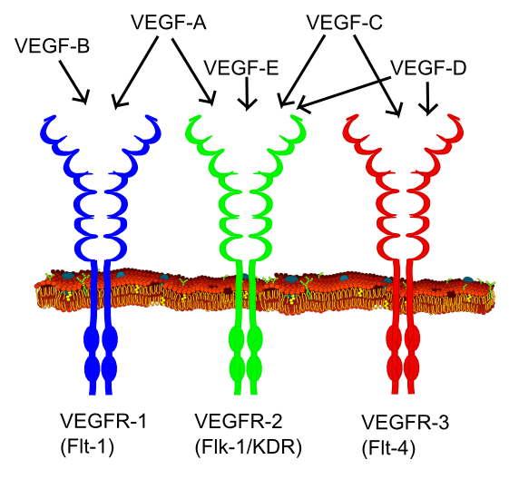
Figure 1: VEGF family members and their respective receptors (Häggström, Mikael (2014). "Medical gallery of Mikael Häggström 2014". WikiJournal of Medicine 1 (2). DOI:10.15347/wjm/2014.008. ISSN 2002-4436. Public Domain. Retrieved 24/05/2017)
How it is Measured or Detected
Methods that have been previously reviewed and approved by a recognized authority should be included in the Overview section above. All other methods, including those well established in the published literature, should be described here. Consider the following criteria when describing each method: 1. Is the assay fit for purpose? 2. Is the assay directly or indirectly (i.e. a surrogate) related to a key event relevant to the final adverse effect in question? 3. Is the assay repeatable? 4. Is the assay reproducible?
VEGF protein can be measured by enzyme-linked immunosorbent assay (Ivnitski-Steele et al. (2005), immunihistochemistry or western blot (Li et al. 2016).
VEGF gene expression, which is directly correlated with protein levels, can be measured by quantitative real-time polymerase chain reaction (QPCR) (Medford et al. 2009).
References
1. Cecilia Y. Cheung (1997) Vascular Endothelial Growth Factor: Possible Role in Fetal Development and Placental Function. J Soc Gynecol Invest. 4: 169-77
2. Ahluwalia, A., and Tarnawski, A. S. (2012). Critical role of hypoxia sensor--HIF-1alpha in VEGF gene activation. Implications for angiogenesis and tissue injury healing. Curr. Med. Chem. 19(1), 90-97.
3. Holmes, K., Roberts, O. L., Thomas, A. M., and Cross, M. J. (2007). Vascular endothelial growth factor receptor-2: structure, function, intracellular signalling and therapeutic inhibition. Cell Signal. 19(10), 2003-2012.
4. Ivnitski-Steele, I. D., Friggens, M., Chavez, M., and Walker, M. K. (2005). 2,3,7,8-tetrachlorodibenzo-p-dioxin (TCDD) inhibition of coronary vasculogenesis is mediated, in part, by reduced responsiveness to endogenous angiogenic stimuli, including vascular endothelial growth factor A (VEGF-A). Birth Defects Res. A Clin Mol. Teratol. 73(6), 440-446.
5. Zhu, D., Fang Y., Gao, K., Shen, J., Zhong, T.P., and Li, F. (2017) Vegfa Impacts Early Myocardium Development in Zebrafish. Int J Mol Sci. 18(2): 444.
6. Eichmann, A., Marcelle, C., Breant, C., and Le Douarin, N.M. (1996). Molecular cloning of Quek 1 and 2, two quail vascular endothelial growth factor (VEGF) receptor-like molecules. Gene 174, 3–8.
7. Masabumi Shibuya (2002) Vascular Endothelial Growth Factor Receptor Family Genes: When Did the Three Genes Phylogenetically Segregate? Biol. Chem., 383: 1573 – 1579.
8. Li, X.; Liu, X.; Guo, H.; Zhao, Z.; Li, Y.S. and Chen, G. (2016) The significance of the increased expression of phosphorylated MeCP2 in the membranes from patients with proliferative diabetic retinopathy. Scientific Reports, volume 6, Article number: 32850. 10.1038/srep32850
9. Medford, A. R., Douglas, S. K., Godinho, S. I., Uppington, K. M., Armstrong, L., Gillespie, K. M., van Zyl, B., Tetley, T.D., Ibrahim, N.B.N. and Millar, A. B. (2009). Vascular Endothelial Growth Factor (VEGF) isoform expression and activity in human and murine lung injury. Respiratory Research, 10(1), 27. http://doi.org/10.1186/1465-9921-10-27
Event: 110: Impairment, Endothelial network
Short Name: Impairment, Endothelial network
Key Event Component
| Process | Object | Action |
|---|---|---|
| endothelium development | abnormal |
AOPs Including This Key Event
| AOP ID and Name | Event Type |
|---|---|
| Aop:43 - Disruption of VEGFR Signaling Leading to Developmental Defects | KeyEvent |
| Aop:150 - Aryl hydrocarbon receptor activation leading to early life stage mortality, via reduced VEGF | KeyEvent |
Biological Context
| Level of Biological Organization |
|---|
| Cellular |
Organ term
| Organ term |
|---|
| embryo |
Domain of Applicability
| Term | Scientific Term | Evidence | Links |
|---|---|---|---|
| Vertebrates | Vertebrates | High | NCBI |
| Life Stage | Evidence |
|---|---|
| Embryo | High |
| Development | High |
| Sex | Evidence |
|---|---|
| Unspecific | High |
Blood vessel development utilizes highly conserved molecular pathways that are active across vertebrate species. Anatomically, however, the molecular toolbox for vasculogenesis/angiogenesis has varied themes for arterial, venous, and lymphatic channels, as well as across different organs and species [Tal et al. 2016]. ToxCast high-throughput screening (HTS) data for 25 assays mapping to targets in embryonic vascular disruption signature [Knudsen and Kleinstreuer, 2011] were used to rank-order 1060 chemicals for their potential to disrupt vascular development. The predictivity of this signature is being evaluated in various angiogenesis assays, including tubulogenesis in endothelial cells from zebrafish, chick, mouse and human species [Tal et al. 2016; Vargesson et al. 2003; Knudsen et al. 2016; McCollum et al. 2016; Nguyen et al. 2016]. As an example, a zebrafish embryo vascular model in conjunction with a mouse endothelial cell model identified 28 potential vascular disruptor compounds (pVDCs) from ToxCast. These exposures invoked a plethora of vascular perturbations in the zebrafish embryo, including malformed intersegmental vessels, uncondensed caudal vein plexus, hemorrhages and cardiac edema; 22 pVDCs inhibited endothelial tubulogenesis in an yolk-sac-derived endothelial cell line [McCollum et al. 2016]. The VEGF pathway was implicated across mouse-zebrafish species. Because gene sequence similarity of the ToxCast pVDC signature is comprised of proteins that primarily map to human in vitro and biochemical assays, the U.S. EPA SeqAPASS tool was used to assess the degree of conservation of signature targets between zebrafish and human, as well as other commonly used model organisms in human health and environmental toxicology research [Tal et al. 2016]. This approach revealed that key nodes in the ontogenetic regulation of angiogenesis have evolved across diverse species.
Key Event Description
In embryological terms the angiogenic cycle entails a stepwise progression of de novo blood vessel morphogenesis (vasculogenesis), maturation and expansion (angiogenesis), and remodeling [Hanahan, 1997; Chung and Ferrara 2011; Coultas et al. 2005]. These events commence as angioblasts migrate, proliferate, and assemble into a tubular network. With maturation, the endothelial tubules co-opt local stromal cells as pericytes and smooth muscle. Local signals acting on receptor tyrosine kinases (RTKs), G-protein coupled receptors (GPCRs), and glycosyl phosphatidyl-inositol (GPI)-anchored receptors, and later vascular flow-mediated signals. The process of endothelial assembly into a tubular network may be disrupted by environmental agents [Sarkanen et al. 2010; Bondesson et al. 2016; Knudsen et al. 2016; Nguyen et al. 2016; Tal et al. 2016].
How it is Measured or Detected
Endothelial tubule formation (tubulogenesis) can be monitored both qualitatively and quantitatively in vitro using different human cell-based angiogenesis assays that score endothelial cell migration and the degree of tubular network formation, including cell counts, tubule counts, tubule length, tubule area, tubule intensity, and node counts [Muller et al. 2002; Masckauchan et al. 2005; Sarkanen et al. 2010; Knudsen et al. 2016; Nguyen et al. 2016]. Standard practice for reproducible in vitro tubule formation uses endothelial cells co-cultured with stromal cells [Bishop et al. 1999]. Cell types commonly employed are human umbilical endothelial cells (HUVECs) or more recently induced pluripotent stem cells (iPSCs) derived to endothelial cells through various differentiation and purification protocols. The assay is run in agonist or antagonist modes to detect chemical enhancement or suppression of tubulogenesis. Synthetic hydrogels are shown to promote robust in vitro network formation by HUVEC or iPSC-ECs as well as their utilization to detect putative vascular disruptive compounds [Nguyen et al. 2016]. Endothelial networks formed on synthetic hydrogels showed superior sensitivity and reproducibility when compared to endothelial networks formed on Matrigel.
References
Bishop ET, Bell GT, Bloor S, Broom IJ, Hendry NFK and Wheatley DN. An in vitro model of angiogenesis: Basic features. Angiogenesis. 1999 3(4): 335-344.
Chung AS, Ferrara N. Developmental and pathological angiogenesis. Annual review of cell and developmental biology. 2011;27:563-84. PubMed PMID: 21756109.
Coultas L, Chawengsaksophak K, Rossant J. Endothelial cells and VEGF in vascular development. Nature. 2005 Dec 15;438(7070):937-45. PubMed PMID: 16355211.
Hanahan D. Signaling vascular morphogenesis and maintenance. Science. 1997 Jul 4;277(5322):48-50. PubMed PMID: 9229772.
Knudsen TB, Kleinstreuer NC. Disruption of embryonic vascular development in predictive toxicology. Birth defects research Part C, Embryo today : reviews. 2011;93(4):312-23.
Knudsen TB, Tomela T, Sarkanen R, Spencer RS, Baker NC, Franzosa J, Tal T, Kleinstreuer NC, Cai B, Berg EL and Heinonen. ToxCast Model-Based Prediction of Human Vascular Disruption: Statistical Correlation to Endothelial Tubulogenesis Assays and Computer Simulation with Agent-Based Models. 2016 (in preparation).
Masckauchan TN, Shawber CJ, Funahashi Y, Li CM, Kitajewski J. Wnt/beta-catenin signaling induces proliferation, survival and interleukin-8 in human endothelial cells. Angiogenesis. 2005;8(1):43-51. PubMed PMID: 16132617.
McCollum CW, Vancells JC, Hans C, Vazquez-Chantada M, Kleinstreuer N, Tal T, Knudsen T, Shah SS, Merchant FA, Finnell RH, Gustafsson JA, Cabrera R and Bondesson M. Identification of vascular disruptor compounds by a tiered analysis in zebrafish embryos and mouse embryonic endothelial cells. 2016 (in preparation).
Muller T, Bain G, Wang X, Papkoff J. Regulation of epithelial cell migration and tumor formation by beta-catenin signaling. Experimental cell research. 2002 Oct 15;280(1):119-33. PubMed PMID: 12372345.
Nguyen EH, Daly WT, Le NNT, Belair DG, Schwartz MP, Lebakken CS, Ananiev GE, Saghiri A, Knudsen TB, Sheibani N and Murphy WL. Identification of a synthetic alternative to matrigel for the screening of anti-angiogenic compounds. 2016 (in preparation).
Tal T, Kilty C, Smith A, LaLone C, Kennedy B, Tennant A, McCollum C, Bondesson M, Knudsen T, Padilla S and Kleinstreuer N. Screening for chemical vascular disruptors in zebrafish to evaluate a predictive model for developmental vascular toxicity. Reprod Toxicol (submitted).
Vargesson N. Vascularization of the developing chick limb bud: role of the TGFβ signalling pathway. J Anat. 2016 Jan, 202(1): 93-103. PMCID: PMC1571066.
Event: 317: Altered, Cardiovascular development/function
Short Name: Altered, Cardiovascular development/function
Key Event Component
| Process | Object | Action |
|---|---|---|
| abnormal cardiovascular system physiology | morphological change | |
| cardiovascular system development | cardiovascular system | abnormal |
AOPs Including This Key Event
Stressors
| Name |
|---|
| 2,3,7,8-tetrachlorodibenzo-p-dioxin (TCDD) |
Biological Context
| Level of Biological Organization |
|---|
| Organ |
Organ term
| Organ term |
|---|
| heart |
Domain of Applicability
| Life Stage | Evidence |
|---|---|
| Embryo | High |
| Sex | Evidence |
|---|---|
| Unspecific | High |
- Some form of cardiovascular system is present in members of the clade Bilateria (Bishopric 2005). This clade includes most animal phyla, except for sponges (Porifera), jellyfishes and corals (Cnidaria), placozoans (Placozoa), and comb jellies (Ctenophora).
- Differences in cardiovascular systems are present among taxa. Vertebrates have closed circulatory systems, while some invertebrate taxa have open circulatory systems (Kardong 2006).
Key Event Description
This key event applies to the disruption of cardiogenesis early enough in embryogenesis to result in gross morphological alterations leading to reduced cardiac function.
How it is Measured or Detected
Altered cardiovascular development/function can be measured in numerous ways:
1) As blood flow in the mesencephalic vein by use of time-lapse recording using a digital video camera (Teraoka et al 2008; 2014). Blood flow is measured as the number of red blood cells passing the mesencephalic vein per second (Teraoka et al 2008; 2014). This method is described in detail by Teraoka et al (2002). However, some studies have assessed blood flow through visualized scoring techniques by use of a microscope as (1) same rate as control, (2) slower rate than control, or (3) no flow (Henry et al 1997).
2) As heart area, pericardial edema area, or yolk sac edema area quantified with area analysis by use of a microscope linked digital camera and conventional image software (Dong et al 2010; Teraoka et al 2008; 2014; Yamauchi et al 2006). Images at the same magnification are used to obtain the area measured as number of pixels (Teraoka et al 2008; 2014). This method can use either live individuals or histologic samples. This method is described in detail by Teraoka et al (2003).
3) As basic physical measurements such as heart weight, heart aspect ratio (horizontal length versus vertical length), heart weight to body weight ratio (Fujisawa et al 2014).
4) As incidence of malformation measured as percent occurrence among individuals (Buckler et al 2015; Dong et al 2010; Park et al 2014; Yamauchi et al 2006).This method is described in detail by Dong et al (2010).
5) As heartbeat rate measured by direct observation by use of a microscope (Park et al 2014). This method is described in detail by Park et al (2014).
References
1. Carro, T., Dean, K., and Ottinger, M. A. (2013a). Effects of an environmentally relevant polychlorinated biphenyl (PCB) mixture on embryonic survival and cardiac development in the domestic chicken. Environ. Toxicol. Chem. 23(6), 1325-1331.
2. Carro, T., Taneyhill, L. A., and Ottinger, M. A. (2013b). The effects of an environmentally relevant 58 congener polychlorinated biphenyl (PCB) mixture on cardiac development in the chick embryo. Environ. Toxicol. Chem.
3. DeWitt, J. C., Millsap, D. S., Yeager, R. L., Heise, S. S., Sparks, D. W., and Henshel, D. S. (2006). External heart deformities in passerine birds exposed to environmental mixtures of polychlorinated biphenyls during development. Environ. Toxicol. Chem. 25(2), 541-551.
4. Heid, S. E., Walker, M. K., and Swanson, H. I. (2001). Correlation of cardiotoxicity mediated by halogenated aromatic hydrocarbons to aryl hydrocarbon receptor activation. Toxicol. Sci 61(1), 187-196.
5. Walker, M. K., and Catron, T. F. (2000). Characterization of cardiotoxicity induced by 2,3,7,8-tetrachlorodibenzo-p-dioxin and related chemicals during early chick embryo development. Toxicol. Appl. Pharmacol. 167(3), 210-221.
6. Walker, M. K., Pollenz, R. S., and Smith, S. M. (1997). Expression of the aryl hydrocarbon receptor (AhR) and AhR nuclear translocator during chick cardiogenesis is consistent with 2,3,7,8-tetrachlorodibenzo-p-dioxin-induced heart defects. Toxicol. Appl. Pharmacol. 143(2), 407-419.
7. Kopf, P. G., and Walker, M. K. (2009). Overview of developmental heart defects by dioxins, PCBs, and pesticides. J. Environ. Sci. Health C. Environ. Carcinog. Ecotoxicol. Rev. 27(4), 276-285.
Bishopric, N.H. (2005). Evolution of the heart from bacteria to man. Ann. N. Y. Acad. Sci. 1047, 13-29.
Buckler J.; Candrl, J.S.; McKee, M.J.; Papoulias, D.M.; Tillitt, D.E.; Galat, D.L. Sensitivity of shovelnose sturgeon (Scaphirhynchus platorynchus) and pallid sturgeon (S. albus) early life stages to PCB-126 and 2,3,7,8-TCDD exposure. Enviro. Toxicol. Chem. 2015, 34(6), 1417-1424.
Carney, S.A.; Prasch, A.L.; Heideman, W.; Peterson, R.E. 2006. Understanding dioxin developmental toxicity using the zebrafish model. Birth Defects Research. A. 76, 7-18.
Cohen-Barnhouse, A.M.; Zwiernik, M.J.; Link, J.E.; Fitzgerald, S.D.; Kennedy, S.W.; Herve, J.C.; Giesy, J.P.; Wiseman, S.; Yang, Y.; Jones, P.D.; Yi, W.; Collins, B.; Newsted, J.L.; Kay, D.; Bursian, S.J. 2011. Sensitivity of Japanese quail (Coturnix japonica), common pheasant (Phasianus colchicus), and white leghorn chicken (Gallus gallus domesticus) embryos to in ovo exposure to TCDD, PeCDF, and TCDF. Toxicol. Sci. 119, 93-102.
Elonen, G.E.; Spehar, R.L.; Holcombe, G.W.; Johnson, R.D.; Fernandez, J.D.; Erickson, R.J.; Tietge, J.E.; Cook, P.M. Comparative toxicity of 2,3,7,8-tetrachlorodibenzo-p-dioxin to seven freshwater fish species during early life-stage development. Enviro. Toxico. Chem. 1998, 17, 472-483.
Goldstone, H.M.H.; Stegeman, J.J. (2008). Molecular mechanisms of 2,3,7,8-tetrachlorodibenzo-p-dioxin cardiovascular embryotoxicity. Drug. Metab. Rev. 38, 261-289.
Heid, S.E.; Walker, M.K.; Swanson, H.I. (2001). Correlation of cardiotoxicity mediated by halogenated aromatic hydrocarbons to aryl hydrocarbon receptor activation. Toxicol. Sci. 61 (1), 187-196.
Huang, L.; Wang, C.; Zhang, Y.; Li, J.; Zhong, Y.; Zhou, Y.; Chen, Y.; Zuo, Z. (2012). Benzo[a]pyrene exposure influences the cardiac development and the expression of cardiovascular relative genes in zebrafish (Daniorerio) embryos. Chemosphere. 87 (4), 369-375.
Johnson, R.D.; Tietge, J.E.; Jensen, K.M.; Fernandez, J.D.; Linnum, A.L.; Lothenbach, D.B.; Holcombe, G.W.; Cook, P.M.; Christ, S.A.; Lattier, D.L.; Gordon, D.A. Toxicity of 2,3,7,8-tetrachlorodibenzo-p-dioxin to early life stage brooke trout (Salvelinus fontinalis) following parental dietary exposure. Enviro. Toxicol. Chem. 1998, 17 (12), 2408-2421.
Kardong, K.V. (2006). Vertebrates: comparative anatomy, function, evolution. McGraw-Hill Higher Eduction. Boston, USA.
Lemly, A.D. (2002). Symptoms and implications of selenium toxicity in fish: the Belews Lake case example. Aquat. Toxicol. 57 (1-2), 39-49.
Park, Y.J.; Lee, M.J.; Kim, H.R.; Chung, K.H.; Oh, S.M. Developmental toxicity of 2,3,7,8-tetrachlorodibenzo-p-dioxin in artificially fertilized crucian carp (Carassius auratus) embryo. Sci. Totl. Enviro. 2014, 491-492, 271-278.
Teraoka, H.; Dong, W.; Hiraga, T. (2003). Zebrafish as a novel experimental model for development toxicology. Congenit. Anom. 43, 123-132.
Teraoka, T.; Dong, W.; Ogawa, S.; Tsukiyama, S.; Okuhara, Y.; Niiyama, M.; Ueno, N.; Peterson, R.E. (2002). 2,3,7,8-tetrachlorodibenzo-p-dioxin toxicity in the zebrafish embryo: Altered regional blood flow and impaired lower jaw development. Toxicol. Sci. 65, 192-199.
Tillitt, D.E.; Buckler, J.A.; Nicks, D.K.; Candrl, J.S.; Claunch, R.A.; Gale, R.W.; Puglis, H.J.; Little, E.E.; Linbo, T.L.; Baker, M. Sensitivity of lake sturgeon (Acipenser fulvescens) early life stages to 2,3,7,8-tetrachlorodibenzo-p-dioxin and 3,3’,4,4’,5-pentachlorobiphenyl. 2015. Enviro. Toxicol. Chem. DOI: 10.1002/etc.3614.
Toomey, B.H.; Bello, S.; Hahn, M.E.; Cantrell, S.; Wright, P.; Tillitt, D.; Di Giulio, R.T. TCDD induces apoptotic cell death and cytochrome P4501A expression in developing Fundulus heteroclitus embryos. Aquat. Toxicol. 2001, 53, 127-138.
Walker, M.K.; Spitsbergen, J.M.; Olson, J.R.; Peterson, R.E. 2,3,7,8-tetrachlorodibenzo-para-dioxin (TCDD) toxicity during early life stage development of lake trout (Salvelinus namaycush). Canad. J. Fisheries Aqua. Sci. 1991, 48, 875-883.
Yamauchi, M.; Kim, E.Y.; Iwata, H.; Shima, Y.; Tanabe, S. Toxic effects of 2,3,7,8-tetrachlorodibenzo-p-dioxin (TCDD) in developing red seabream (Pagrus major) embryos: an association of morphological deformities with AHR1, AHR2 and CYP1A expressions. Aquat. Toxicol. 2006, 16, 166-179.
Zabel, E.W; Cook, P.M.; Peterson, R.E. Toxic equivalency factors of polychlorinated dibenzo-p-dioxin, dibenzofuran and biphenyl congeners based on early-life stage mortality in rainbow trout (Oncorhynchus mykiss). Aquat Toxicol. 1995. 31, 315-328.
List of Adverse Outcomes in this AOP
Event: 947: Increase, Early Life Stage Mortality
Short Name: Increase, Early Life Stage Mortality
Key Event Component
| Process | Object | Action |
|---|---|---|
| embryonic lethality | increased | |
| mortality | increased |
AOPs Including This Key Event
| AOP ID and Name | Event Type |
|---|---|
| Aop:150 - Aryl hydrocarbon receptor activation leading to early life stage mortality, via reduced VEGF | AdverseOutcome |
| Aop:21 - aryl hydrocarbon receptor activation leading to early life stage mortality, via increased COX-2 | KeyEvent |
Stressors
| Name |
|---|
| 2,3,7,8-tetrachlorodibenzo-p-dioxin (TCDD) |
Biological Context
| Level of Biological Organization |
|---|
| Individual |
Evidence for Perturbation by Stressor
2,3,7,8-tetrachlorodibenzo-p-dioxin (TCDD)
Exposure of embryos to 2,3,7,8-TCDD causes early life stage mortality in all studied species of fishes (Doering et al 2013).
Domain of Applicability
| Term | Scientific Term | Evidence | Links |
|---|---|---|---|
| Vertebrates | Vertebrates | High | NCBI |
| Life Stage | Evidence |
|---|---|
| Embryo | High |
| Foetal | High |
| Development | High |
| Sex | Evidence |
|---|---|
| Unspecific | High |
All members of the subphylum vertebrata are susceptible to early life stage death (Weinstein 1999).
Key Event Description
Increased early life stage mortality refers to an increase in the number of individuals dying in an experimental replicate group or in a population over a specific period of time.
In Birds:
Early life stage mortality occurs at any stage in development prior to birth/hatch and is considered embryolethal.
In Fishes:
Early Life Stage Mortality refers to death prior to yolk sac adsorption and swim-up.
How it is Measured or Detected
In birds it may be identified as failure to hatch or lack of movement within the egg when candled; heartbeat monitors are available for identifying viable avian and reptillian eggs (ex. Avitronic's Buddy monitor). In mammals, stillborn or mummified offspring, or an increased rate of resorptions early in pregnancy are all considered embryolethal, and can be detected using ultra-high frequency ultrasound (30-70 MHz; a.k.a. ultrasound biomicroscopy) (Flores et al. 2014). In fishes, mortality is typically measured by observation. Lack of any heart beat, gill movement, and body movement are typical signs of death used in the evaluation of mortality.
Regulatory Significance of the AO
Poor early life stage survival is an endpoint of major relevance to environmental regulators, as it is likely to lead to population decline. Early-life stage, acute and chronic test guidelines have been established by the Organisation for Economic Co-operation and Development (OECD), U.S. Environmental Protection Agency (EPA) and Environment and Climate Change Canada (ECCC), and are currently used in risk assessments to set limits for safe exposures. Aquatic test guidlines are most prevalent and include OECD210, OECD229, EPA850.1400 and ECCC EPS 1/RM/28 for fish and OECD241 for frogs.
References
1. Flores, L.E., Hildebrandt, T.B., Kuhl, A.A., and Drews, B. (2014) Early detection and staging of spontaneous embryo resorption by ultrasound biomicroscopy in murine pregnancy. Reproductive Biology and Endocrinology 12(38). DOI: 10.1186/1477-7827-12-38
2. Weinstein, B. M. (1999). What guides early embryonic blood vessel formation? Dev. Dyn. 215(1), 2-11.
Doering, J.A.; Giesy, J.P.; Wiseman S.; Hecker, M. (2013). Predicting the sensivity of fishes to dioxin-like compounds: possible role of the aryl hydrocarbon receptor (AhR) ligand binding domain. Environmental Science and Pollution Research. 20 (3), 1219-1224.
Appendix 2
List of Key Event Relationships in the AOP
List of Adjacent Key Event Relationships
Relationship: 972: Activation, AhR leads to dimerization, AHR/ARNT
AOPs Referencing Relationship
| AOP Name | Adjacency | Weight of Evidence | Quantitative Understanding |
|---|---|---|---|
| Aryl hydrocarbon receptor activation leading to early life stage mortality, via reduced VEGF | adjacent | High | Moderate |
| aryl hydrocarbon receptor activation leading to early life stage mortality, via increased COX-2 | adjacent | High | Moderate |
Evidence Supporting Applicability of this Relationship
| Term | Scientific Term | Evidence | Links |
|---|---|---|---|
| Mus musculus | Mus musculus | High | NCBI |
| Danio rerio | Danio rerio | High | NCBI |
| rainbow trout | Oncorhynchus mykiss | High | NCBI |
| Pagrus major | Pagrus major | High | NCBI |
| Acipenser fulvescens | Acipenser fulvescens | High | NCBI |
| Salmo salar | Salmo salar | High | NCBI |
| Acipenser transmontanus | Acipenser transmontanus | High | NCBI |
| Xenopus laevis | Xenopus laevis | High | NCBI |
| Ambystoma mexicanum | Ambystoma mexicanum | High | NCBI |
| Microgadus tomcod | Microgadus tomcod | High | NCBI |
| human | Homo sapiens | High | NCBI |
| Gallus gallus | Gallus gallus | High | NCBI |
| Phasianus colchicus | Phasianus colchicus | High | NCBI |
| Coturnix japonica | Coturnix japonica | High | NCBI |
| Life Stage | Evidence |
|---|---|
| All life stages | High |
| Sex | Evidence |
|---|---|
| Unspecific | High |
- The aryl hydrocarbon receptor (AhR) and aryl hydrocarbon receptor nuclear translocator (ARNT) are highly conserved and ancient proteins with homologs having been identified in most major animal groups, apart from the most ancient lineages, such as sponges (Porifera) (Hahn et al 2002).
- In vitro dimerization of AhRs and ARNTs have been demonstrated in mammals, birds, reptiles, amphibians, teleost and non-teleost fishes, and some invertebrates (Butler et al 2001; Emmons et al 1999; Hahn et al 2002; Powell-Coffman et al 1998).
Key Event Relationship Description
In its unliganded form, the AHR is part of a cytosolic complex containing heat shock protein 90 (HSP90), the HSP90 co-chaperone p23 and AHR-interacting protein (AIP) (Fujii-Kuriyama et al. 2010). Upon ligand binding, the aryl hydrocarbon receptor (AHR) migrates to the nucleus where it dissociates from the cytosolic complex and forms a heterodimer with AHR nuclear translocator (ARNT) (Mimura and Fujii-Kuriyama 2003).
AhRs can heterodimerize with ARNT1 and ARNT2 isoforms in order to activate reporter constructs in transfected cells and recognize response elements in gel shift assays in all investigated vertebrates, including birds, fishes, and reptiles (Abnet et al 1999; Andreasen et al 2002a; 2002b; Bak et al 2013; Doering et al 2014; Doering et al 2015; Farmahin et al 2012; 2013; Hansson & Hahn 2008; Karchner et al 1999; 2006; Lavine et al 2005; Shoots et al 2015; Tanguay et al 1999; 2000; Wirgin et al 2011).
Evidence Supporting this KER
Biological PlausibilityThe mechanism of AHR-mediated transcriptional regulation is well understood (Fujii-Kuriyama and Kawajiri 2010).
Numerous PAS proteins are known to interact with each other in response to environmental and developmental cues through dimerization at their PAS domains (Pohjanvirta 2012).
Empirical EvidenceARNT is a necessary dimerization partner for the transcriptional activation of AHR regulated genes (Hoffman et al. 1991; Poland et al. 1976). The AHR/ARNT complex was confirmed following in vitro exposure to halogenated aromatic hydrocarbons using an electrophoretic mobility shift assay; a dose-dependent supershift in DNA-binding was observed using specific antibodies in chicken and human cell lines (Heid et al. 2001).
- Unliganded AhR exists as a cytosolic 9S form, while in the presence of a ligand the AhR exists as a nuclear 6S form. ARNT exists as a nuclear 6S form (Okey 2007).
- The 6S form of AhR is approximately 210 kDa. Ligated AhR is approximately 100 kDa and ARNT is approximately 110 kDa (Elferink et al 1990; Swanson et al 1993).
- Dimerization of AhRs with ARNTs has been demonstrated in all invertebrate and vertebrate species so far investigated (Butler et al 2001; Emmons et al 1999; Hahn et al 2002; Powell-Coffman et al 1998).
- Heterodimers are not formed on response elements in gel shift assays in the absence of AhR and/or ARNT (Tanguay et al 2000).
Uncertainties and Inconsistencies
- There are uncertainties in the precise physiological and toxicological roles of different AhR clades (AhR1, AhR2, AhR3) and isoforms (α, β, δ, γ).
- There are uncertainties in the precise physiological and toxicological roles of different ARNT clades (ARNT1, ARNT2, ARNT3) and isoforms (a, b, c).
- Nothing is known about differences in binding affinity of AhR for ARNT and of the AhR/ARNT heterodimer for DNA among species and taxa.
- There is uncertainty in whether anthropogenic contaminants that act as ligands of the AhR and lead to dimerization of AhR with ARNT in vertebrates also act as ligands in invertebrates.
References
1. Fujii-Kuriyama, Y., and Kawajiri, K. (2010). Molecular mechanisms of the physiological functions of the aryl hydrocarbon (dioxin) receptor, a multifunctional regulator that senses and responds to environmental stimuli. Proc. Jpn. Acad. Ser. B Phys. Biol. Sci. 86(1), 40-53.
2. Giesy, J. P., Kannan, K., Blankenship, A. L., Jones, P. D., and Newsted, J. L. (2006). Toxicology of PCBs and related compounds. In Endocrine Disruption Biological Bases for Health Effects in Wildlife and Humans (D.O.Norris and J.A.Carr, Eds.), pp. 245-331. Oxford University Press, New York.
3. Heid, S. E., Walker, M. K., and Swanson, H. I. (2001). Correlation of cardiotoxicity mediated by halogenated aromatic hydrocarbons to aryl hydrocarbon receptor activation. Toxicol. Sci 61(1), 187-196.
4. Hoffman, E. C., Reyes, H., Chu, F. F., Sander, F., Conley, L. H., Brooks, B. A., and Hankinson, O. (1991). Cloning of a factor required for activity of the Ah (dioxin) receptor. Science 252(5008), 954-958.
5. Mimura, J., and Fujii-Kuriyama, Y. (2003). Functional role of AhR in the expression of toxic effects by TCDD. Biochimica et Biophysica Acta - General Subjects 1619(3), 263-268.
6. Poland, A., Glover, E., and Kende, A. S. (1976). Stereospecific, high affinity binding of 2,3,7,8-tetrachlorodibenzo-p-dioxin by hepatic cytosol. Evidence that the binding species is receptor for induction of aryl hydrocarbon hydroxylase. J. Biol. Chem. 251(16), 4936-4946.
7. Safe, S. (1994). Polychlorinated biphenyls (PCBs): Environmental impact, biochemical and toxic responses, and implications for risk assessment. Critical Reviews in Toxicology 24(2), 87-149.
Andreasen, E.A.; Tanguay, R.L.; Peterson, R.E.; Heideman, W. 2002. Identification of a critical amino acid in the aryl hydrocarbon receptor. J. Biol. Chem. 277 (15), 13210-13218.
Bak, S.M.; Lida, M.; Hirano, M.; Iwata, H.; Kim, E.Y. 2013. Potencies of red seabream AHR1- and AHR2-mediated transactivation by dioxins: implications of both AHRs in dioxin toxicity. Environ. Sci. Technol. 47 (6), 2877-2885.
Butler, R.A.; Kelley, M.L.; Powell, W.H.; Hahn, M.E.; Van Beneden, R.J. (2001). An aryl hydrocarbon receptor (AHR) homologue from the soft-shelled clam, Mya arenaria: evidence that invertebrate AHR homologues lack 2,3,7,8-tetrachlorodibenzo-p-dioxin and beta-naphthoflavone binding. Gene. 278, 223-234.
Doering, J.A.; Farmahin, R.; Wiseman, S.; Beitel, S.C.; Kennedy, S.W.; Giesy, J.P.; Hecker, M. 2015. Differences in activation of aryl hydrocarbon receptors of white sturgeon relative to lake sturgeon are predicted by identities of key amino acids in the ligand binding domain. Enviro. Sci. Technol. 49, 4681-4689.
Doering, J.A.; Farmahin, R.; Wiseman, S.; Kennedy, S.; Giesy J.P.; Hecker, M. 2014. Functionality of aryl hydrocarbon receptors (AhR1 and AhR2) of white sturgeon (Acipenser transmontanus) and implications for the risk assessment of dioxin-like compounds. Enviro. Sci. Technol. 48, 8219-8226.
Elfrink, C.; Gasiewicz, T.; Whitlock, J. (1990). Protein-DNA interactions at a dioxin-responsive enhancer. Evidence that the transformed Ah receptor is heteromeric. J. Biol. Chem. 265, 20708-20712.
Emmons, R.B.; Duncan, D.; Estes, P.A.; Kiefel, P.; Mosher, J.T.; Sonnenfeld, M.; Ward, M.P.; Duncan, I.; Crews, S.T. (1999). The spineless-aristapedia and tango bHLH-PAS proteins interact and control antennal and tarsal development in Drosophilia. Dev. 126, 3937-3945.
Farmahin, R.; Manning, G.E.; Crump, D.; Wu, D.; Mundy, L.J.; Jones, S.P.; Hahn, M.E.; Karchner, S.I.; Giesy, J.P.; Bursian, S.J.; Zwiernik, M.J.; Fredricks, T.B.; Kennedy, S.W. 2013. Amino acid sequence of the ligand-binding domain of the aryl hydrocarbon receptor 1 predicts sensitivity of wild birds to effects of dioxin-like compounds. Toxicol. Sci. 131 (1), 139-152.
Farmahin, R.; Wu, D.; Crump, D.; Herve, J.C.; Jones, S.P.; Hahn, M.E.; Karchner, S.I.; Giesy, J.P.; Bursian, S.J.; Zwiernik, M.J.; Kennedy, S.W. 2012. Sequence and in vitro function of chicken, ring-necked pheasant, and Japanese quail AHR1 predict in vivo sensitivity to dioxins. Enviro. Sci. Toxicol. 46 (5), 2967-2975.
Farmahin, R.; Crump, D.; O’Brien, J.M.; Jones, S.P.; Kennedy, S.W. (2016). Time-dependent transcriptomic and biochemical responses of 6-formylindolo[3,2-b]carbazole (FICZ) and 2,3,7,8-tetrachlorodibenzo-p-dioxin (TCDD) are explained by AHR activation time. Biochem. Pharmacol. 115 (1), 134-143.
Hahn, M.E. 2002. Aryl hydrocarbon receptors: diversity and evolution. Chemico-Biol. Interact. 141, 131-160.
Hansson, M.C.; Hahn, M.E. 2008. Functional properties of the four Atlantic salmon (Salmo salar) aryl hydrocarbon receptor type 2 (AHR2) isoforms. Aquat. Toxicol. 86, 121-130.
Karchner, S.I.; Franks, D.G.; Kennedy, S.W.; Hahn, M.E. 2006. The molecular basis for differential dioxin sensitivity in birds: Role of the aryl hydrocarbon receptor. Proc. Natl. Acad. Sci. USA. 103, 6252-6257.
Karchner, S.I.; Powell, W.H.; Hahn, M.E. 1999. Identification and functional characterization of two highly divergent aryl hydrocarbon receptors (AHR1 and AHR2) in the Teleost Fundulus heteroclitus. Evidence for a novel subfamily of ligand-binding basic helix loop helix-Per-ARNT-Sim (bHLH-PAS) factors. J. Biol. Chem. 274, 33814-33824.
Lavine, J.A.; Rowatt, A.J.; Klimova, T.; Whitington, A.J.; Dengler, E.; Beck, C.; Powell, W.H. 2005. Aryl hydrocarbon receptors in the frog Xenopus laevis: two AhR1 paralogs exhibit low affinity for 2,3,7,8-tetrachlorodibenzo-p-dioxin (TCDD). Toxicol. Sci. 88 (1), 60-72.
Manning G.E.; Farmahin, R.; Crump, D.; Jones, S.P.; Klein, J.; Konstantinov, A.; Potter, D.; Kennedy, S.W. 2012. A luciferase reporter gene assay and aryl hydrocarbon receptor 1 genotype predict the LD50 of polychlorinated biphenyls in avian species. Toxicol. Appl. Pharm. 263, 390-401.
Ohi, H.; Fujita, Y.; Miyao, M.; Saguchi, K.; Murayama, N.; Higuchi, S. 2003. Molecular cloning and expression analysis of the aryl hydrocarbon receptor of Xenopus laevis. Biochem. Biophysic. Res. Comm. 307 (3), 595-599.
Powell-Coffman, J.A.; Bradfield, C.A.; Wood, W.B. (1998). Caenorhabditis elgans orthologs of the aryl hydrocarbon receptor and its dimerization partner the aryl hydrocarbon receptor nuclear translocator. Proceedings of the National Academy of Sciences of the United States of America. 95, 2844-2449.
Shoots, J.; Fraccalvieri, D.; Franks, D.G.; Denison, M.S.; Hahn, M.E.; Bonati, L.; Powell, W.H. 2015. An aryl hydrocarbon receptor from the salamander Ambystoma mexicanum exhibits low sensitivity to 2,3,7,8-tetrachlorodibenzo-p-dioxin. Enviro. Sci. Technol. 49, 6993-7001.
Swanson, H.; Tullis, K.; Denison, M. (1993). Binding of transformed Ah receptor complex to a dioxin responsive transcriptional enhancer: evidence for two distinct heterodimeric DNA-binding forms. Biochem. 32, 12841-12849.
Tanguay, R.L.; Abnet, C.C.; Heideman, W. Peterson, R.E. (1999). Cloning and characterization of the zebrafish (Danio rerio) aryl hydrocarbon receptor1. Biochimica et Biophysica Act 1444, 35-48.
Tanguay, R.L.; Andreasen, E.; Heideman, W.; Peterson, R.E. (2000). Identification and expression of alternatively spliced aryl hydrocarbon nuclear translocator 2 (ARNT2) cDNAs from zebrafish with distinct functions. BBA. 1494 (1-2), 117-128.
Okey, A. (2007). An aryl hydrocarbon receptor odyssey to the shores of toxicology: the deichmann Lecture, International Congress of Toxicology-XI. Toxicol. Sci. 98, 5-38.
Wirgin, I.; Roy, N.K.; Loftus, M.; Chambers, R.C.; Franks, D.G.; Hahn, M.E. 2011. Mechanistic basis of resistance to PCBs in Atlantic tomcod from the Hudson River. Science. 331, 1322-1324.
Relationship: 973: dimerization, AHR/ARNT leads to reduced dimerization, ARNT/HIF1-alpha
AOPs Referencing Relationship
| AOP Name | Adjacency | Weight of Evidence | Quantitative Understanding |
|---|---|---|---|
| Aryl hydrocarbon receptor activation leading to early life stage mortality, via reduced VEGF | adjacent | Moderate | Low |
Evidence Supporting Applicability of this Relationship
| Life Stage | Evidence |
|---|---|
| Embryo | High |
| During development and at adulthood | High |
| Sex | Evidence |
|---|---|
| Unspecific | High |
The cross-talk between AHR and HIF1α has been demonstrated in chicken embryos (Ivnitski-Steele et al. 2004) mice (Ichihara et al. 2007) Atlantic killifish and zebrafish (McElroy et al. 2012), Mummichog (Kraemer et al. 2004) and a number of human cell lines (Chan et al. 1999; Seifert et al. 2008; Vorrink et al. 2014a, Vorrink et al. 2014b).
Key Event Relationship Description
The aryl hydrocarbon receptor nuclear translocator (ARNT) is common dimerization partner for both the aryl hydrocarbon receptor (AHR) and hypoxia inducible factor alpha (HIF-1α). There is considerable cross talk between the two nuclear receptors, leading to the hypothesis that AHR activation leads to sustained AHR/ARNT dimerization and reduced HIF-1α/ARNT dimerization, assuming ARNT is not available in excess (Chan et al. 1999; Vorrink et al 2014b).
Evidence Supporting this KER
Biological PlausibilityThe ARNT serves as a dimerization partner for multiple transcription factors including the xenobiotic sensing AHR and HIF1α; therefore, it is plausible that sequestration of ARNT by one receptor would reduce the responsiveness of the other, assuming that ARNT is available in limited quantity (Vorrink et al. 2014b). Gel-shift and coimmunoprecipitation experiments have shown that the AHR and HIF1α compete for ARNT in vitro, with approximately equal dimerization efficiencies (Schmidt and Bradfield 1996).
Empirical EvidenceInclude consideration of temporal concordance here
- Activation of either AHR (by 2,3,7,8-tetrachlorodibenzo-p-dioxin) or HIF1 (by hypoxia) inhibits the activity of the other, in Hep3B cells (Chan et al. 1999)
- TCDD and hypoxia together reduced the stabilization of HIF1α and HRE-mediated promoter activity when compared to hypoxia alone, in MCF-7 and HepG2 cells (Seifert et al. 2008).
- Hypoxia increased EF5 binding (hypoxic tissue marker) in chicken embryos, whereas it was decreased by TCDD relative to controls (D10 of incubation) (Ivnitski-Steele et al. 2004)
- TCDD reduces the expression of cardiac HIF1α mRNA in chicken embryos (Ivnitski-Steele et al. 2004)
- ARNT overexpression rescued human HepG2 and HaCaT cells from inhibitory effect of hypoxia on XRE-luciferase reporter activity. This indicates that the mechanism of interference between the AHR and HIF1α pathways at least partially dependent on ARNT availability (Vorrink et al. 2014)
- Ischemia-induced upregulation of the expression of HIF1α and ARNT and DNA binding activity of the HIF1α-ARNT complex were enhanced in AHR-null mice (Ichihara et al. 2007).
- Vorrink et al (2014b) provides a thorough summary of supporting evidence as well as contradictions and uncertainties in the literature.
Although crosstalk between AHR and HIF1α clearly exists, the nature of the relationship is still not clearly defined (Vorrink et al 2014). It has been suggested that HIF1α and AHR do not competitively regulate each other for hetero-dimerization with ARNT, as ARNT is constitutively and abundantly expressed in cells and does not deplete due to hypoxia or AHR activation (Chan et al. 1999; Pollenz et al. 1999). Nie et al. (2001) hypothesized that the degree of interaction among ARNT-dependent pathways depends on the abundance of ARNT in the cells. They observed crosstalk in Hepa 1 cells but not H4IIE cells, and attributed this to the ratio of AhR to ARNT of 0.3 (i.e. excess ARNT), compared to a ratio of 10 in Hepa 1 cells (Holmes and Pollenz, 1997)
Some studies have shown that the effect of hypoxia on AHR mediated pathways is stronger than effects of a AHR-mediated xenobiotic response on the HIF1α pathway (Gassmann et al. 1997; Gradin et al. 1996; Nie et al. 2001; Prasch et al. 2004); this has been attributed to the stronger binding affinity of HIF1α to ARNT relative to AHR (Gradin et al. 1996).
References
1. Chan, W. K., Yao, G., Gu, Y. Z., and Bradfield, C. A. (1999). Cross-talk between the aryl hydrocarbon receptor and hypoxia inducible factor signaling pathways. Demonstration of competition and compensation. J Biol. Chem. 274(17), 12115-12123.
2. Ichihara, S., Yamada, Y., Ichihara, G., Nakajima, T., Li, P., Kondo, T., Gonzalez, F. J., and Murohara, T. (2007). A role for the aryl hydrocarbon receptor in regulation of ischemia-induced angiogenesis. Arterioscler. Thromb. Vasc. Biol. 27(6), 1297-1304.
3. Ivnitski-Steele, I. D., Sanchez, A., and Walker, M. K. (2004). 2,3,7,8-tetrachlorodibenzo-p-dioxin reduces myocardial hypoxia and vascular endothelial growth factor expression during chick embryo development. Birth Defects Res. A Clin. Mol. Teratol. 70(2), 51-58.
4. Nie, M., Blankenship, A. L., and Giesy, J. P. (2001). Interactions between aryl hydrocarbon receptor (AhR) and hypoxia signaling pathways. Environ. Toxicol. Pharmacol. 10(1-2), 17-27.
5. Pollenz, R. S., Davarinos, N. A., and Shearer, T. P. (1999). Analysis of aryl hydrocarbon receptor-mediated signaling during physiological hypoxia reveals lack of competition for the aryl hydrocarbon nuclear translocator transcription factor. Mol. Pharmacol. 56(6), 1127-1137.
6. Schmidt, J. V., and Bradfield, C. A. (1996). Ah receptor signaling pathways. Annu. Rev. Cell Dev. Biol. 12, 55-89.
7. Seifert, A., Katschinski, D. M., Tonack, S., Fischer, B., and Navarrete, S. A. (2008). Significance of prolyl hydroxylase 2 in the interference of aryl hydrocarbon receptor and hypoxia-inducible factor-1 alpha signaling. Chem Res. Toxicol. 21(2), 341-348.
8. Vorrink, S. U., Severson, P. L., Kulak, M. V., Futscher, B. W., and Domann, F. E. (2014a). Hypoxia perturbs aryl hydrocarbon receptor signaling and CYP1A1 expression induced by PCB 126 in human skin and liver-derived cell lines. Toxicol. Appl. Pharmacol. 274(3), 408-416.
9. McElroy, A., Clark, C., Duffy, T., Cheng, B., Gondek, J., Fast, M., Cooper, K., and White, L. (2012). Interactions between hypoxia and sewage-derived contaminants on gene expression in fish embryos. Aquat. Toxicol. 108, 60-69.
10. L.D. Kraemer and P.M. Schulte, (2004) Prior PCB exposure suppresses hypoxia-induced up-regulation of glycolytic enzymes in Fundulus heteroclitus, Comp. Biochem. Physiol. C Toxicol. Pharmacol. 139: 23–29.
11. Vorrink, S.A. and Domann, F.E. (2014b) Regulatory crosstalk and interference between the xenobiotic and hypoxia sensing pathways at the AhR-ARNT-HIF1a signaling node. Chemico-Biological Interactions 218: 82–88
Relationship: 974: reduced dimerization, ARNT/HIF1-alpha leads to reduced production, VEGF
AOPs Referencing Relationship
| AOP Name | Adjacency | Weight of Evidence | Quantitative Understanding |
|---|---|---|---|
| Aryl hydrocarbon receptor activation leading to early life stage mortality, via reduced VEGF | adjacent | Moderate | Moderate |
Evidence Supporting Applicability of this Relationship
| Life Stage | Evidence |
|---|---|
| Embryo | High |
| During development and at adulthood | High |
| Sex | Evidence |
|---|---|
| Unspecific | High |
Transcriptional regulation of VEGF by the HIF-1 complex has been demonstrated in chicken embryos (Cheung 1997; Ivnitski-Steele et al. 2004), Baltic salmon (Vuori et al. 2004), mice (Maltepe et al. 1997) and rats (Levy et al.1995). This KER is likely applicable in general to birds, fish and mammals based on the conserved nature of the VEGF gene (Masabumi Shibuya 2002).
Key Event Relationship Description
Dimerization between AHR nuclear translocator (ARNT) and hypoxia inducible factor 1 alpha (HIF-1α) forms a transcription factor complex (HIF-1) that binds to hypoxia response enhancer sequences on DNA to activate the expression of angiogenic factors including vascular endothelial growth factor (VEGF) (Fong 2009). The HIF-1 complex binds to the VEGF gene promoter, then recruits additional transcriptional factors such as P-CREB and P-STAT3, to the promoter and initiates VEGF transcription (Ahluwalia and Tarnawski 2012). In the absence of HIF-1, VEGF expression and secretion is diminished.
Evidence Supporting this KER
Biological PlausibilityThe transcriptional control of VEGF by HIF-1 is well understood (Ahluwalia and Tarnawski 2012; Fong 2009)
Empirical EvidenceInclude consideration of temporal concordance here
- In chick embryo development, the oxygen gradient within myocardium induces VEGF mRNA in cardiac myocytes (Cheung 1997).
- ARNT- and HIF-1α- null mice cannot survive gestation due to defects in vasculature development (Iyer et al. 1998; Maltepe et al. 1997)
- Hypoxia increased VEGF expression in AHR+/+ aortic endothelial cells (MAECs) but not in AHR-/- MAECs, suggesting that HIF-1α modulates endothelial VEGF expression in an AHR-dependent manner (Roman et al. 2009)
- HIF-1α protein degradation by 2-methoxyestradio blocked hypoxia induced VEGF expression in AHR+/+ but not AHR-/- MAECs (Roman et al. 2009)
- Exogenous hypoxia significantly increased cardiac VEGF-A mRNA expression and expanded its spatial expression in the myocardium of developing chicks; in contrast, AHR activation (which competes with HIF1α for ARNT) tended to limit the spatial expression of VEGF-A to ventricular trabeculae (Ivnitski-Steele et al. 2004)
- AHR activation reduced myocardial VEGF-A expression in chick embryos and reduced explant VEGF-A secretion (Ivnitski-Steele et al. 2005)
Uncertainties and Inconsistencies
- ARNT knock-out in mice (effectively null for HIF-1) show disrupted angiogenesis and reduced VEGF expression (Maltepe et al. 1997); however, HIF-1α null mice (also effectively null for HIF-1) show disrupted angiogenesis with a slight increase in VEGF expression (Compernolle et al. 2003). This may indicate that alternate, compensatory mechanisms for transcriptional regulation of VEGF exist, which are HIF-1α-independent but ARNT dependent.
- There is also the potential for HIF-1-independent regulation of VEGF, as illustrated in an ARNT-deficient mutant cell line (Hepa1 C4) in which VEGF expression was only partially abrogated (Gassmann et al. 1997).
- It has been reported that the AHR/ARNT heterodimer binds to estrogen response elements, with mediation of the estrogen receptor (ER), and activates transcription of VEGF-A (Ohtake et al. 2003). The potential involvement of AHR in opposing regulatory cascades (directly inducing VEGF through ER and indirectly suppressing it by ARNT sequestration) also helps explain conflicting results found in the literature.
References
1. Ahluwalia, A., and Tarnawski, A. S. (2012). Critical role of hypoxia sensor--HIF-1alpha in VEGF gene activation. Implications for angiogenesis and tissue injury healing. Curr. Med. Chem 19(1), 90-97.
2. Cheung, C. Y. (1997). Vascular endothelial growth factor: possible role in fetal development and placental function. J Soc. Gynecol. Investig. 4(4), 169-177.
3. Fong, G. H. (2009). Regulation of angiogenesis by oxygen sensing mechanisms. J Mol. Med. (Berl) 87(6), 549-560.
4. Ivnitski-Steele, I. D., Friggens, M., Chavez, M., and Walker, M. K. (2005). 2,3,7,8-tetrachlorodibenzo-p-dioxin (TCDD) inhibition of coronary vasculogenesis is mediated, in part, by reduced responsiveness to endogenous angiogenic stimuli, including vascular endothelial growth factor A (VEGF-A). Birth Defects Res. A Clin Mol. Teratol. 73(6), 440-446.
5. Ivnitski-Steele, I. D., Sanchez, A., and Walker, M. K. (2004). 2,3,7,8-tetrachlorodibenzo-p-dioxin reduces myocardial hypoxia and vascular endothelial growth factor expression during chick embryo development. Birth Defects Res. A Clin. Mol. Teratol. 70(2), 51-58.
6. Iyer, N. V., Kotch, L. E., Agani, F., Leung, S. W., Laughner, E., Wenger, R. H., Gassmann, M., Gearhart, J. D., Lawler, A. M., Yu, A. Y., and Semenza, G. L. (1998). Cellular and developmental control of O2 homeostasis by hypoxia-inducible factor 1 alpha. Genes Dev. 12(2), 149-162.
7. Maltepe, E., Schmidt, J. V., Baunoch, D., Bradfield, C. A., and Simon, M. C. (1997). Abnormal angiogenesis and responses to glucose and oxygen deprivation in mice lacking the protein ARNT. Nature 386(6623), 403-407.
8. Roman, A. C., Carvajal-Gonzalez, J. M., Rico-Leo, E. M., and Fernandez-Salguero, P. M. (2009). Dioxin receptor deficiency impairs angiogenesis by a mechanism involving VEGF-A depletion in the endothelium and transforming growth factor-beta overexpression in the stroma. J Biol. Chem 284(37), 25135-25148.
9. Vuori, K.A.M., Soitamo, A., Vuorinen, P.J., and Nikinmaa, M. (2004) Baltic salmon (Salmo salar) yolk-sac fry mortality is associated with disturbances in the function of hypoxia-inducible transcription factor (HIF-1α) and consecutive gene expression. Aquatic Toxicology 68: 301–313
10. Maltepe, E., Achmidt, J.V., Baunoch, D, Bradfield, C.A., ad Simon, M.C. (1997) Abnormal angiogenesis and responses to glucose and oxygen deprivation in mice lacking the protein ARNT. Nature 386 (6623). p.403 - 407.
11. Levy, A. P., Levy, N. S., Wegner, S., and Goldberg, M. A. (1995). Transcriptional regulation of the rat vascular endothelial growth factor gene by hypoxia. J. Biol. Chem. 270(22), 13333-13340.
12. Compernolle, V., Brusselmans, K., Franco, D., Moorman, A., Dewerchin, M., Collen, D., and Carmeliet, P. (2003). Cardia bifida, defective heart development and abnormal neural crest migration in embryos lacking hypoxia-inducible factor-1alpha. Cardiovasc. Res. 60(3), 569-579.
13. Gassmann, M., Kvietikova, I., Rolfs, A., and Wenger, R. H. (1997). Oxygen- and dioxin-regulated gene expression in mouse hepatoma cells. Kidney Int. 51(2), 567-574.
14. Ohtake, F., Takeyama, K., Matsumoto, T., Kitagawa, H., Yamamoto, Y., Nohara, K., Tohyama, C., Krust, A., Mimura, J., Chambon, P., Yanagisawa, J., Fujii-Kuriyama, Y., and Kato, S. (2003). Modulation of oestrogen receptor signalling by association with the activated dioxin receptor. Nature 423(6939), 545-550.
15. Masabumi Shibuya (2002) Vascular Endothelial Growth Factor Receptor Family Genes: When Did the Three Genes Phylogenetically Segregate? Biol. Chem., 383: 1573 – 1579
Relationship: 975: reduced production, VEGF leads to Impairment, Endothelial network
AOPs Referencing Relationship
| AOP Name | Adjacency | Weight of Evidence | Quantitative Understanding |
|---|---|---|---|
| Aryl hydrocarbon receptor activation leading to early life stage mortality, via reduced VEGF | adjacent | High | Low |
Evidence Supporting Applicability of this Relationship
| Life Stage | Evidence |
|---|---|
| Embryo | High |
| Development | High |
| Sex | Evidence |
|---|---|
| Unspecific | High |
The role of VEGF in vasculogenesis and angiogenesis (which include endothelial cell formation, migration and assemply) has been demostrated in chicken[4], zebrafish[8], Baltic salmon[9] and mammals[7].
Key Event Relationship Description
During vasculogenesis, angioblasts, which express vascular endothelial growth factor (VEGF) receptor 2 (fetal liver kinase; Flk-1), are stimulated to proliferate and differentiate into endothelial cells by VEGF-A. These endothelial cells then assemble into patent capillary tubes via stimulation of VEGF receptor 1 (fms-like tyrosine kinase; Flt-1) by VEGF-A. The endothelial cells then are activated by angiogenic stimuli (such as basic fibroblast growth factor and VEGF-A) to migrate and proliferate, producing new capillary sprouts (Ivnitski-Steele and Walker 2005).
Evidence Supporting this KER
Biological PlausibilityThe importance of VEGF for endothelial network formation and integrity is clear (Ivnitski-Steele and Walker 2005); loss of a single VEGF-A allele results in defective vascularization and early embryonic lethality (Carmeliet et al. 1996; Ferrara et al. 1996).
Empirical EvidenceInclude consideration of temporal concordance here
- Chick explants (cell culture derive from treated embryos) with reduced endothelial tube length (40%±1.7%) and number (36%±3%) relative to controls, were rescued by exogenous VEGF treatment or hypoxia (i.e. endothelial tube length and number were increased). The increase by hypoxia was prevented by VEGF neutralizing antibody (Ivnitski-Steele and Walker 2003)
- Hearts from TCDD treated embryos, which exhibited altered cardiovascular growth, showed sig. reduction in VEGF mRNA and protein (Ivnitski-Steele and Walker 2003)
- Reduced coronary artery number in chick embryos and reduced tube outgrowth were associated with reduced VEGF-A secretion (43±3%) in vitro (Ivnitski-Steele et al. 2005)
- In the absence of VEGF-A, human primary umbilical vein endothelial cells (HUVECs) from control cultures elongate and form linear attachments, while addition of VEGF-A stimulates formation of complex interconnected networks (Ivnitski-Steele and Walker 2005).
Uncertainties and Inconsistencies
Reduced secretion of VEGF is not the sole mechanism responsible for reduced coronary vasculogenesis as TCDD caused a dose-related reduction in tube outgrowth in vitro but all doses reduced VEGF-A secretion equally (Ivnitski-Steele et al. 2005).
References
1. Carmeliet, P., Ferreira, V., Breier, G., Pollefeyt, S., Kieckens, L., Gertsenstein, M., Fahrig, M., Vandenhoeck, A., Harpal, K., Eberhardt, C., Declercq, C., Pawling, J., Moons, L., Collen, D., Risau, W., and Nagy, A. (1996). Abnormal blood vessel development and lethality in embryos lacking a single VEGF allele. Nature 380(6573), 435-439.
2. Ferrara, N., Carver-Moore, K., Chen, H., Dowd, M., Lu, L., O'Shea, K. S., Powell-Braxton, L., Hillan, K. J., and Moore, M. W. (1996). Heterozygous embryonic lethality induced by targeted inactivation of the VEGF gene. Nature 380(6573), 439-442.
3. Ivnitski-Steele, I., and Walker, M. K. (2005). Inhibition of neovascularization by environmental agents. Cardiovasc. Toxicol. 5(2), 215-226.
4. Ivnitski-Steele, I. D., Friggens, M., Chavez, M., and Walker, M. K. (2005). 2,3,7,8-tetrachlorodibenzo-p-dioxin (TCDD) inhibition of coronary vasculogenesis is mediated, in part, by reduced responsiveness to endogenous angiogenic stimuli, including vascular endothelial growth factor A (VEGF-A). Birth Defects Res. A Clin Mol. Teratol. 73(6), 440-446.
5. Ivnitski-Steele, I. D., and Walker, M. K. (2003). Vascular endothelial growth factor rescues 2,3,7,8-tetrachlorodibenzo-p-dioxin inhibition of coronary vasculogenesis. Birth Defects Res. A Clin Mol. Teratol. 67(7), 496-503.
6. Cecilia Y. Cheung (1997) Vascular Endothelial Growth Factor: Possible Role in Fetal Development and Placental Function. J Soc Gynecol Invest. 4: 169-77
7. Ahluwalia, A., and Tarnawski, A. S. (2012). Critical role of hypoxia sensor--HIF-1alpha in VEGF gene activation. Implications for angiogenesis and tissue injury healing. Curr. Med. Chem. 19(1), 90-97.
8. Zhu, D., Fang Y., Gao, K., Shen, J., Zhong, T.P., and Li, F. (2017) Vegfa Impacts Early Myocardium Development in Zebrafish. Int J Mol Sci. 18(2): 444.
9. Vuori, K.A.M., Soitamo, A., Vuorinen, P.J., and Nikinmaa, M. (2004) Baltic salmon (Salmo salar) yolk-sac fry mortality is associated with disturbances in the function of hypoxia-inducible transcription factor (HIF-1α) and consecutive gene expression. Aquatic Toxicology 68: 301–313
Relationship: 976: Impairment, Endothelial network leads to Altered, Cardiovascular development/function
AOPs Referencing Relationship
| AOP Name | Adjacency | Weight of Evidence | Quantitative Understanding |
|---|---|---|---|
| Aryl hydrocarbon receptor activation leading to early life stage mortality, via reduced VEGF | adjacent | Moderate | Low |
Evidence Supporting Applicability of this Relationship
| Life Stage | Evidence |
|---|---|
| Embryo | High |
| Development | High |
| Adult | Moderate |
| Sex | Evidence |
|---|---|
| Unspecific | High |
The importance of endothelial integrity for normal cardiac function has been demonstrated in zebrafish (Plavicki et al. 2013) and chicken (Carro et al. 2013; Ivnitski et al. 2001) embryos as well as mammals (Kopf et al 2008; Paulus 1994).
Key Event Relationship Description
The formation of new blood vessels during development occurs via de novo assembly of blood vessels from angioblast precursors (vasculogenesis) and formation of new capillary sprouts from preexisting vessels (angiogenesis) (Ivnitski-Steele and Walker 2005). The epicardium is a single cell layer that spreads over the surface of the heart during embryo development and is the source of angioblasts, which penetrate into the myocardium, providing the endothelial and mural cell progenitor populations that eventually form the entire coronary vasculature (Ivnitski-Steele and Walker 2005; Viragh et al. 1993; Vrancken Peeters et al. 1999). The development of the vasculature into highly branched conduits needs to occur in numerous sites and in precise patterns to supply oxygen and nutrients to the rapidly expanding tissue of the embryo; aberrant regulation and coordination of angiogenic signals during development result in impaired organ development (Chung and Ferrara 2011).
Evidence Supporting this KER
Biological PlausibilityThe importance of endothelial cell migration, proliferation and integrity in neovascularization and organogenesis is well documented (Chung and Ferrara 2011; Ivnitski-Steele and Walker 2005).
Empirical EvidenceInclude consideration of temporal concordance here
- 2,3,7,8-tetrachlorodibenzo-p-dioxin (TCDD) induced cardiotoxicity in zebrafish coincides with epicardium formation. Cardiotoxicity begins at 48 hours post fertilization (hpf; start of pre-epicardium formation) and starts to decline at 5 days post fertilization, which is about the time the initial epicardial cell layer is complete. Cardiotoxicity disappears at 2 weeks, after epicardium formation is complete. TCDD prevented the formation of the epicardial cell layer when exposed 4hpf, and blocked epicardial expansion from the ventricle to the atrium following exposure at 96hpf. These effects ultimately result in valve malformation, reduced heart size, impaired development of the bulbus arteriosus, decreased cardiac output, reduced end diastolic volume, decreased peripheral blood flow, edema and death (Plavicki et al. 2013).
- Significant decreases in cardiomyocyte proliferation and thinning of the ventricular wall were observed in chicken embryos exposed to PCB58 (Carro et al. 2013).
- TCDD inhibition of coronary development is preceded by a decrease in myocyte proliferation and an increase in cardiac apoptosis (Ivnitski et al. 2001)
- Sectioned and stained heart samples from patients with ischemic heart disease lack epicardial cells (Di et al. 2010)
- Juvenile mice with induced cardiovascular disease show altered heart morphology and function, including epithelial dysfunction (Kopf et al. 2008).
No uncertainties or inconsistencies to report.
References
1. Carro, T., Taneyhill, L. A., and Ottinger, M. A. (2013). The effects of an environmentally relevant 58 congener polychlorinated biphenyl (PCB) mixture on cardiac development in the chick embryo. Environ. Toxicol. Chem.
2. Chung, A. S., and Ferrara, N. (2011). Developmental and pathological angiogenesis. Annu. Rev. Cell Dev. Biol. 27, 563-584.
3. Ivnitski, I., Elmaoued, R., and Walker, M. K. (2001). 2,3,7,8-tetrachlorodibenzo-p-dioxin (TCDD) inhibition of coronary development is preceded by a decrease in myocyte proliferation and an increase in cardiac apoptosis. Teratology 64(4), 201-212.
4. Ivnitski-Steele, I., and Walker, M. K. (2005). Inhibition of neovascularization by environmental agents. Cardiovasc. Toxicol. 5(2), 215-226.
5. Plavicki, J., Hofsteen, P., Peterson, R. E., and Heideman, W. (2013). Dioxin inhibits zebrafish epicardium and proepicardium development. Toxicol. Sci. 131(2), 558-567.
6. Viragh, S., Gittenberger-de Groot, A. C., Poelmann, R. E., and Kalman, F. (1993). Early development of quail heart epicardium and associated vascular and glandular structures. Anat. Embryol. (Berl) 188(4), 381-393.
7. Vrancken Peeters, M. P., Gittenberger-de Groot, A. C., Mentink, M. M., and Poelmann, R. E. (1999). Smooth muscle cells and fibroblasts of the coronary arteries derive from epithelial-mesenchymal transformation of the epicardium. Anat. Embryol. (Berl) 199(4), 367-378.
8. Di, M. F., Castaldo, C., Nurzynska, D., Romano, V., Miraglia, R., and Montagnani, S. (2010). Epicardial cells are missing from the surface of hearts with ischemic cardiomyopathy: a useful clue about the self-renewal potential of the adult human heart? Int. J Cardiol. 145(2), e44-e46.
9. Kopf, P. G., Huwe, J. K., and Walker, M. K. (2008). Hypertension, cardiac hypertrophy, and impaired vascular relaxation induced by 2,3,7,8-tetrachlorodibenzo-p-dioxin are associated with increased superoxide. Cardiovasc. Toxicol. 8(4), 181-193.
10. Paulus, W. J. (1994). Endothelial control of vascular and myocardial function in heart failure. Cardiovasc. Drugs Ther. 8(3), 437-446.
Relationship: 1567: Altered, Cardiovascular development/function leads to Increase, Early Life Stage Mortality
AOPs Referencing Relationship
| AOP Name | Adjacency | Weight of Evidence | Quantitative Understanding |
|---|---|---|---|
| Aryl hydrocarbon receptor activation leading to early life stage mortality, via reduced VEGF | adjacent | High | Low |
| aryl hydrocarbon receptor activation leading to early life stage mortality, via increased COX-2 | adjacent | High | Low |
Evidence Supporting Applicability of this Relationship
| Life Stage | Evidence |
|---|---|
| Embryo | High |
| Sex | Evidence |
|---|---|
| Unspecific | High |
Cardiovasular remodelling and cardiac failure leading to embryo death has been observed in mammals (kopf and Walker 2009, Thakur et al.2013), fish (kopf and Walker 2009) and chickens (kopf and Walker 2009). Although the chick is preferenrially used as a lab model for developemental studies, this KER likely extends to other avian species aswell.
Key Event Relationship Description
Changes in heart morphology can result in decreased cardiac output and are associated with myocardial disease, abnormalities in cardiac loading, rhythm disorders, ischemia (restriction in blood supply to tissues, causing a shortage of oxygen and glucose needed for cellular metabolism), and cardiac compression. Severe cardiac dysfunction can result in congestive fetal heart failure (inability of the heart to deliver adequate blood flow to organs) leading to fluid build-up in tissues and cavities (edema and effusion, respectively). Fluid buildup exerts a positive pressure on fetal cardiac chambers, which further limits the diastolic ventricular filling reserve, potentiating the diminished cardiac output and leading to fetal death (Thakur et al. 2013).
It remains unclear whether edema plays an essential role in causing fetal death, or whether it simply accelerates the rate of deterioration; nonetheless, it is a reliable indicator of cardiotoxicity.
Evidence Supporting this KER
Biological PlausibilityThe connection between altered cardiovascular developement during embryogenesis, diminished cardiac output and embryonic death have been well studied (Thakur et al. 2013; kopf and Walker 2009)
Empirical Evidence- The most common cause of infant death due to birth defects is congenital cardiovascular malformation (Kopf and Walker 2009)
- At low doses of dioxin-like compounds, disrupted heart looping (Henshel et al. 1993), congenital heart defects, (Cheung et al. 1981) and impaired contraction of cardiac myocytes (Canga et al. 1993) were observed in chick embryos without the onset of edema. Whereas at higher doses edema and embryo death are increased (Walker et al. 1997).
- Changes in heart morphology consistent with dilated cardiomyopathy (decreased cardiac output and ventricular cavity expansion) were observed in chick embryos exposed to 2,3,7,8-tetrachlorodibenzo-p-dioxin (TCDD) followed by progression to congestive heart failure.
- Changes in heart morphology and decreases in cardiac output and peripheral blood flow precede heart failure in Zebrafish (Antkiewicz et al. 2005; Belair et al. 2001; Henry et al. 1997; Plavicki et al. 2013)
- When mannitol is used as a protective agent against chemical-induced edema in zebrafish, cardiotoxic effects are still observed; therefore, edema is secondary to cardiotoxicity (Antkiewicz et al. 2005; Plavicki et al. 2013)
- Edema is a hallmark sign of cardio-developmental toxicity in fish, chick, and mammalian species exposed to strong AHR agonists early in embryogenesis (Carney et al. 2006)
- Note that it presents as pericardial and yolk sac edema in fish, pericardial, peritoneal and subcutaneous edema on chicks, and peritoneal and subcutaneous edema in mice.
There is no doubt that severely altered cardiovascular development early in embryogenesis causes embryonic death, however the precise sequence of events leading to heart failure remains to be elucidated.
References
1. Thakur, V., Fouron, J. C., Mertens, L., and Jaeggi, E. T. (2013). Diagnosis and management of fetal heart failure. Can. J Cardiol. 29(7), 759-767.
2. Kopf, P. G., and Walker, M. K. (2009). Overview of developmental heart defects by dioxins, PCBs, and pesticides. J. Environ. Sci. Health C. Environ. Carcinog. Ecotoxicol. Rev. 27(4), 276-285.
3. Antkiewicz, D. S., Burns, C. G., Carney, S. A., Peterson, R. E., and Heideman, W. (2005). Heart malformation is an early response to TCDD in embryonic zebrafish. Toxicol. Sci. 84(2), 368-377.
4. Belair, C. D., Peterson, R. E., and Heideman, W. (2001). Disruption of erythropoiesis by dioxin in the zebrafish. Dev. Dyn. 222(4), 581-594.
5. Canga, L., Paroli, L., Blanck, T. J., Silver, R. B., and Rifkind, A. B. (1993). 2,3,7,8-tetrachlorodibenzo-p-dioxin increases cardiac myocyte intracellular calcium and progressively impairs ventricular contractile responses to isoproterenol and to calcium in chick embryo hearts. Mol. Pharmacol. 44(6), 1142-1151.
6. Cheung, M. O., Gilbert, E. F., and Peterson, R. E. (1981). Cardiovascular teratogenicity of 2, 3, 7, 8-tetrachlorodibenzo-p-dioxin in the chick embryo. Toxicol. Appl. Pharmacol. 61(2), 197-204.
7. Henry, T. R., Spitsbergen, J. M., Hornung, M. W., Abnet, C. C., and Peterson, R. E. (1997). Early life stage toxicity of 2,3,7,8-tetrachlorodibenzo-p-dioxin in zebrafish (Danio rerio). Toxicol. Appl. Pharmacol. 142(1), 56-68.
8. Henshel, D. S., Hehn, B. M., Vo, M. T., and Steeves, J. D. (1993). A short-term test for dioxin teratogenicity using chicken embryos. In Environmental Toxicology and Risk Assessment: Volume 2 (J.W.Gorsuch, F.J.Dwyer, C.G.Ingersoll, and T.W.La Point, Eds.), pp. 159-174. American Society of Testing and materials, Philedalphia.
9. Plavicki, J., Hofsteen, P., Peterson, R. E., and Heideman, W. (2013). Dioxin inhibits zebrafish epicardium and proepicardium development. Toxicol. Sci. 131(2), 558-567.
10. Carney, S. A., Prasch, A. L., Heideman, W., and Peterson, R. E. (2006). Understanding dioxin developmental toxicity using the zebrafish model. Birth Defects Res. A Clin Mol. Teratol. 76(1), 7-18.
11. Walker, M. K., and Catron, T. F. (2000). Characterization of cardiotoxicity induced by 2,3,7,8-tetrachlorodibenzo-p-dioxin and related chemicals during early chick embryo development. Toxicol. Appl. Pharmacol. 167(3), 210-221.
12. Walker, M. K., Pollenz, R. S., and Smith, S. M. (1997). Expression of the aryl hydrocarbon receptor (AhR) and AhR nuclear translocator during chick cardiogenesis is consistent with 2,3,7,8-tetrachlorodibenzo-p-dioxin-induced heart defects. Toxicol. Appl. Pharmacol. 143(2), 407-419.
List of Non Adjacent Key Event Relationships
Relationship: 984: Activation, AhR leads to Increase, Early Life Stage Mortality
AOPs Referencing Relationship
| AOP Name | Adjacency | Weight of Evidence | Quantitative Understanding |
|---|---|---|---|
| Aryl hydrocarbon receptor activation leading to early life stage mortality, via reduced VEGF | non-adjacent | High | Moderate |
| aryl hydrocarbon receptor activation leading to early life stage mortality, via increased COX-2 | non-adjacent | High | Moderate |
Evidence Supporting Applicability of this Relationship
| Term | Scientific Term | Evidence | Links |
|---|---|---|---|
| chicken | Gallus gallus | High | NCBI |
| Japanese quail | Coturnix japonica | High | NCBI |
| Ring-necked pheasant | Phasianus colchicus | High | NCBI |
| turkey | Meleagris gallopavo | High | NCBI |
| bobwhite quail | Colinus virginianus | High | NCBI |
| American kestrel | Falco sparverius | High | NCBI |
| Double-crested cormorant | Double-crested cormorant | High | NCBI |
| Eastern bluebird | Eastern bluebird | High | NCBI |
| zebrafish | Danio rerio | High | NCBI |
| Fundulus heteroclitus | Fundulus heteroclitus | High | NCBI |
| Mus musculus | Mus musculus | High | NCBI |
| Oncorhynchus mykiss | Oncorhynchus mykiss | Moderate | NCBI |
| Xenopus laevis | Xenopus laevis | Low | NCBI |
| rat | Rattus norvegicus | High | NCBI |
| Life Stage | Evidence |
|---|---|
| Embryo | High |
| Development | High |
| Sex | Evidence |
|---|---|
| Unspecific | High |
- Overall, this KER is believed to be applicable to all vertebrates based on mortality as a result of exposure to known agonists of the AhR (Buckler et al 2015; Cohen-Barnhouse et al 2011; Elonen et al 1998; Johnson et al 1998; Jung et al 1997; Kopf & Walker 2009; Park et al 2014; Tillitt et al 2016; Toomey et al 2001; Walker et al 1991; Wang et al 2013; Yamauchi et al 2006; Zabel et al 1995).
- The correlation between AHR-mediated reporter gene activity and embryo death has been demonstrated in avian species as described above.
- Little is known about differences in binding affinity of AhRs and how this relates to sensitivity in non-avian taxa.
- Low binding affinity for DLCs of AhR1s of African clawed frog (Xenopus laevis) and axolotl (Ambystoma mexicanum) has been suggested as a mechanism for tolerance of these amphibians to DLCs (Lavine et al 2005; Shoots et al 2015).
- Among reptiles, only AhRs of American alligator (Alligator mississippiensis) have been investigated and little is known about the sensitivity of American alligator or other reptiles to DLCs (Oka et al 2016).
- Among fishes, great differences in sensitivity to DLCs are known both for AhRs and for embryos among species that have been tested (Doering et al 2013; 2014).
- Differences in binding affinity of the AhR2 have been demonstrated to explain differences in sensitivity to DLCs between sensitive and tolerant populations of Atlantic Tomcod (Microgadus tomcod) (Wirgin et al 2011).
- Information is not yet available regarding whether differences in binding affinity of AhRs of fishes are predictive of differences in sensitivity of embryos, juveniles, or adults (Doering et al 2013).
Key Event Relationship Description
The aryl hydrocarbon receptor is commonly known for its involvement in xenobiotic metabolism and clearance, but it also regulates a number of endogenous processes including angiogenesis, immune responses, neuronal processes, metabolism, and development of numerous organ systems (Duncan et al., 1998; Emmons et al., 1999; Hahn et al 2002; Lahvis and Bradfield, 1998). Strong AHR agonists that cause sustained AHR activation interfere with the receptor's endogenous role in embryogenesis, which causes numerous developmental abnormalities and ultimately leads to embryonic death (Kopf and Walker 2009; Carreira et al 2015).
It's important to note that his relationship only applies to AHR agonists that cause sustained AHR activation. Strong AHR agonists that are rapidly metabolized, such as polycyclic aromatic hydrocarbons, only cause transient AHR activation leading to an alternate mode of toxicity.
This Key Event Relationship describes the indirect link between the Molecular Initiating Event (activation of the AhR) and the Adverse Outcome (increased early life stage mortality).
Evidence Supporting this KER
Biological PlausibilityAHR Ligand Binding Domain
- Mammalian and avian sensitivity to DLCs ultimately comes down to the identity of two particular amino acids in the ligand binding domain (LBD) of the AHR: positions 375 and 319 in mice and 380 and 324 in birds.
- A 10-fold difference between two strains of mice (non-responsive DBA/2 mouse, and responsive C57BL/6 14 mouse) in CYP1A induction, lethality and teratogenicity following TCDD exposure (Poland et al. 1976), was attributed to a single nucleotide polymorphism at position 375 (Ema et al. 1994; Poland et al. 1994; Poland and Knutson 1982).
- Several other studies reported the importance of this amino acid in birds and mammals (Backlund and Ingelman-Sundberg 2004; Ema et al. 1994; Karchner et al. 2006; Murray et al. 2005; Pandini et al. 2007; Pandini et al. 2009; Poland et al. 1994; Ramadoss and Perdew 2004).
- The amino acid at position 319 plays an important role in ligand-binding affinity to the AHR and transactivation ability of the AHR, due to its involvement in LBD cavity volume and its steric effect (Pandini et al. 2009).
- Mutation at position 319 in the mouse eliminated AHR DNA binding (Pandini et al. 2009).
Using AHR LBD Constructs to Determine Avian Sensitivity
- Using chimeric AHR1 constructs combining three AHR1 domains (DBD, LBD and TAD) from the chicken (highly sensitive to DLC toxicity) and common tern (resistant to DLC toxicity), Karchner and colleagues (2006), showed that amino acid differences within the LBD were responsible for differences in TCDD sensitivity between the chicken and common tern.
- They specifically attributed positions 324 and 380 with differences in TCDD binding affinity and transactivation between the chicken (Ile324_Ser380) and common tern (Val324_Ala380) receptors.
- The LBD of over 85 bird species have since been analyzed to find that 6 amino acid residues differed among species (Farmahin et al. 2013; Head et al. 2008), but only positions 324 and 380 in the AHR1 LBD were associated with differences in DLC toxicity in ovo and AHR1-mediated gene expression in vitro (Farmahin et al. 2013; Head et al. 2008; Manning et al. 2012).
- Based on these results, avian species can be divided into one of three AHR1 types based on the amino acids found at positions 324 and 380 of the AHR1 LBD: type 1 (Ile324_Ser380; most sensitive), type 2 (Ile324_Ala380; moderately sensitive) and type 3 (Val324_Ala380; least sensitive) (Farmahin et al. 2013; Head et al. 2008; Manning et al. 2012).
- A sampling of bird species and their AHR LBD category is described in table 1. A list of 86 species and their subtype can be found in Farmahin et al. (2013).

Mammals:
- AhR deficient strains of mice (Mus musculus) are unaffected by exposure to agonists of the AhR (Fernandez-Salguero et al 1996).
- Strains of mice that express AhRs with lesser affinity for agonists are more tolerant to adverse effects of exposure relative to strains of mice that express AhRs with greater affinity for agonists (Bisson et al 2009; Ema et al 1993).
Birds:
Binding of dioxin-like compounds (DLCs) to avian AHR1 (Farmahin et al. 2014; Karchner et al. 2006) and AHR1-mediated transactivation measured using luciferase reporter gene (LRG) assays have been demonstrated in domestic and wild species of birds (Farmahin et al. 2012; Farmahin et al. 2013b; Fujisawa et al. 2012; Lee et al. 2009; Manning et al. 2012; Mol et al. 2012), and binding affinity was found to be strongly correlated with embryotoxicity (Manning et al. 2012) .
Fish:
- Knockdown of the AhR2 prevents mortality following exposure to agonist of the AhR in fishes (Clark et al 2010; Hanno et al 2010; Prasch et al 2003; Van Tiem & Di Giulio 2011). Relative potencies of dioxin-like compounds for activation of AHR2 alpha of rainbow trout (Oncorhynchus mykiss) is predictive of relative potencies for early life stage mortality (Abnet et al 1999).
Amphibians:
- AhR1s of amphibians studied to date are insensitive to activation by dioxin-like compounds in vitro, while amphibians studies to date are extremely tolerant to adverse effects of exposure to dioxin-like compounds in vivo (Jung et al 1997; Lavine et al 2005; Shoots et al 2015).
Invertebrates:
- Chemicals that activate the AhR of vertebrates are not known to bind AhRs of invertebrates and increased mortality in invertebrates has never been observed as a result of exposure to these agonists (Hahn 2002; Hahn et al 1994).
Interestingly, interference with endogenous AHR functions, either by knock-out or by agonist exposure during early development, causes similar cardiac abnormalities (Carreira et al 2015). Although this is counterintuitive, it demonstrates that the AHR has an optimal window of activity, and deviation either above or below this range results in toxicity.
Uncertainites:
- Only limited AhR activation information is currently available for fishes.
- Only limited AhR activation information and mortality information is currently available for reptiles and amphibians.
- Despite decades of research into the molecular initiating event (i.e., binding of chemicals to the AhR) and resulting adverse outcomes (i.e. mortality), less is known about the precise cascade of key events that link activation of the AhR to the adverse outcome (Doering et al 2016).
- However, hundreds to thousands of different genes are regulated, either directly or indirectly, by activation of the AhR, which presents major uncertainties in the precise pathway of key events or whether perturbation to multiple pathways is the cause of mortality (Brinkmann et al 2016; Doering et al 2016; Huang et al 2014; Li et al 2013; Whitehead et al 2010).
- Despite these uncertainties in the AOP, considerable research has investigated the indirect relationship between activation of the AhR and increased mortality among different chemicals, species, and taxa (Doering et al 2013).
Inconsistencies:
- There are no currently known inconsistencies between AhR activation and increased mortality among vertebrates.
References
1. Backlund, M., and Ingelman-Sundberg, M. (2004). Different structural requirements of the ligand binding domain of the aryl hydrocarbon receptor for high- and low-affinity ligand binding and receptor activation. Mol. Pharmacol. 65(2), 416-425.
2. Ema, M., Ohe, N., Suzuki, M., Mimura, J., Sogawa, K., Ikawa, S., and Fujii-Kuriyama, Y. (1994). Dioxin binding activities of polymorphic forms of mouse and human arylhydrocarbon receptors. J. Biol. Chem. 269(44), 27337-27343.
3. Farmahin, R., Manning, G. E., Crump, D., Wu, D., Mundy, L. J., Jones, S. P., Hahn, M. E., Karchner, S. I., Giesy, J. P., Bursian, S. J., Zwiernik, M. J., Fredricks, T. B., and Kennedy, S. W. (2013). Amino acid sequence of the ligand-binding domain of the aryl hydrocarbon receptor 1 predicts sensitivity of wild birds to effects of dioxin-like compounds. Toxicol. Sci. 131(1), 139-152.
4. Head, J. A., Hahn, M. E., and Kennedy, S. W. (2008). Key amino acids in the aryl hydrocarbon receptor predict dioxin sensitivity in avian species. Environ. Sci. Technol. 42(19), 7535-7541.
5. Karchner, S. I., Franks, D. G., Kennedy, S. W., and Hahn, M. E. (2006). The molecular basis for differential dioxin sensitivity in birds: Role of the aryl hydrocarbon receptor. Proc. Natl. Acad. Sci. U. S. A 103(16), 6252-6257.
6. Manning, G. E., Farmahin, R., Crump, D., Jones, S. P., Klein, J., Konstantinov, A., Potter, D., and Kennedy, S. W. (2012). A luciferase reporter gene assay and aryl hydrocarbon receptor 1 genotype predict the embryolethality of polychlorinated biphenyls in avian species. Toxicol. Appl. Pharmacol. 263(3), 390-399.
7. Murray, I. A., Reen, R. K., Leathery, N., Ramadoss, P., Bonati, L., Gonzalez, F. J., Peters, J. M., and Perdew, G. H. (2005). Evidence that ligand binding is a key determinant of Ah receptor-mediated transcriptional activity. Arch. Biochem. Biophys. 442(1), 59-71.
8. Pandini, A., Denison, M. S., Song, Y., Soshilov, A. A., and Bonati, L. (2007). Structural and functional characterization of the aryl hydrocarbon receptor ligand binding domain by homology modeling and mutational analysis. Biochemistry 46(3), 696-708.
9. Pandini, A., Soshilov, A. A., Song, Y., Zhao, J., Bonati, L., and Denison, M. S. (2009). Detection of the TCDD binding-fingerprint within the Ah receptor ligand binding domain by structurally driven mutagenesis and functional analysis. Biochemistry 48(25), 5972-5983.
10. Poland, A., Glover, E., and Kende, A. S. (1976). Stereospecific, high affinity binding of 2,3,7,8-tetrachlorodibenzo-p-dioxin by hepatic cytosol. Evidence that the binding species is receptor for induction of aryl hydrocarbon hydroxylase. J. Biol. Chem. 251(16), 4936-4946.
11. Poland, A., and Knutson, J. C. (1982). 2,3,7,8-tetrachlorodibenzo-p-dioxin and related halogenated aromatic hydrocarbons: examination of the mechanism of toxicity. Annu. Rev. Pharmacol. Toxicol. 22, 517-554. 12. Poland, A., Palen, D., and Glover, E. (1994). Analysis of the four alleles of the murine aryl hydrocarbon receptor. Mol. Pharmacol. 46(5), 915-921.
13. Ramadoss, P., and Perdew, G. H. (2004). Use of 2-azido-3-[125I]iodo-7,8-dibromodibenzo-p-dioxin as a probe to determine the relative ligand affinity of human versus mouse aryl hydrocarbon receptor in cultured cells. Mol. Pharmacol. 66(1), 129-136.
14. Farmahin, R., Wu, D., Crump, D., Hervé, J.C., Jones, S.P., Hahn, M.E., Karchner, S.I., Giesy, J.P., Bursian, S.J., Zwiernik, M.J., Kennedy, S.W. (2012) Sequence and in vitro function of chicken, ring-necked pheasant, and Japanese quail AHR1 predict in vivo sensitivity to dioxins. Environ Sci Technol. 46(5), 2967-75.
15. Mimura, J., and Fujii-Kuriyama, Y. (2003). Functional role of AhR in the expression of toxic effects by TCDD. Biochimica et Biophysica Acta - General Subjects 1619, 263-268.
16. Wirgin, I., Roy, N. K., Loftus, M., Chambers, R. C., Franks, D. G., and Hahn, M. E. (2011). Mechanistic basis of resistance to PCBs in Atlantic tomcod from the Hudson River. Science 331, 1322-1325
17. Kopf, P. G., and Walker, M. K. (2009). Overview of developmental heart defects by dioxins, PCBs, and pesticides. J. Environ. Sci. Health C. Environ. Carcinog. Ecotoxicol. Rev. 27(4), 276-285.
18. Lavine, J.A.; Rowatt, A.J.; Klimova, T.; Whitington, A.J.; Dengler, E.; Beck, C.; Powell, W.H. 2005. Aryl hydrocarbon receptors in the frog Xenopus laevis: two AhR1 paralogs exhibit low affinity for 2,3,7,8-tetrachlorodibenzo-p-dioxin (TCDD). Toxicol. Sci. 88 (1), 60-72.
19. Shoots, J.; Fraccalvieri, D.; Franks, D.G.; Denison, M.S.; Hahn, M.E.; Bonati, L.; Powell, W.H. 2015. An aryl hydrocarbon receptor from the salamander Ambystoma mexicanum exhibits low sensitivity to 2,3,7,8-tetrachlorodibenzo-p-dioxin. Enviro. Sci. Technol. 49, 6993-7001.
20. Oka, K.; Kohno, S.; Ohta, Y.; Guillette, L.J.; Iguchi, T.; Katsu, Y. (2016). Molecular cloning and characterization of the aryl hydrocarbon receptors and aryl hydrocarbon receptor nuclear translocators in the American alligator. Gen. Comp. Endo. 238, 13-22.
21. Doering, J.A.; Giesy, J.P.; Wiseman, S.; Hecker, M. Predicting the sensitivity of fishes to dioxin-like compounds: possible role of the aryl hydrocarbon receptor (AhR) ligand binding domain. Environ. Sci. Pollut. Res. Int. 2013, 20(3), 1219-1224.
22. Doering, J.A.; Farmahin, R.; Wiseman, S.; Kennedy, S.; Giesy J.P.; Hecker, M. 2014. Functionality of aryl hydrocarbon receptors (AhR1 and AhR2) of white sturgeon (Acipenser transmontanus) and implications for the risk assessment of dioxin-like compounds. Enviro. Sci. Technol. 48, 8219-8226.
Abnet, C.C.; Tanguay, R.L.; Heideman, W.; Peterson, R.E. 1999. Transactivation activity of human, zebrafish, and rainbow trout aryl hydrocarbon receptors expressed in COS-7 cells: Greater insight into species differences in toxic potency of polychlorinated dibenzo-p-dioxin, dibenzofuran, and biphenyl congeners. Toxicol. Appl. Pharmacol. 159, 41-51.
Bisson, W.H.; Koch, D.C.; O’Donnell, E.F.; Khalil, S.M.; Kerkvliet, N.I.; Tanguay, R.L.; Abagyan, R.; Kolluri, S.K. 2009. Modeling of the aryl hydrocarbon receptor (AhR) ligand binding domain and its utility in virtual ligand screening to predict new AhR ligands. J. Med. Chem. 52, 5635-5641.
Brinkmann, M.; Koglin, S.; Eisner, B.; Wiseman, S.; Hecker, M.; Eichbaum, K.; Thalmann, B.; Buchinger, S.; Reifferscheid, G.; Hollert, H. 2016. Characterization of transcriptional responses to dioxins and dioxin-like contaminants in roach (Rutilus rutilus) using whole transcriptome analysis. Sci. Totl. Enviro. 541, 412-423.
Buckler J.; Candrl, J.S.; McKee, M.J.; Papoulias, D.M.; Tillitt, D.E.; Galat, D.L. Sensitivity of shovelnose sturgeon (Scaphirhynchus platorynchus) and pallid sturgeon (S. albus) early life stages to PCB-126 and 2,3,7,8-TCDD exposure. Enviro. Toxicol. Chem. 2015, 34(6), 1417-1424.
Clark, B.W.; Matson, C.W.; Jung, D.; Di Giulio, R.T. 2010. AHR2 mediates cardiac teratogenesis of polycyclic aromatic hydrocarbons and PCB-126 in Atlantic killifish (Fundulus heteroclitus). Aquat. Toxicol. 99, 232-240.
Doering, J.A.; Tang, S.; Peng, H.; Eisner, B.K.; Sun, J.; Giesy, J.P.; Wiseman, S.; Hecker, M. 2016. High conservation in transcriptomic and proteomic response of white sturgeon to equipotent concentrations of 2,3,7,8-TCDD, PCB 77, and benzo[a]pyrene. Enviro. Sci. Technol. 50 (9), 4826-4835.
Doering, J.A.; Giesy, J.P.; Wiseman, S.; Hecker, M. Predicting the sensitivity of fishes to dioxin-like compounds: possible role of the aryl hydrocarbon receptor (AhR) ligand binding domain. Environ. Sci. Pollut. Res. Int. 2013, 20(3), 1219-1224.
Dong, W.; Matsumura, F.; Kullman, S.W. (2010). TCDD induced pericardial edema and relative COX-2 expression in medaka (Oryzias latipes) embryos. Toxicol. Sci. 118 (1), 213-223.
Duncan, D.M.; Burgess, E.A.; Duncan, I. 1998. Control of distal antennal identity and tarsal development in Drosophila by spineless-aristapedia, a homolog of the mammalian dioxin receptor. Genes Dev. 12, 1290-1303.
Elonen, G.E.; Spehar, R.L.; Holcombe, G.W.; Johnson, R.D.; Fernandez, J.D.; Erickson, R.J.; Tietge, J.E.; Cook, P.M. Comparative toxicity of 2,3,7,8-tetrachlorodibenzo-p-dioxin to seven freshwater fish species during early life-stage development. Enviro. Toxico. Chem. 1998, 17, 472-483.
Ema, M.; Ohe, N.; Suzuki, M.; Mimura, J.; Sogawa, K.; Ikawa, S.; Fujii-Kuriyama, Y. 1993. Dioxin binding activities of polymorphic forms of mouse and human aryl hydrocarbon receptors. J. Biol. Chem. 269 (44), 27337-27343
Emmons, R.B.; Duncan, D.; Estes, P.A.; Kiefel, P.; Mosher, J.T.; Sonnenfeld, M.; Ward, M.P.; Duncan, I.; Crews, S.T. 1999. The spineless-aristapedia and tango bHLH-PAS proteins interact to control antennal and tarsal development in Drosophila. Development. 126, 3937-3945.
Farmahin, R.; Manning, G.E.; Crump, D.; Wu, D.; Mundy, L.J.; Jones, S.P.; Hahn, M.E.; Karchner, S.I.; Giesy, J.P.; Bursian, S.J.; Zwiernik, M.J.; Fredricks, T.B.; Kennedy, S.W. 2013. Amino acid sequence of the ligand-binding domain of the aryl hydrocarbon receptor 1 predicts sensitivity of wild birds to effects of dioxin-like compounds. Toxicol. Sci. 131 (1), 139-152.
Farmahin, R.; Wu, D.; Crump, D.; Herve, J.C.; Jones, S.P.; Hahn, M.E.; Karchner, S.I.; Giesy, J.P.; Bursian, S.J.; Zwiernik, M.J.; Kennedy, S.W. 2012. Sequence and in vitro function of chicken, ring-necked pheasant, and Japanese quail AHR1 predict in vivo sensitivity to dioxins. Enviro. Sci. Toxicol. 46 (5), 2967-2975.
Hahn, M.E. 2002. Aryl hydrocarbon receptors: diversity and evolution. Chemico-Biol. Interact. 141, 131-160.
Hahn, M.E.; Poland, A.; Glover, E.; Stegeman, J.J. 1994. Photoaffinity labeling of the Ah receptor: phylogenetic survey of diverse vertebrate and invertebrate species. Arch. Biochem. Biophys. 310, 218-228.
Huang, L.; Zuo, Z.; Zhang, Y.; Wu, M.; Lin, J.J.; Wang, C. 2014. Use of toxicogenomics to predict the potential toxic effects of benzo(a)pyrene on zebrafish embryos: Ocular developmental toxicity. Chemosphere. 108, 55-61.
Lahvis, G.P.; Bradfield, C.A. 1998. Ahr null alleles: distinctive or different? Biochem. Pharmacol. 56, 781-787.
Li, Z.; Xu, H.; Zheng, W.; Lam, S.H.; Gong, Z. 2013. RNA-sequencing analysis of TCDD-induced responses in zebrafish liver reveals high relatedness to in vivo mammalian models and conserved biological pathways. PLOS ONE. 8 (10), e77292.
Jung, R.E.; Walker, M.K. (1997). Effects of 2,3,7,8-tetrachlorodibenzo-p-dioxin (TCDD) on development of anuran amphibians. Enviro. Toxicol. Chem. 16 (2), 230-240.
Fernandez-Salquero, P.M.; Hilbert, D.M.; Rudikoff, S.; Ward, J.M.; Gonzalez, F.J. (1996). Aryl-hydrocarbon receptor-deficient mice are resistant to 2,3,7,8-tetrachlorodibenzo-p-dioxin-induced toxicity. Toxicol. Appl. Pharmacol. 140 (1), 173-179.
Karchner, S.I.; Franks, D.G.; Kennedy, S.W.; Hahn, M.E. 2006. The molecular basis for differential dioxin sensitivity in birds: Role of the aryl hydrocarbon receptor. Proc. Natl. Acad. Sci. USA. 103, 6252-6257.
Lavine, J.A.; Rowatt, A.J.; Klimova, T.; Whitington, A.J.; Dengler, E.; Beck, C.; Powell, W.H. 2005. Aryl hydrocarbon receptors in the frog Xenopus laevis: two AhR1 paralogs exhibit low affinity for 2,3,7,8-tetrachlorodibenzo-p-dioxin (TCDD). Toxicol. Sci. 88 (1), 60-72.
Johnson, R.D.; Tietge, J.E.; Jensen, K.M.; Fernandez, J.D.; Linnum, A.L.; Lothenbach, D.B.; Holcombe, G.W.; Cook, P.M.; Christ, S.A.; Lattier, D.L.; Gordon, D.A. Toxicity of 2,3,7,8-tetrachlorodibenzo-p-dioxin to early life stage brooke trout (Salvelinus fontinalis) following parental dietary exposure. Enviro. Toxicol. Chem. 1998, 17 (12), 2408-2421.
Kopf, P.G.; Walker, M.K. (2009). Overview of developmental heart defects by dioxins, PCBs, and pesticides. J. Environ. Sci. Health C. Environ. Carcinog. Ecotoxicol. Rev. 27 94), 276-285.
Manning G.E.; Farmahin, R.; Crump, D.; Jones, S.P.; Klein, J.; Konstantinov, A.; Potter, D.; Kennedy, S.W. 2012. A luciferase reporter gene assay and aryl hydrocarbon receptor 1 genotype predict the LD50 of polychlorinated biphenyls in avian species. Toxicol. Appl. Pharm. 263, 390-401.
Park, Y.J.; Lee, M.J.; Kim, H.R.; Chung, K.H.; Oh, S.M. Developmental toxicity of 2,3,7,8-tetrachlorodibenzo-p-dioxin in artificially fertilized crucian carp (Carassius auratus) embryo. Sci. Totl. Enviro. 2014, 491-492, 271-278.
Prasch, A.L.; Teraoka, H.; Carney, S.A.; Dong, W.; Hiraga, T.; Stegeman, J.J.; Heideman, W.; Peterson, R.E. 2003. Toxicol. Sci. Aryl hydrocarbon receptor 2 mediated 2,3,7,8-tetrachlorodibenzo-p-dioxin developmental toxicity in zebrafish. 76 (1), 138-150.
Shoots, J.; Fraccalvieri, D.; Franks, D.G.; Denison, M.S.; Hahn, M.E.; Bonati, L.; Powell, W.H. 2015. An aryl hydrocarbon receptor from the salamander Ambystoma mexicanum exhibits low sensitivity to 2,3,7,8-tetrachlorodibenzo-p-dioxin. Enviro. Sci. Technol. 49, 6993-7001.
Teraoka, H.; Kubota, A.; Kawai, Y.; Hiraga, T. (2008). Prostanoid signaling mediates circulation failure caused by TCDD in developing zebrafish. Interdis. Studies Environ. Chem. Biol. Resp. Chem. Pollut. 61-80.
Teraoka, H.; Okuno, Y.; Nijoukubo, D.; Yamakoshi, A.; Peterson, R.E.; Stegeman, J.J.; Kitazawa, T.; Hiraga, T.; Kubota, A. (2014). Involvement of COX2-thromboxane pathway in TCDD-induced precardiac edema in developing zebrafish. Aquat. Toxicol. 154, 19-25.
Tillitt, D.E.; Buckler, J.A.; Nicks, D.K.; Candrl, J.S.; Claunch, R.A.; Gale, R.W.; Puglis, H.J.; Little, E.E.; Linbo, T.L.; Baker, M. Sensitivity of lake sturgeon (Acipenser fulvescens) early life stages to 2,3,7,8-tetrachlorodibenzo-p-dioxin and 3,3’,4,4’,5-pentachlorobiphenyl. 2015. Enviro. Toxicol. Chem. DOI: 10.1002/etc.3614.
Van den Berg, M.; Birnbaum, L.; Bosveld, A.T.C.; Brunstrom, B.; Cook, P.; Feeley, M.; Giesy, J.P.; Hanberg, A.; Hasegawa, R.; Kennedy, S.W.; Kubiak, T.; Larsen, J.C.; van Leeuwen, R.X.R.; Liem, A.K.D.; Nolt, C.; Peterson, R.E.; Poellinger, L.; Safe, S.; Schrenk, D.; Tillitt, D.; Tysklind, M.; Younes, M.; Waern, F.; Zacharewski, T. Toxic equivalency factors (TEFs) for PCBs, PCDDs, PECDFs for human and wildlife. Enviro. Hlth. Persp. 1998, 106, 775-792.
Van Tiem, L.A.; Di Giulio, R.T. 2011. AHR2 knockdown prevents PAH-mediated cardiac toxicity and XRE- and ARE-associated gene induction in zebrafish (Danio rerio). Toxicol. Appl. Pharmacol. 254 (3), 280-287.
Wang, Y.; Wang, Q.; Wu, B.; Li, Y.; Lu, G. (2013). Correlation between TCDD acute toxicity and aryl hydrocarbon receptor structure for different mammals. Ecotox. Enviro. Saf. 89, 84-88.
Whitehead, A.; Triant, D.A.; Champlin, D.; Nacci, D. 2010. Comparative transcriptomics implicates mechanisms of evolved pollution tolerance in a killifish population. Molec. Ecol. 19, 5186-5203.
Carreira VS, Fan Y, Kurita H, Wang Q, Ko C-I, Naticchioni M, et al. (2015) Disruption of Ah Receptor Signaling during Mouse Development Leads to Abnormal Cardiac Structure and Function in the Adult. PLoS ONE 10(11): e0142440. doi:10.1371/journal.pone.0142440