
This Key Event Relationship is licensed under the Creative Commons BY-SA license. This license allows reusers to distribute, remix, adapt, and build upon the material in any medium or format, so long as attribution is given to the creator. The license allows for commercial use. If you remix, adapt, or build upon the material, you must license the modified material under identical terms.
Relationship: 3488
Title
Decrease, intratesticular testosterone leads to Hypospadias
Upstream event
Downstream event
Key Event Relationship Overview
AOPs Referencing Relationship
| AOP Name | Adjacency | Weight of Evidence | Quantitative Understanding | Point of Contact | Author Status | OECD Status |
|---|---|---|---|---|---|---|
| Decreased testosterone synthesis leading to hypospadias in male (mammalian) offspring | non-adjacent | Moderate | Terje Svingen (send email) | Under development: Not open for comment. Do not cite |
Taxonomic Applicability
Sex Applicability
| Sex | Evidence |
|---|---|
| Male | High |
Life Stage Applicability
| Term | Evidence |
|---|---|
| Foetal | High |
Key Event Relationship Description
This non-adjacent KER describes a fetal decrease in testis testosterone leading to hypospadias in male offspring. In this KER, intratesticular testosterone levels can both be measured in whole testes homogenates or by measuring ex vivo testosterone production from cultured testes.
In male mammals, the testes differentiate in early fetal life and begin steroidogenesis to synthesize testosterone. Testosterone is secreted from the fetal testes for initiation of differentiation of the male reproductive tissues. Testosterone acts at the androgen receptor (AR) or is converted by 5α-reductase to the more potent androgen dihydrotestosterone (DHT). Activation of AR in the bipotential genital tubercle starts differentiation into a penis. While penis differentiation is a longer process, programming of the genital tubercle is largely constrained to a fixed period (gestational day (GD) 16-20 in rats, presumably gestational week (GW) 8-14 in humans), when testicular testosterone production is high (Sharpe, 2020; Welsh et al., 2014). Failure of proper penis differentiation can cause genital malformations, of which the most common is hypospadias, where the urethral opening is on the underside of the penis.
A decrease in intratesticular testosterone levels may therefore lead to hypospadias in male offspring.
Evidence Collection Strategy
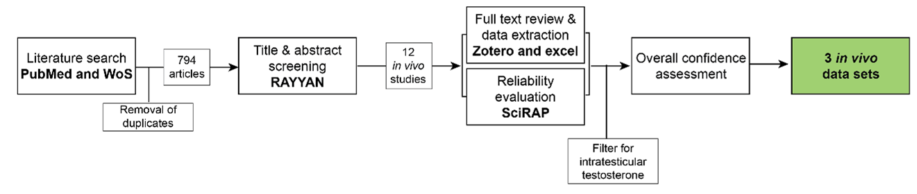
A systematic approach (fig. 1, 1s9lo2a792_KER_3488_Figure_1.png (1321×280)) was used to collect evidence based on the methodology described in (Holmer et al., 2024). The evidence collection for this KER was done concurrently with the evidence collection for KER 3350 ‘decrease, circulating testosterone leads to hypospadias’, for which the same search string was used.
Search strategy
Search strings were synthesized for PubMed and Web of Science Core Collection based on the review question ‘Does decreased testosterone during fetal development lead to decreased hypospadias in male mammals?’
Search string in PubMed: ("testosterone*") AND ("genital malformation*" OR "hypospadias"[MeSH terms] OR “hypospadia*”)
Search string in Web of Science Core Collection: ("testosterone*”) AND ("hypospadia*" OR "genital malformation*")
Title & abstract screening:
Retrieved articles were screened in the online tool RAYYAN https://www.rayyan.ai/ After removal of duplicates, the titles and abstracts of the remaining 793 articles were screened according to pre-defined inclusion and exclusion criteria:
Inclusion criteria:
- In vivo studies in male mammals where fetal testosterone is reduced and hypospadias is evaluated*
- Mechanistic reviews on hypospadias
- Epidemiologic and human case studies with measurement of testosterone levels and hypospadias as an outcome. Chromosomal abnormalities were excluded from human case studies.
- In vitro, ex vivo, and in vivo mechanistic studies on hypospadias or testosterone production
- Non-clinical reviews on hypospadias
Exclusion criteria:
- Papers not in English
- Abstracts and other non-full text publications
*In cases where this criterion could not be determined by reading the abstract, they were included for full text review
Full text review, data extraction and reliability evaluation of animal studies:
For the in vivo studies, the full text papers were reviewed using the same exclusion criteria as in the title & abstract screening, and data were extracted from the included papers into an Excel template. In parallel, methodological reliability was assessed using the online tool Science in Risk Assessment and Policy (SciRAP; http://www.scirap.org, see Appendix 1, 85hky9n0dm_KER_3488__appendix_1.pdf). Based on the SciRAP evaluations, animal studies were assigned a reliability category using the principles outlined in Table 1. Studies were divided into different datasets, if multiple different chemicals, different exposure windows, or different time points of measurement of hypospadias were included.
Moreover, as this KER was made in parallel with several other KERs for other male reproductive endpoints (nipple retention and anogenital distance), three studies retrieved in the searches for these KERs which also evaluated hypospadias, but were not detected in the search for this KER, were also data extracted and evaluated for reliability.
The collected data were filtered to only include data sets measuring intratesticular testosterone, either in testes homogenates or in ex vivo testes cultures. Mixture studies were excluded to avoid different chemicals having different modes-of-action.
Overall assessment of the empirical evidence was performed according the AOP handbook. Only studies in reliability categories 1 (reliable without restriction) and 2 (reliable with restriction) were included.
Table 1 Principles for translation SciRAP evaluations into reliability categories.
|
Reliability Category |
Principles for Categorization |
|
1.Reliable without restriction |
SciRAP methodological quality Score > 80 and all key criteriaa are “Fulfilled” and there are no deficiencies in the non-key criteria that might affect study reliability. |
|
2. Reliable with restriction |
SciRAP methodological quality Score > 65 and one or several of the key criteria are “Partially Fulfilled” or there are minor deficiencies in the non-key criteria that might affect study reliability. |
|
3. Not reliable |
SciRAP methodological quality Score < 65 or one or several of the key criteria are “Not Fulfilled” or there are major deficiencies in the non-key criteria that affect study reliability. |
|
4. Not assignable |
Two or more of the key criteria are “Not Determined” |
aKey criteria were criteria judged as specifically critical for reliability of the data for this KER and were determined a priori. The key criteria for this data collection are outlined in Appendix 1, 85hky9n0dm_KER_3488__appendix_1.pdf.
Human studies
Studies in humans were regarded as supporting evidence, and study quality was not systematically evaluated. All studies were assessed for any major errors in study design or interpretation.
Evidence Supporting this KER
Biological Plausibility
The biological plausibility for this KER is judged to be high given the canonical biological knowledge on normal reproductive development.
Differentiation of the penis is programmed during fetal development. Once the testes have formed around GW8 in humans and GD16 in rats, they synthesize testosterone through the steroidogenesis pathway (Murashima et al., 2015). Although the adrenal glands may also produce testosterone, the testes are the main site of testosterone production (Naamneh Elzenaty et al., 2022). Testosterone is secreted from the testes and is transported to the peripheral tissues, including the genital tubercle. Testosterone may act directly on the AR or be converted to the more potent androgen DHT.
The genital tubercle is the bipotential structure that upon hormonal cues differentiates to either penis or clitoris. Both human and rodent genital tubercles express AR (C. M. Amato & Yao, 2021; Baskin et al., 2020). Upon activation of AR, the genital tubercle differentiates to a penis by elongation and formation of a central urethra which terminates at the tip of the penis (C. Amato et al., 2022). The programming of the genital tubercle happens in the masculinization programming window (GD 16-20 in rats, GW 8-14 in humans) (Welsh et al., 2014), although elongation and growth of the penis is also programmed later, at least in rats (Welsh et al., 2008). Hypospadias is one of the most common genital malformations caused by disruptions to penis development (Baskin & Ebbers, 2006; Yu et al., 2019).
Given the dependency of testosterone for penis differentiation, either through direct AR activation or conversion to DHT, it is plausible that a decrease in intratesticular testosterone will cause hypospadias.
Empirical Evidence
The empirical evidence from studies in animals for this KER is overall judged as moderate
From the data collection, three data sets were extracted. The data sets included different stressors causing reduced fetal levels of testosterone, all in rats (Table 2 and Appendix 2, 2s48n3874x_KER_3488_appendix_2.pdf). All studies showed concurrent hypospadias in the male offspring.
Table 2 Empirical evidence for KER 3488 LOAEL: Lowest observed adverse effect level; see Appendix 2 for specifications: 2s48n3874x_KER_3488_appendix_2.pdf
|
Stressors(s) |
Effect on upstream event (intratesticular testosterone) |
Effect on downstream event hypospadias |
Reference |
|
|
Rat |
Dibutyl phthalate |
LOAEL 750 mg/kg bw/day |
LOAEL 750 mg/kg bw/day |
(van den Driesche et al., 2020) |
|
Rat |
Dibutyl phthalate |
LOAEL 500 mg/kg bw/day |
LOAEL 500 mg/kg bw/day |
(Drake et al., 2009) |
|
Rat |
Diisooctyl phthalate |
LOAEL 0.1 g/kg bw/day |
LOAEL 1 g/kg bw/day |
(Saillenfait et al., 2013) |
Supporting human studies
Supporting the empirical evidence are human cases of patients with less testicular tissue (i.e. partial gonadal dysgenesis) which can cause severe hypospadias (Boehmer et al., 2001; Crone et al., 2002).
Dose concordance
One study informs and supports dose concordance for this KER. In this study, diisocytol phthalate caused reduced ex vivo testosterone production at a dose of 0.1 g/kg bw/day, while hypospadias was observed in male offspring at 1 g/kg bw/day (Saillenfait et al., 2013).
Temporal concordance
Empirical evidence indirectly supports temporal concordance. In all studies, the exposure window was prenatal, and low testosterone levels were detected at GD18-22, while hypospadias was assessed in adult rats.
Incidence concordance
Incidence concordance cannot be directly informed from the empirical evidence, because testosterone levels are reported as means of all values and is a continuous variable. However, given that hypospadias was not registered in all males in any of the studies, this could suggest a higher incidence of lower testosterone levels than the incidence of hypospadias.
Uncertainties and Inconsistencies
In one study (Drake et al., 2009), testosterone levels were only reduced when performing statistical analysis on individual values and not on litter means. Hypospadias was observed in 30% of males. The difference in statistical significance between litter means and individual values is an uncertainty to this study.
In (Saillenfait et al., 2013), intratesticular testosterone was only measured in ex vivo testes cultures, which are assumed to be a good proxy for intratesticular testosterone levels, although it should be kept in mind as an uncertainty.
Another uncertainty for this KER from the literature is the observation that humans with 5α-reductase deficiency have hypospadias due to low DHT levels despite normal or higher testosterone levels (Mendonca et al., 1996). This indicates that the effects of low testosterone may be more through reduced conversion to DHT than due to a direct loss of testosterone action on AR. To this, there is also the existence of a “backdoor pathway” to DHT in humans. This pathway in peripheral tissues (i.e. not testes) can circumvent testosterone as a precursor for DHT by synthesis of DHT is from reduction of androsterone by 17β-HSD (Miller & Auchus, 2019). This would create the possibility that testosterone is not required for DHT production and ultimately AR activation.
Known modulating factors
There are no known modulating factors for this KER
Quantitative Understanding of the Linkage
The quantitative understanding of this KER is low.
Response-response Relationship
A model for phthalate-induced malformations has been developed which aims to predict the frequency of hypospadias related to a phthalate’s reduction in ex vivo testosterone production. The model predicted that a 60% reduction in testosterone levels would induce hypospadias, although the predictivity of this model was not good when tested for one phthalate (Earl Gray et al., 2024). Thus, this model should be improved and extended to include other substances.
Time-scale
The time-scale of this KER largely depends on species, but is likely weeks. In humans, the masculinization programming window is weeks long, while in rodents it is days (Sharpe, 2020; Welsh et al., 2014). Hypospadias is diagnosed at birth in humans (Yu et al., 2019) and can also be observed at birth in rodents, but as development of the penis continues after birth in rodents, hypospadias may be more optimally evaluated later in juvenile or adult male rats (Schlomer et al., 2013; Sinclair et al., 2017).
Known Feedforward/Feedback loops influencing this KER
Local disruption of AR activation in the genital tubercle irreversibly disrupts development, so there are no known feedback/feedforward loops for this KER.
Domain of Applicability
Taxonomic applicability
The development and differentiation of the penis is driven by androgen hormones, mainly produced by fetal testes, in all mammals. It is therefore biologically plausible that this KER is applicable to all mammals (Murashima et al., 2015). The empirical evidence in this KER provides support that reduced intratesticular testosterone levels in fetal life can cause hypospadias in rats. Studies in humans with gonadal dysgenesis and concurrent hypospadias support this KER’s applicability to humans (Boehmer et al., 2001; Crone et al., 2002).
Sex applicability
This KER is applicable to males, where the testis is the primary sex organ.
Life stage applicability
The genital tubercle is programmed by androgen hormones in the masculinization programming window (GD16-20 in rats, and GW8-14 in humans ), when the testes produce high levels of testosterone (Sharpe, 2020; Welsh et al., 2014). The genital tubercle starts differentiating in fetal life, and in humans the penis is fully formed at birth, where hypospadias is usually diagnosed (Yu et al., 2019). In rats and mice, penis development continues postnatally for around 20-25 days, and hypospadias is optimally diagnosed after this timepoint, although it may also be observed earlier (Schlomer et al., 2013; Sinclair et al., 2017).
References
Amato, C., Fricke, A., Marella, S., Mogus, J., Bereman, M., & McCoy, K. (2022). An experimental evaluation of the efficacy of perinatal sulforaphane supplementation to decrease the incidence and severity of vinclozolin-induced hypospadias in the mouse model. Toxicology and Applied Pharmacology, 451, 116177. https://doi.org/10.1016/j.taap.2022.116177
Amato, C. M., & Yao, H. H.-C. (2021). Developmental and sexual dimorphic atlas of the prenatal mouse external genitalia at the single-cell level. Proceedings of the National Academy of Sciences of the United States of America, 118(25). https://doi.org/10.1073/pnas.2103856118
Baskin, L., Cao, M., Sinclair, A., Li, Y., Overland, M., Isaacson, D., & Cunha, G. R. (2020). Androgen and estrogen receptor expression in the developing human penis and clitoris. Differentiation; Research in Biological Diversity, 111, 41–59. https://doi.org/10.1016/j.diff.2019.08.005
Baskin, L., & Ebbers, M. (2006). Hypospadias: Anatomy, etiology, and technique. Journal of Pediatric Surgery, 41(3), 463–472. https://doi.org/10.1016/j.jpedsurg.2005.11.059
Boehmer, A., Nijman, R., Lammers, B., de Coninck, S., Van Hemel, J., Themmen, A., Mureau, M., de Jong, F., Brinkmann, A., Niermeijer, M., & Drop, S. (2001). Etiological studies of severe or familial hypospadias. The Journal of Urology, 165(4), 1246–1254.
Crone, J., Amann, G., Gheradini, R., Kirchlechner, V., & Fékété, C. (2002). Management of 46, XY partial gonadal dysgenesis—Revisited. Wiener Klinische Wochenschrift, 114(12), 462–467.
Drake, A., van den Driesche, S., Scott, H., Hutchison, G., Seckl, J., & Sharpe, R. (2009). Glucocorticoids Amplify Dibutyl Phthalate-Induced Disruption of Testosterone Production and Male Reproductive Development. ENDOCRINOLOGY, 150(11), 5055–5064. https://doi.org/10.1210/en.2009-0700
Earl Gray, L. J., Lambright, C., Evans, N., Ford, J., & Conley, M. (2024). Using targeted fetal rat testis genomic and endocrine alterations to predict the effects of a phthalate mixture on the male reproductive tract. Current Research in Toxicology, 7, 100180. https://doi.org/10.1016/j.crtox.2024.100180
Holmer, M. L., Zilliacus, J., Draskau, M. K., Hlisníková, H., Beronius, A., & Svingen, T. (2024). Methodology for developing data-rich Key Event Relationships for Adverse Outcome Pathways exemplified by linking decreased androgen receptor activity with decreased anogenital distance. Reproductive Toxicology, 128, 108662. https://doi.org/10.1016/j.reprotox.2024.108662
Murashima, A., Kishigami, S., Thomson, A., & Yamada, G. (2015). Androgens and mammalian male reproductive tract development. Biochimica et Biophysica Acta (BBA) - Gene Regulatory Mechanisms, 1849(2), 163–170. https://doi.org/10.1016/j.bbagrm.2014.05.020
Naamneh Elzenaty, R., Du Toit, T., & Flück, C. E. (2022). Basics of androgen synthesis and action. Best Practice & Research Clinical Endocrinology & Metabolism, 36(4), 101665. https://doi.org/10.1016/j.beem.2022.101665
Saillenfait, A., Sabaté, J., Robert, A., Cossec, B., Roudot, A., Denis, F., & Burgart, M. (2013). Adverse effects of diisooctyl phthalate on the male rat reproductive development following prenatal exposure. Reproductive Toxicology (Elmsford, N.Y.), 42, 192–202. https://doi.org/10.1016/j.reprotox.2013.09.004
Schlomer, B. J., Feretti, M., Rodriguez, E. J., Blaschko, S., Cunha, G., & Baskin, L. (2013). Sexual differentiation in the male and female mouse from days 0 to 21: A detailed and novel morphometric description. The Journal of Urology, 190(4 Suppl), 1610–1617. https://doi.org/10.1016/j.juro.2013.02.3198
Sharpe, R. (2020). Androgens and the masculinization programming window: Human-rodent differences. Biochemical Society Transactions, 48(4), 1725–1735. https://doi.org/10.1042/BST20200200
Sinclair, A., Cao, M., Pask, A., Baskin, L., & Cunha, G. (2017). Flutamide-induced hypospadias in rats: A critical assessment. Differentiation; Research in Biological Diversity, 94, 37–57. https://doi.org/10.1016/j.diff.2016.12.001
van den Driesche, S., Shoker, S., Inglis, F., Palermo, C., Langsch, A., & Otter, R. (2020). Systematic comparison of the male reproductive tract in fetal and adult Wistar rats exposed to DBP and DINP in utero during the masculinisation programming window. Toxicology Letters, 335, 37–50. https://doi.org/10.1016/j.toxlet.2020.10.006
Welsh, M., Saunders, P., Fisken, M., Scott, H., Hutchison, G., Smith, L., & Sharpe, R. (2008). Identification in rats of a programming window for reproductive tract masculinization, disruption of which leads to hypospadias and cryptorchidism. The Journal of Clinical Investigation, 118(4), 1479–1490. https://doi.org/10.1172/JCI34241
Welsh, M., Suzuki, H., & Yamada, G. (2014). The masculinization programming window. Endocrine Development, 27, 17–27. https://doi.org/10.1159/000363609
Yu, X., Nassar, N., Mastroiacovo, P., Canfield, M., Groisman, B., Bermejo-Sánchez, E., Ritvanen, A., Kiuru-Kuhlefelt, S., Benavides, A., Sipek, A., Pierini, A., Bianchi, F., Källén, K., Gatt, M., Morgan, M., Tucker, D., Canessa, M. A., Gajardo, R., Mutchinick, O. M., … Agopian, A. J. (2019). Hypospadias Prevalence and Trends in International Birth Defect Surveillance Systems, 1980-2010. European Urology, 76(4), 482–490. https://doi.org/10.1016/j.eururo.2019.06.027