
This AOP is licensed under the BY-SA license. This license allows reusers to distribute, remix, adapt, and build upon the material in any medium or format, so long as attribution is given to the creator. The license allows for commercial use. If you remix, adapt, or build upon the material, you must license the modified material under identical terms.
AOP: 237
Title
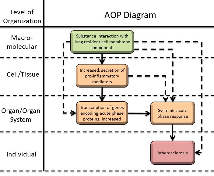
Substance interaction with lung resident cell membrane components leading to atherosclerosis via acute phase response
Short name
Graphical Representation
Additional AOP Exploration Options
Click links below to explore AOP 237, Substance interaction with lung resident cell membrane components leading to atherosclerosis via acute phase response in tools offered by third parties.

Point of Contact
Contributors
- Sarah Søs Poulsen
- Ulla Vogel
- Claudia Torero Gutierrez
- Jorid Birkelund Sørli
- Sabina Halappanavar
Coaches
- Sabina Halappanavar
- Shihori Tanabe
OECD Information Table
| OECD Project # | OECD Status | Reviewer's Reports | Journal-format Article | OECD iLibrary Published Version |
|---|---|---|---|---|
| 1.55 | Under Development | Scientific Review |
This AOP was last modified on August 11, 2026 03:14
Revision dates for related pages
| Page | Revision Date/Time |
|---|---|
| Transcription of genes encoding acute phase proteins, Increased | August 11, 2026 03:15 |
| Systemic acute phase response | August 11, 2026 03:18 |
| Atherosclerosis | August 11, 2026 03:19 |
| Substance interaction with the lung resident cell membrane components | June 22, 2026 18:07 |
| Increased, secretion of proinflammatory mediators | June 22, 2026 19:07 |
| Interaction with the lung cell membrane leads to Increased proinflammatory mediators | June 22, 2026 21:25 |
| Interaction with the lung cell membrane leads to Increased transcription of genes encoding acute phase proteins | August 11, 2026 03:08 |
| Increased proinflammatory mediators leads to Increased transcription of genes encoding acute phase proteins | August 11, 2026 03:03 |
| Interaction with the lung cell membrane leads to Systemic acute phase response | August 11, 2026 03:10 |
| Increased proinflammatory mediators leads to Systemic acute phase response | August 11, 2026 03:25 |
| Increased transcription of genes encoding acute phase proteins leads to Systemic acute phase response | August 11, 2026 03:00 |
| Interaction with the lung cell membrane leads to Atherosclerosis | August 11, 2026 03:22 |
| Systemic acute phase response leads to Atherosclerosis | August 11, 2026 02:57 |
| Lipopolysaccharride | May 29, 2018 07:05 |
| Graphene oxide nanoparticles | February 15, 2017 04:41 |
| Carbon nanotubes | August 09, 2017 08:03 |
| Insoluble nano-sized particles | May 29, 2018 07:09 |
| Virus | May 29, 2018 07:10 |
Abstract
AOP237 describes key events initiated with the interaction of substances with the membrane components of the pulmonary cells, and leading to atherosclerosis in humans. Atherosclerosis is defined as the thickening of the wall of an artery due to plaque deposition, and this condition can lead to severe events as myocardial infarction and stroke. This AOP presents the induction of acute phase response as a pathway for atherosclerosis progression. The interaction between a substance and the lung resident cell membrane components is the molecular initiating event (MIE; Event 1495) for this AOP; this interaction leads to an increased secretion of proinflammatory mediators [Key event (KE)1; Event 1496]. The release of proinflammatory factors triggers an increase in transcription of genes encoding acute phase proteins (KE2; Event 1438), leading to systemic acute phase response (KE3; Event 1439) once the acute phase proteins are translated and released into the systemic circulation. A continuous acute phase response leads to atherosclerosis, the adverse outcome (AO) of this AOP (Event 1443).
AOP 237 mainly focus on particles or particulate matter as stressors, however other compounds or inflammatory conditions that induce acute phase response, can be consider stressors and lead to atherosclerosis. In addition, most of the evidence is based on animal studies (mice) as a model for the human system, however the adverse outcome of the present AOP, atherosclerosis, is only applicable to humans. The AOP presents the biological plausibility, evidence and quantitative understanding for the relationship between KEs. In addition, evidence that KE2, KE3 and AO occur after the MIE is presented as non-adjacent relationships. This AOP presents a mechanism of substance-induced acute phase response leading to atherosclerosis, and it can be used for regulatory purposes and health-based risk assessments of inhalable materials.
AOP Development Strategy
Context
Cardiovascular disease (CVD) is the leading cause of death worldwide, being responsible for 32% of all deaths in 2019 (WHO; http://www.who.int). The term CVD covers all diseases of the cardiovascular system, including atherosclerosis, which is manifested as increased plaque deposition or build-up in the arteries. Although, atherosclerosis is not a cause of death, it can lead to fatal conditions as stroke and myocardial infarction. Atherosclerosis is normally an asymptotic disease and is initiated by a biological, chemical or physical insult to the artery walls. This leads to the expression of cell adhesion molecules on the endothelial lining of the arteries, which facilitates the activation, recruitment, and migration of monocytes through the endothelial monolayer (Cybulsky et al., 2001; Hansson & Libby, 2006). Inside the intima layer, the monocytes differentiate into macrophages and internalize fatty deposits (mainly oxidized low-density lipoprotein). This results in them transforming into foam cells, which is a major component of the atherosclerotic fatty streaks. The fatty streaks reduce the elasticity of the artery walls and the foam cells promote a pro-inflammatory environment by secretion of cytokines and reactive oxidative species. In addition, foam cells also induce the recruitment of smooth muscle cells to the intima. Added together, these changes lead to the formation of plaques on the artery walls. A fibrous cap of collagen and vascular smooth muscle cells protects the necrotic core and stabilizes the plaque (Libby, 2012; Virmani et al., 2005). However, blood clots can be formed if the plaque ruptures. These may travel with the bloodstream and obstruct the blood flow of smaller vessels, e.g. the coronary arteries, which ultimately can lead to myocardial infarction.
Inhalation of particulate matter, chemicals and pathogens have been related to increased pulmonary inflammation. Whereas a normal immune reaction is crucial for effective elimination of threats to the body, chronic and unresolved inflammation has been linked to both adverse pulmonary and adverse systemic effects in humans. In concordance with this, various retrospective and prospective epidemiological studies have linked pulmonary exposure to respirable air particulates with increased the risk of developing CVD (Clancy, Goodman, Sinclair, & Dockery, 2002; Dockery et al., 1993; Pope et al., 2004; Pope et al., 1995). Inhalation of particles has been proposed to affect the cardiovascular system in several different ways, including through disruption of vasomotor function and through acceleration of plaque progression in atherosclerosis (Cao et al., 2014; Moller et al., 2016).
Acute phase response is characterized by the change in plasma concentration of acute phase proteins (APP), along with other physiological changes during inflammatory conditions (Gabay & Kushner, 1999; Mantovani & Garlanda, 2023). Serum amyloid A (SAA) and C-reactive protein (CRP) are the major acute phase proteins in humans and are considered risk factors for CVDs (Table 1 presents acute phase response characteristics in humans and mice). In particular, SAA restricts the transport of cholesterol to the liver, allowing the accumulation of cholesterol in arteries and the formation of foam cells.
Table 1. Selected differences in APR between humans and mice.
|
Characteristic |
Humans |
Mice |
|
Number of identified genes involved in acute phase response |
61 |
62 |
|
Major acute phase proteins |
CRP, SAA |
Haptoglobin, SAA, serum amyloid P |
|
Moderate and minor acute phase proteins |
Haptoglobin, fibrinogen, α1 acid glycoprotein |
CRP, fibrinogen |
|
SAA isoforms |
Saa1, Saa2 and Saa4 |
Saa1, Saa2, Saa3 and Saa4 |
References: (Cray, 2012; Gabay & Kushner, 1999; NCBI, 2023; Tannock et al., 2018).
Atherosclerosis is a disease influenced by multiple factors including high levels of lipoproteins in blood, elevated blood pressure, smoking, obesity, type 2 diabetes, diet, and physical activity (Herrington, Lacey, Sherliker, Armitage, & Lewington, 2016; Libby et al., 2019; Raitakari, Pahkala, & Magnussen, 2022). Inflammation is also involved in atherosclerosis, providing pathways via which risk factors might cause the development and advancement of atherosclerotic plaques (Libby, 2021a, 2021b). Therefore, although inflammation and acute phase response are not the only causes of atherosclerosis, the early key events (KE1, KE2 and KE3) can be used to evaluate the particle-induced risk of developing atherosclerosis.
For the development of AOP 237, the MIE and KE1 from AOP 173 have been used (AOP 173: Substance interaction with the pulmonary resident cell membrane components leading to pulmonary fibrosis). The information presented in AOP 173 has not been modified for AOP 237.
The development of the present AOP was supported by the EU project NanoPASS (Grant number: 101092741) and the Focused Research Effort on Chemicals in the Working Environment (FFIKA) form the Danish Government.
Strategy
Summary of the AOP
Events:
Molecular Initiating Events (MIE)
Key Events (KE)
Adverse Outcomes (AO)
| Type | Event ID | Title | Short name |
|---|
| MIE | 1495 | Substance interaction with the lung resident cell membrane components | Interaction with the lung cell membrane |
| KE | 1496 | Increased, secretion of proinflammatory mediators | Increased proinflammatory mediators |
| KE | 1438 | Transcription of genes encoding acute phase proteins, Increased | Increased transcription of genes encoding acute phase proteins |
| KE | 1439 | Systemic acute phase response | Systemic acute phase response |
| AO | 1443 | Atherosclerosis | Atherosclerosis |
Relationships Between Two Key Events (Including MIEs and AOs)
| Title | Adjacency | Evidence | Quantitative Understanding |
|---|
| Interaction with the lung cell membrane leads to Increased proinflammatory mediators | adjacent | Moderate | Moderate |
| Increased proinflammatory mediators leads to Increased transcription of genes encoding acute phase proteins | adjacent | High | Moderate |
| Increased transcription of genes encoding acute phase proteins leads to Systemic acute phase response | adjacent | High | Moderate |
| Systemic acute phase response leads to Atherosclerosis | adjacent | High | High |
| Interaction with the lung cell membrane leads to Increased transcription of genes encoding acute phase proteins | non-adjacent | High | Moderate |
| Interaction with the lung cell membrane leads to Systemic acute phase response | non-adjacent | High | Moderate |
| Increased proinflammatory mediators leads to Systemic acute phase response | non-adjacent | High | Moderate |
| Interaction with the lung cell membrane leads to Atherosclerosis | non-adjacent | High | Moderate |
Network View
Prototypical Stressors
Life Stage Applicability
| Life stage | Evidence |
|---|---|
| Adult | High |
Taxonomic Applicability
Sex Applicability
| Sex | Evidence |
|---|---|
| Male | High |
| Female | High |
Overall Assessment of the AOP

Domain of Applicability
This AOP is applicable to adult humans of both sexes. Although atherosclerosis is a condition that begins during childhood and progresses through life, its clinical manifestation is mostly observed in older individuals (Raitakari, Pahkala, & Magnussen, 2022).
Susceptible populations include smokers, dyslipidaemic individuals, and people with pre-existing CVD and diabetes who have increased baseline levels of CRP (Emerging Risk Factors et al., 2010; Rizo-Tellez, Sekheri, & Filep, 2023).
The AOP is applicable to inhaled stressors, which can interact with the pulmonary system, and induce pulmonary inflammation.
Essentiality of the Key Events
For the development of AOP 237, the molecular initiating event (MIE) and key event (KE) 1 from AOP 173 have been reused (AOP 173: Substance interaction with the pulmonary resident cell membrane components leading to pulmonary fibrosis). As part of the scientific review of AOP 237, the reused KEs and KER, MIE, KE1 and KER1 have been slightly revised.
|
Support for essentiality of KEs |
Defining question |
High |
Moderate |
Low |
|
What is the impact on downstream KEs and/or the AO if an upstream KE is modified or prevented? |
Direct evidence from specifically designed experimental studies illustrating prevention or impact on downstream KEs and/or the AO if upstream KEs are blocked or modified |
Indirect evidence that modification of one or more upstream KEs is associated with a corresponding (increase or decrease) in the magnitude or frequency of downstream KEs |
No or contradictory experimental evidence of the essentiality of any of the KEs. |
|
|
MIE: Substance interaction with the lung resident cell membrane components (Event 1495) |
Moderate. Although not routinely measured in experimental models, it is known that the interaction of a substance with the components of pulmonary system is a prerequisite for stressor-induced lung toxicity. This is especially true for particle-induced toxicity. For example, binding of toll-like receptor (Tlr) 2 and Tlr4 by the stressors is shown to activate the downstream pro-inflammatory response (KE1) in vivo. Knockout of Tlr2 results in inhibition of Saa1 mRNA expression in liver tissue (KE2) and in plasma (KE3) following pulmonary exposure to nanoparticles of carbon black and graphene oxide, and carbon nanotubes in mice (Danielsen et al., 2021). Knockout of Tlr4 prevents the lipopolysaccharide induced increase of cytokine/chemokines mRNA levels in lung tissue and the systemic acute phase response (KE3) in mice (Danielsen et al., 2021). Stressors such as graphene oxide, reduced graphene oxide, nanoclay, ZnO particles, single-walled and multiwalled nanotubes, titanium dioxide nanoparticles, carbon black nanoparticles have a dose-response relationship with transcription of genes encoding acute phase proteins (KE2: Transcription of genes encoding acute phase proteins, Increased) and the systemic acute phase response (KE3: Systemic acute phase response ) (Bengtson et al., 2017; Di Ianni et al., 2020; Monse et al., 2018; Poulsen et al., 2017; Saber et al., 2013). |
|||
|
KE1: Increased, secretion of proinflammatory mediators |
High. Disruption of IL-6 gene leads to reduced mRNA (KE2: Transcription of genes encoding acute phase proteins, Increased ) and protein levels of acute phase proteins haptoglobin, α1-acid glycoprotein and Saa in mice liver and in serum (KE3: Systemic acute phase response) (Kopf et al., 1994). Blocking the IL-6 receptors reduced SAA1 mRNA, while blocking the IL-1β and tumor necrosis factor α receptors partially reduced the expression of SAA1 mRNA (KE2: Transcription of genes encoding acute phase proteins, Increased), in hepatic cell lines (Hagihara et al., 2004). Administration of monoclonal antibodies against IL-1β reduced blood levels of CRP (KE2: Transcription of genes encoding acute phase proteins, Increased and KE3: Systemic acute phase response), and decreased the incidence rates of recurrent cardiovascular events (AO), in patients with a history of myocardial infarction (Ridker et al., 2017). |
|||
|
KE2: Transcription of genes encoding acute phase proteins, Increased (Event 1438) |
High. Gene transcription is necessary for the synthesis of proteins (KE3: Systemic acute phase response) (Alberts, 2017). Suppression of SAA3 and double knockout of SAA1/SAA2 reduces atherosclerotic plaque area (AO), in ApoE-/- mice, whereas overexpression of SAA3 or pulmonary dosing of SAA increases the atherosclerotic plaque area. |
|||
|
KE3: Systemic acute phase response (Event 1439) |
High. Elevated levels of SAA (measured in blood) induce plaque progression (AO) (Christophersen et al., 2021; Dong et al., 2011; Thompson et al., 2018). Suppression of SAA3 and double knockout of SAA1/SAA2 reduces atherosclerotic plaque area (AO), in ApoE-/- mice (Thompson et al., 2018). CRP and SAA levels in blood are predictive of risk of cardiovascular disease (Emerging Risk Factors et al., 2010; Pai et al., 2004; Ridker, Hennekens, Buring, & Rifai, 2000). Anti-inflammatory therapy targeting IL-1β innate immunity pathway for 48 months via IL-1β antibody led to a significantly lowered CRP levels and the rate of recurrent cardiovascular events among patients with previous myocardial infarction as compared to subjects treated with placebo (Ridker et al., 2017). |
|||
|
AO: Atherosclerosis (Event 1443) |
N/A. This is the AO and it is essential. |
|||
Uncertainties or Inconsistencies
- Physicochemical characteristics of nanomaterials such as size, surface area, surface functionalization, shape, composition, among others, affect the magnitude and duration of inflammation and acute phase response in mice (Bengtson et al., 2017; Danielsen et al., 2024; Gutierrez et al., 2023; Miszczak et al., 2025; Poulsen et al., 2017). In animal models, both inflammatory and acute phase response are predicted by the total surface area of the retained insoluble particles (Cosnier et al., 2021; Gutierrez et al., 2023).
- Carbon nanoparticle-induced proinflammatory transcriptional response and neutrophil influx in the lung occur independently of TLR2 and TLR4 following pulmonary exposure in mice (Danielsen et al., 2021).
- CRP and SAA are risk factors for cardiovascular disease (Ridker et al., 2000). However, Mendelian randomization studies have shown that CRP genotypes are not associated with risk of coronary heart disease and that genetically elevated levels of CRP are not associated with coronary heart disease risk (Collaboration et al., 2011; Elliott et al., 2009).
- In mice studies, it is possible to measure both SAA gene expression and protein levels, however the dynamic range for Saa gene expression is larger. In humans, measuring gene expression of acute phase proteins is not very common, as a tissue sample is needed, while measuring acute phase protein in blood is more common.
- It is suggested that acute phase proteins are mainly produced in the liver (Gabay & Kushner, 1999), however in mice the liver has subtle upregulation of Saa genes after exposure to ultrafine carbon particles or diesel exhaust particle. On the other hand, the lung shows a marked expression of Saa1, Saa2 and Saa3 mRNA as well as increased SAA3 levels in bronchoalveolar lavage fluid and lung tissue (Halappanavar et al., 2011; Saber et al., 2009; Saber et al., 2013).
- A level of inconsistency between the human study results exists. It has been observed that in most controlled human studies, an increase in CRP and/or SAA was observed after exposure to particulate matter (Baumann et al., 2018; Haase et al., 2022; Monse et al., 2018; Monse et al., 2021; Walker et al., 2022; Wyatt, Devlin, Rappold, Case, & Diaz-Sanchez, 2020). However, in other studies the exposure did not induce acute phase response (Andersen, Saber, Clausen, et al., 2018; Andersen, Saber, Pedersen, et al., 2018), maybe due to low levels of exposure (Andersen et al., 2019) and/or limited statistical power.
Evidence Assessment
Biological plausibility of each KER
Please also refer to AOP173: Substance interaction with the pulmonary resident cell membrane components leading to pulmonary fibrosis, which shares MIE and KE1 with the present AOP.
|
Support for Biological Plasuibility of KERs |
Defining question |
High |
Moderate |
Low |
|
Is there a mechanistic (i.e., structural or functional) relationship between KEup and KEdown consistent with established biological knowledge? |
Extensive understanding based on extensive previous documentation and broad acceptance -Established mechanistic basis |
The KER is plausible based on analogy to accepted biological relationships but scientific understanding is not completely established. |
There is empirical support for a statistical association between KEs (See 3.), but the structural or functional relationship between them is not understood. |
|
|
MIE => KE1: Interaction with the lung cell membrane leads to Increased proinflammatory mediators (Relationship 1702) |
Biological Plausibility of the MIE => KE1 is High. It is important to note that, traditionally, lung tissue inflammation in vivo is assessed by evaluating changes in the immune cell population within bronchoalveolar lavage fluid (i.e. Key event 1497). In contrast, in vitro studies typically assess tissue inflammation by measuring changes in the expression or levels of pro-inflammatory mediators (i.e. Key event 1496). Recent studies have established a link between elevated pro-inflammatory mediator levels (at the gene and/or protein level) and the influx of pro-inflammatory cells (e.g., neutrophil influx) into lung tissue (i.e. KER1703). Accordingly, throughout this document, neutrophil influx is used as proxy evidence to support KE1 (increased pro-inflammatory mediators). In clinical settings, CRP is one of the prominent markers of infection and inflammation, which is routinely assessed along the side of complete blood count for immune cells. Rationale: There is extensive evidence showing that interaction of stressors with the respiratory system induces the release of proinflammatory markers (Behzadi et al., 2017; Denholm & Phan, 1990; Dostert et al., 2008; Mossman & Churg, 1998). In addition, in vivo, recruitment of pro-inflammatory cells (example studies are referenced below) is measured as a marker of lung inflammation, increase of which is experimentally linked to increased pro-inflammatory mediators (KE1). Lipopolysaccharide (LPS)-induced pro-inflammatory response in terms of Il6 and Tnf transcription and neutrophil influx in bronchoalveolar lavage fluid was dependent on TLR2 receptor binding and activation (Danielsen et al., 2021). The neutrophil influx induced by the long and rigid carbon nanotubes was lower in TLR4 knockout mice compared to TLR4 wildtype mice. Similarly, graphene oxide-induced neutrophil influx was reduced in TLR2 knockout mice (Danielsen et al., 2021). In vitro, single-walled carbon nanotube-induced activation of primary human macrophages was shown to depend on both TLR2 and TLR4 (Mukherjee et al., 2018). Neutrophil influx was also shown to depend on macrophage motility and phagocytic activity following inhalation exposure to carbon black nanoparticles and quantum dots (Q. Liu et al., 2025). Following inhalation exposure, neutrophil influx in lungs of rats correlated with the total surface area of the pulmonary deposited nanomaterials for carbon black nanoparticles, titanium dioxide nanoparticles and different types of carbon nanotubes at all assessed post-exposure time points (Cosnier et al., 2021), suggesting that increased particle surface area can lead to increased particle-cell interaction and consequently, to inflammatory response in lungs. Different particles may interact with different lung resident cells following inhalation. In mice, cell-specific pro-inflammatory responses was observed after 12 h post-single intratracheal instillation to carbon black nanoparticles, double-walled carbon nanotubes, multiwalled carbon nanotube or lipopolysaccharide (Voss et al., 2025). The carbon-based materials elicited distinct inflammatory cytokine and cell responses via different modes of action. Carbon black nanoparticles triggered neutrophilia through alveolar epithelial activation and Cxcl1 and Csf2 expression but without apparent cell damage or macrophage activation. In contrast, the carbon nanotubes induced epithelial and macrophage damage, with alarmin release (IL-1α, IL-33) dominating the multiwalled carbon nanotube response. Double-walled carbon nanotubes caused alveolar epithelial injury, and pro-inflammatory macrophage and fibroblast-derived monocyte attractant (Ccl2, Ccl7) activation. All three carbon-based nanomaterials induced neutrophil influx in the bronchoalveolar lavage fluid. In an in vivo study of pulmonary exposure to diesel engine exhaust, dose-dependent production of pro-inflammatory cytokines in bronchoalveolar lavage fluid were observed 24 h postexposure to 5 different types of diesel engine exhaust (6, 18 and 54 ug/animal) (Bendtsen et al., 2020; McCarrick et al., 2025). Dose-dependent increases were reported for the following cytokines: CCL2, CCL20, CCL3L3, CSF2, CXCL1, GDNF, IL-1A, ITGB6, LGMN, TPP1 and PDGFB . Furthermore, CCL2, CXCL1, CCL3L3, CSF2, IL-1A were reported to correlate positively with neutrophil influx in bronchoalveolar lavage fluid (McCarrick et al., 2025). |
|||
|
KE1 => KE2: Increased proinflammatory mediators leads to Increased transcription of genes encoding acute phase proteins (Relationship 2053) |
Biological Plausibility of the KE1 => KE2 is High. Rationale: Acute phase proteins are induced by pro-inflammatory cytokines. These cytokines are produced at sites of inflammation mainly by monocytes and macrophages (Gabay & Kushner, 1999; Mantovani & Garlanda, 2023; Uhlar & Whitehead, 1999; Venteclef, Jakobsson, Steffensen, & Treuter, 2011). |
|||
|
KE2 => KE3: Increased transcription of genes encoding acute phase proteins leads to Systemic acute phase response (Relationship 1589) |
Biological Plausibility of the KE2 => KE3 is High. Rationale: After gene expression of acute phase proteins in tissues during inflammatory conditions, mRNA is translated and folded into proteins (Alberts, 2017). These proteins are then release to the systemic circulation (Van Eeden, Leipsic, Paul Man, & Sin, 2012). |
|||
|
KE3 => AO: Systemic acute phase response leads to Atherosclerosis (Relationship 2860) |
Biological Plausibility of the KE3 => KE2 is High. Rationale: During acute phase response, SAA, one of the major acute phase proteins, replaces apolipoprotein A-1 from high density lipoprotein (HDL). This replacement obstructs the reverse transport of cholesterol to the liver, allowing the accumulation of cholesterol in cells (Lindhorst, Young, Bagshaw, Hyland, & Kisilevsky, 1997; McGillicuddy et al., 2009; Meek, Urieli-Shoval, & Benditt, 1994). Blood levels of CRP correlate with risk of cardiovascular disease in a large meta-analysis of 48 studies (Emerging Risk Factors et al., 2010). |
|||
|
Non-adjacent MIE => KE2: Interaction with the lung cell membrane leads to Increased transcription of genes encoding acute phase proteins (Relationship 2958) |
Biological Plausibility of the MIE => KE2 is High. Rationale: After cells sense pathogens, tissue damage or dysmetabolism, production of acute phase proteins is triggered by cellular pattern-recognition molecules, through a cytokine cascade (Mantovani & Garlanda, 2023). Pulmonary exposure to nanomaterials induces expression of acute phase response genes in mice (Bengtson et al., 2017; Di Ianni et al., 2020; Erdely, Liston, et al., 2011; Gliga et al., 2026; Gutierrez et al., 2023; Hadrup et al., 2019; Hadrup et al., 2020; Halappanavar et al., 2015; Poulsen, Saber, Mortensen, et al., 2015; Saber et al., 2013). |
|||
|
Non-adjacent MIE => KE3: Interaction with the lung cell membrane leads to Systemic acute phase response (Relationship 2959) |
Biological Plausibility of the MIE => KE3 is High. Rationale: Pulmonary inflammation occurs when stressors interact with the airways (Moldoveanu et al., 2009) and acute phase response is induced during inflammatory conditions (Gabay & Kushner, 1999). There is plenty of evidence showing that inhalation or instillation of stressors induces systemic acute phase response in controlled human exposure studies and in animal studies (Baumann et al., 2016; Bendtsen et al., 2019; Bengtson et al., 2017; Bourdon et al., 2012; Erdely, Liston, et al., 2011; Kim, Chen, Boyce, & Christiani, 2005; Monse et al., 2018; Monse et al., 2021; Poulsen et al., 2017; Poulsen, Saber, Williams, et al., 2015; Westberg et al., 2016). |
|||
|
Non-adjacent KE1 => KE3: Increased proinflammatory mediators leads to Systemic APR (Relationship 3052) |
Biological Plausibility of the KE1 => KE3 is High. Rationale: Pro-inflammatory cytokines induce the release of acute phase proteins. These proteins are released from inflammatory sites to the systemic circulation (Gabay & Kushner, 1999; Mantovani & Garlanda, 2023). |
|||
|
Non-adjacent MIE => AO: Interaction with the lung cell membrane leads to Atherosclerosis (Relationship 2960) |
Biological Plausibility of the MIE => AO is Moderate. Rationale: There is evidence that the interaction of the lungs with stressor induces atherosclerotic plaque progression; however, the mechanistic relationship has not been clarified and many different pathways may contribute (Christophersen et al., 2021; Erdely, Hulderman, et al., 2011; M. R. Miller et al., 2013; M. R. Miller & Newby, 2020; Van Eeden et al., 2012). |
|||
Empirical support for each KER
Please also refer to AOP173: Substance interaction with the pulmonary resident cell membrane components leading to pulmonary fibrosis, which shares MIE and KE1 with the present AOP.
|
Empirical Support |
Defining question |
High |
Moderate |
Low |
|
Does KEup occur at lower doses and earlier time points than KE down and at the same dose of prototypical stressor, is the incidence of KEup > than that for KEdown? Are there inconsistencies in empirical support across taxa, species and prototypical stressor that don’t align with expected pattern for hypothesised AOP? |
Multiple studies showing dependent change in both events following exposure to a wide range of specific prototypical stressors. (Extensive evidence for temporal, dose- response and incidence concordance) and no or few critical data gaps or conflicting data |
Demonstrated dependent change in both events following exposure to a small number of specific prototypical stressors and some evidence inconsistent with expected pattern that can be explained by factors such as experimental design, technical considerations, differences among laboratories, etc.
|
Limited or no studies reporting dependent change in both events following exposure to a specific prototypical stressor (i.e., endpoints never measured in the same study or not at all); and/or significant inconsistencies in empirical support across taxa and species that don’t align with expected pattern for hypothesised AOP |
|
|
MIE => KE1: Interaction with the lung cell membrane leads to Increased proinflammatory mediators (Relationship 1702) |
Empirical Support of the MIE => KE1 is Moderate.
Rationale: There are limited in vitro studies which show a temporal and dose-dependent relationship between these two events (Chan et al., 2018; Denholm & Phan, 1990; Roy, Singh, Das, Tripathi, & Dwivedi, 2014). |
|||
|
KE1 => KE2: Increased proinflammatory mediators leads to Increased transcription of genes encoding acute phase proteins |
Empirical Support of the KE1 => KE2 is High. Rationale: There are several studies showing dose concordance and temporal concordance between KEs (Bendtsen et al., 2019; Bendtsen et al., 2020; Di Ianni et al., 2020; Gliga et al., 2026; Kyjovska et al., 2015; McCarrick et al., 2025; Saber et al., 2012; Saber et al., 2013; Wallin et al., 2017). |
|||
|
KE2 => KE3: Increased transcription of genes encoding acute phase proteins leads to Systemic acute phase response (Relationship 1589) |
Empirical Support of the KE2 => KE3 is High. Rationale: There are studies showing a dose concordance and temporal concordance between KE (Bengtson et al., 2017; Gutierrez et al., 2023; Poulsen et al., 2017; Saber et al., 2013). Increased transcription of acute phase response genes can be detected at lower dose levels than increased levels of acute phase proteins (Gutierrez et al., 2023; Saber et al., 2013). |
|||
|
KE3 => AO: Systemic acute phase response leads to Atherosclerosis (Relationship 2860) |
Empirical Support of the KE3 => AO is Moderate. Rationale: There is a limited number of animal studies showing the relationship between the KEs, in addition of epidemiological studies showing association between the KEs (Christophersen et al., 2021; Dong et al., 2011; Emerging Risk Factors et al., 2010; Pai et al., 2004; Rivera et al., 2013; Thompson et al., 2015; Thompson et al., 2018). Anti-inflammatory therapy targeting IL-1β innate immunity pathway for 48 months via IL-1β antibody led to a significantly lowered CRP levels and the rate of recurrent cardiovascular events among patients with previous myocardial infarction as compared to subjects treated with placebo (Ridker et al., 2017). |
|||
|
Non-adjacent MIE => KE2: Interaction with the lung cell membrane leads to Increased transcription of genes encoding acute phase proteins (Relationship 2958) |
Empirical Support of the MIE => KE2 is Moderate. Rationale: There are several studies showing a dose concordance and temporal concordance in animal studies. However, in the case of nanomaterials it has been shown that physicochemical characteristics affect the magnitude and duration of the expression of acute phase proteins in mice (Bengtson et al., 2017; Bourdon et al., 2012; Gutierrez et al., 2023; Kyjovska et al., 2015; Poulsen et al., 2017; Saber et al., 2013; Wallin et al., 2017). |
|||
|
Non-adjacent MIE => KE3: Interaction with the lung cell membrane leads to Systemic acute phase response (Relationship 2959) |
Empirical Support of the MIE => KE3 is Moderate. Rationale: There are plenty of studies showing a dose concordance and temporal concordance in animal and controlled human studies (Brand et al., 2014; Erdely, Liston, et al., 2011; Kim et al., 2005; Monse et al., 2018; Monse et al., 2021; Poulsen et al., 2017; Walker et al., 2022; Wyatt et al., 2020). It has been observed that systemic acute phase response is not always observed after exposure. |
|||
|
Non-adjacent KE1 => KE3: Increased proinflammatory mediators leads to Systemic APR (Relationship 3052) |
Empirical Support of the KE1 => KE3 is High. Rationale: There are several studies showing a dose concordance and temporal concordance. However, there are inconsistencies between changes in blood levels of pro-inflammatory mediators and systemic APR (Baumann et al., 2016; Kim et al., 2005; Monse et al., 2018; Monse et al., 2021; Poulsen et al., 2017). |
|||
|
Non-adjacent MIE => AO: Interaction with the lung cell membrane leads to Atherosclerosis (Relationship 2960) |
Empirical Support of the MIE => AO is High. Rationale: There are several studies showing the relationship between the key events (Christophersen et al., 2021; Li et al., 2007; Mikkelsen et al., 2011; M. R. Miller et al., 2013). In a large meta-analysis, strong association was seen between particulate ambient air pollution and cardiovascular disease (C. Liu et al., 2019). Reduction of exposure to black smoke in Dublin, Ireland resulted in reduced cardiovascular mortality (Clancy, Goodman, Sinclair, & Dockery, 2002). |
|||
Known Modulating Factors
|
Quantitative Understanding
The table below presents the quantitative understanding of every KER.
When assessing stressors in mice , it is possible to measure the expression of genes coding for acute phase proteins (KE2) in different tissues, whereas in humans this is not likely as a tissue sample would be required. On the other hand, in humans it is much more common and easier to measure systemic acute phase response (KE3) through a blood sample. In mice, it has been shown that Saa3 mRNA in lung tissue and blood levels of serum amyloid A (SAA)3 are correlated (Gutierrez et al., 2023). In addition, SAA levels in mice and humans show changes of similar magnitude after exposure to zinc oxide nanoparticles (Gutierrez et al., 2023). This suggests that systemic acute phase response in humans or no-effect-levels may be estimated from studies in mice (Gutierrez et al., 2023).
Saa3 mRNA levels in mouse lung tissue is correlated with pulmonary inflammation measured as neutrophil influx in broncheoalveolar lavage fluid (i.e. indirect marker of the release of pro-inflammatory mediators) apost-pulmonary exposure to nanomaterials (Danielsen et al., 2024; Gutierrez et al., 2023). In the context of particle exposure, both of these endpoints can be estimated by calculating the dosed particle surface area (specific surface area multiplied by dose level) (Gutierrez et al., 2023).
The relative risk of people developing a cardiovascular disease can be calculated from blood levels of acute phase proteins in epidemiological studies (Emerging Risk Factors et al., 2010).
|
KER |
Quantitative understanding |
|
MIE => KE1: Interaction with the lung cell membrane leads to Increased proinflammatory mediators (Relationship 1702) |
The quantitative understanding of MIE => KE1 is Low. Rationale: The quantitative prediction of the release of proinflammatory factors can be made from the interaction of the stressors with the pulmonary system. In the case of some stressors (nanomaterials), it is possible to make a prediction using the dosed surface area of the materials and neutrophil numbers in broncheoalveolar lavage (BALF) as an indirect marker of the release of pro-inflammatory factors (Gutierrez et al., 2023; Oberdorster, Ferin, Gelein, Soderholm, & Finkelstein, 1992; Oberdorster, Ferin, & Lehnert, 1994; Schmid & Stoeger, 2016; Stoeger et al., 2006). A dose-response relationship between pulmonary exposure to particles and increased levels of proinflammatory mediators in bronchoalveolar lavage fluid is observed in several studies involving animals (McCarrick et al., 2025; Saber et al., 2014) |
|
KE1 => KE2: Increased proinflammatory mediators leads to Increased transcription of genes encoding acute phase proteins (Relationship 2053) |
The quantitative understanding is of KE1 => KE2 is Moderate. Rationale: In mice, the gene expression of the acute phase protein SAA after exposure to various nanomaterials including metal oxide nanomaterials is closely correlated to pulmonary inflammation measured as neutrophil numbers in BALF (Danielsen et al., 2024; Gutierrez et al., 2023; Saber et al., 2013). |
|
KE2 => KE3: Increased transcription of genes encoding acute phase proteins leads to Systemic acute phase response (Relationship 1589) |
The quantitative understanding of KE2 => KE3 is Moderate. Rationale: In mice, the systemic levels of the acute phase protein SAA after exposure to metal oxide nanomaterials is closely correlated with gene expression of SAA in lung tissue (Gutierrez et al., 2023). |
|
KE3 => AO: Systemic acute phase response leads to Atherosclerosis (Relationship 2860) |
The quantitative understanding of KE3 => AO is High. Rationale: The risk of developing a cardiovascular disease at the population level is associated with blood levels of acute phase proteins in epidemiological studies (KER 2860) (Emerging Risk Factors et al., 2010). |
|
Non-adjacent MIE => KE2: Interaction with the lung cell membrane leads to Increased transcription of genes encoding acute phase proteins (Relationship 2958) |
The quantitative understanding of MIE => KE2 is Moderate. Rationale: In mice, the gene expression of the acute phase protein SAA after exposure to metal oxide nanomaterials is correlated with the dosed surface area (Gutierrez et al., 2023; Saber et al., 2014). |
|
Non-adjacent MIE => KE3: Interaction with the lung cell membrane leads to Systemic acute phase response |
The quantitative understanding of MIE => KE3 is Moderate. Rationale: In mice, the blood levels of the acute phase protein SAA after exposure to metal oxide nanomaterials is correlated to the dosed surface area (Gutierrez et al., 2023). In humans, systemic CRP levels correlate positively with increasing levels of air pollution measured as PM2.5 (Zhang et al., 2017). |
|
Non-adjacent KE1 => KE3: Increased proinflammatory mediators leads to Systemic APR (Relationship 3052) |
The quantitative understanding of KE1 => KE3 is Moderate. Rationale: In mice, the blood levels of the acute phase protein SAA after exposure to metal oxide nanomaterials and multiwalled carbon nanotubes can be estimated from neutrophil numbers in broncheoalveolar lavage fluid (Gutierrez et al., 2023; Poulsen et al., 2017). |
|
Non-adjacent MIE => AO: Interaction with the lung cell membrane leads to Atherosclerosis (Relationship 2960) |
The quantitative understanding of MIE => AO is Moderate. Rationale: Epidemiological studies have shown the risk ratios of having a cardiovascular event per increase or decrease of exposure to particulate matter (Beelen et al., 2014; Cesaroni et al., 2014; Clancy et al., 2002; K. A. Miller et al., 2007). A multicenter meta-analysis provides quantitative risk estimates for ambient air pollution measured as PM2.5 and PM10 (C. Liu et al., 2019). In the report ‘Documentation for health-based occupational exposure limits for ZnO’ the critical effect was based on identifying the no-effect-levels for ZnO-induced systemic acute phase response leading to cardiovascular disease (Hadrup et al., 2021). |
Considerations for Potential Applications of the AOP (optional)
Particle-induced acute phase response can be regarded as a critical effect linking particle-exposure to cardiovascular disease. Dose-response relationships can be used to establish no-observed-adverse-effect levels (NOAEL) for regulatory purposes and occupational exposure limits for inhalable materials can be determined through health-based risk assessments. This approach was taken by the Danish National Research Centre for the Working Environment at request of the Danish Working Environment Authority and an occupational exposure limit for zinc oxide was proposed based on the induction of acute phase response as the critical effect (the report can be found in: Dokumentation for helbredsbaserede grænseværdier for kemiske stoffer i arbejdsmiljøet (nfa.dk)).
As mentioned previously, not all KEs can easily be measured in humans, therefore animal studies can be used to measure early KEs and perform a risk assessment of different stressors. Additionally, physicochemical properties, such as specific surface area and dissolution, are important predictors of particle-induced acute phase response that can be used for hazard assessment (Gutierrez et al., 2023).
References
References
Alberts, B. (2017). Molecular biology of the cell (Sixth edition. ed.). Boca Raton, FL: CRC Press, an imprint of Garland Science.
Andersen, M. H. G., Frederiksen, M., Saber, A. T., Wils, R. S., Fonseca, A. S., Koponen, I. K., . . . Vogel, U. (2019). Health effects of exposure to diesel exhaust in diesel-powered trains. Part Fibre Toxicol, 16(1), 21. doi:10.1186/s12989-019-0306-4
Andersen, M. H. G., Saber, A. T., Clausen, P. A., Pedersen, J. E., Lohr, M., Kermanizadeh, A., . . . Vogel, U. (2018). Association between polycyclic aromatic hydrocarbon exposure and peripheral blood mononuclear cell DNA damage in human volunteers during fire extinction exercises. Mutagenesis, 33(1), 105-115. doi:10.1093/mutage/gex021
Andersen, M. H. G., Saber, A. T., Pedersen, J. E., Pedersen, P. B., Clausen, P. A., Lohr, M., . . . Moller, P. (2018). Assessment of polycyclic aromatic hydrocarbon exposure, lung function, systemic inflammation, and genotoxicity in peripheral blood mononuclear cells from firefighters before and after a work shift. Environ Mol Mutagen, 59(6), 539-548. doi:10.1002/em.22193
Baumann, R., Gube, M., Markert, A., Davatgarbenam, S., Kossack, V., Gerhards, B., . . . Brand, P. (2018). Systemic serum amyloid A as a biomarker for exposure to zinc and/or copper-containing metal fumes. J Expo Sci Environ Epidemiol, 28(1), 84-91. doi:10.1038/jes.2016.86
Baumann, R., Joraslafsky, S., Markert, A., Rack, I., Davatgarbenam, S., Kossack, V., . . . Gube, M. (2016). IL-6, a central acute-phase mediator, as an early biomarker for exposure to zinc-based metal fumes. Toxicology, 373, 63-73. doi:10.1016/j.tox.2016.11.001
Beelen, R., Stafoggia, M., Raaschou-Nielsen, O., Andersen, Z. J., Xun, W. W., Katsouyanni, K., . . . Hoek, G. (2014). Long-term exposure to air pollution and cardiovascular mortality: an analysis of 22 European cohorts. Epidemiology, 25(3), 368-378. doi:10.1097/EDE.0000000000000076
Behzadi, S., Serpooshan, V., Tao, W., Hamaly, M. A., Alkawareek, M. Y., Dreaden, E. C., . . . Mahmoudi, M. (2017). Cellular uptake of nanoparticles: journey inside the cell. Chem Soc Rev, 46(14), 4218-4244. doi:10.1039/c6cs00636a
Bendtsen, K. M., Brostrom, A., Koivisto, A. J., Koponen, I., Berthing, T., Bertram, N., . . . Vogel, U. (2019). Airport emission particles: exposure characterization and toxicity following intratracheal instillation in mice. Part Fibre Toxicol, 16(1), 23. doi:10.1186/s12989-019-0305-5
Bendtsen, K. M., Gren, L., Malmborg, V. B., Shukla, P. C., Tunér, M., Essig, Y. J., . . . Vogel, U. B. (2020). Particle characterization and toxicity in C57BL/6 mice following instillation of five different diesel exhaust particles designed to differ in physicochemical properties. Part Fibre Toxicol, 17(1), 38. doi:10.1186/s12989-020-00369-9
Bengtson, S., Knudsen, K. B., Kyjovska, Z. O., Berthing, T., Skaug, V., Levin, M., . . . Vogel, U. (2017). Differences in inflammation and acute phase response but similar genotoxicity in mice following pulmonary exposure to graphene oxide and reduced graphene oxide. PLoS One, 12(6), e0178355. doi:10.1371/journal.pone.0178355
Bourdon, J. A., Halappanavar, S., Saber, A. T., Jacobsen, N. R., Williams, A., Wallin, H., . . . Yauk, C. L. (2012). Hepatic and pulmonary toxicogenomic profiles in mice intratracheally instilled with carbon black nanoparticles reveal pulmonary inflammation, acute phase response, and alterations in lipid homeostasis. Toxicol Sci, 127(2), 474-484. doi:10.1093/toxsci/kfs119
Brand, P., Bauer, M., Gube, M., Lenz, K., Reisgen, U., Spiegel-Ciobanu, V. E., & Kraus, T. (2014). Relationship between welding fume concentration and systemic inflammation after controlled exposure of human subjects with welding fumes from metal inert gas brazing of zinc-coated materials. J Occup Environ Med, 56(1), 1-5. doi:10.1097/JOM.0000000000000061
Cesaroni, G., Forastiere, F., Stafoggia, M., Andersen, Z. J., Badaloni, C., Beelen, R., . . . Peters, A. (2014). Long term exposure to ambient air pollution and incidence of acute coronary events: prospective cohort study and meta-analysis in 11 European cohorts from the ESCAPE Project. BMJ, 348, f7412. doi:10.1136/bmj.f7412
Chan, J. Y. W., Tsui, J. C. C., Law, P. T. W., So, W. K. W., Leung, D. Y. P., Sham, M. M. K., . . . Chan, C. W. H. (2018). Regulation of TLR4 in silica-induced inflammation: An underlying mechanism of silicosis. Int J Med Sci, 15(10), 986-991. doi:10.7150/ijms.24715
Christophersen, D. V., Moller, P., Thomsen, M. B., Lykkesfeldt, J., Loft, S., Wallin, H., . . . Jacobsen, N. R. (2021). Accelerated atherosclerosis caused by serum amyloid A response in lungs of ApoE(-/-) mice. FASEB J, 35(3), e21307. doi:10.1096/fj.202002017R
Clancy, L., Goodman, P., Sinclair, H., & Dockery, D. W. (2002). Effect of air-pollution control on death rates in Dublin, Ireland: an intervention study. Lancet, 360(9341), 1210-1214. doi:10.1016/S0140-6736(02)11281-5
Collaboration, C. R. P. C. H. D. G., Wensley, F., Gao, P., Burgess, S., Kaptoge, S., Di Angelantonio, E., . . . Danesh, J. (2011). Association between C reactive protein and coronary heart disease: mendelian randomisation analysis based on individual participant data. BMJ, 342, d548. doi:10.1136/bmj.d548
Cosnier, F., Seidel, C., Valentino, S., Schmid, O., Bau, S., Vogel, U., . . . Gate, L. (2021). Retained particle surface area dose drives inflammation in rat lungs following acute, subacute, and subchronic inhalation of nanomaterials. Part Fibre Toxicol, 18(1), 29. doi:10.1186/s12989-021-00419-w
Danielsen, P. H., Bendtsen, K. M., Knudsen, K. B., Poulsen, S. S., Stoeger, T., & Vogel, U. (2021). Nanomaterial- and shape-dependency of TLR2 and TLR4 mediated signaling following pulmonary exposure to carbonaceous nanomaterials in mice. Part Fibre Toxicol, 18(1), 40. doi:10.1186/s12989-021-00432-z
Danielsen, P. H., Poulsen, S. S., Knudsen, K. B., Clausen, P. A., Jensen, K. A., Wallin, H., & Vogel, U. (2024). Physicochemical properties of 26 carbon nanotubes as predictors for pulmonary inflammation and acute phase response in mice following intratracheal lung exposure. Environ Toxicol Pharmacol, 107, 104413. doi:10.1016/j.etap.2024.104413
Denholm, E. M., & Phan, S. H. (1990). Bleomycin binding sites on alveolar macrophages. J Leukoc Biol, 48(6), 519-523. doi:10.1002/jlb.48.6.519
Di Ianni, E., Moller, P., Mortensen, A., Szarek, J., Clausen, P. A., Saber, A. T., . . . Jacobsen, N. R. (2020). Organomodified nanoclays induce less inflammation, acute phase response, and genotoxicity than pristine nanoclays in mice lungs. Nanotoxicology, 14(7), 869-892. doi:10.1080/17435390.2020.1771786
Dong, Z., Wu, T., Qin, W., An, C., Wang, Z., Zhang, M., . . . An, F. (2011). Serum amyloid A directly accelerates the progression of atherosclerosis in apolipoprotein E-deficient mice. Mol Med, 17(11-12), 1357-1364. doi:10.2119/molmed.2011.00186
Dostert, C., Petrilli, V., Van Bruggen, R., Steele, C., Mossman, B. T., & Tschopp, J. (2008). Innate immune activation through Nalp3 inflammasome sensing of asbestos and silica. Science, 320(5876), 674-677. doi:10.1126/science.1156995
Elliott, P., Chambers, J. C., Zhang, W., Clarke, R., Hopewell, J. C., Peden, J. F., . . . Kooner, J. S. (2009). Genetic Loci associated with C-reactive protein levels and risk of coronary heart disease. JAMA, 302(1), 37-48. doi:10.1001/jama.2009.954
Emerging Risk Factors, C., Kaptoge, S., Di Angelantonio, E., Lowe, G., Pepys, M. B., Thompson, S. G., . . . Danesh, J. (2010). C-reactive protein concentration and risk of coronary heart disease, stroke, and mortality: an individual participant meta-analysis. Lancet, 375(9709), 132-140. doi:10.1016/S0140-6736(09)61717-7
Erdely, A., Hulderman, T., Salmen-Muniz, R., Liston, A., Zeidler-Erdely, P. C., Chen, B. T., . . . Simeonova, P. P. (2011). Inhalation exposure of gas-metal arc stainless steel welding fume increased atherosclerotic lesions in apolipoprotein E knockout mice. Toxicol Lett, 204(1), 12-16. doi:10.1016/j.toxlet.2011.03.030
Erdely, A., Liston, A., Salmen-Muniz, R., Hulderman, T., Young, S. H., Zeidler-Erdely, P. C., . . . Simeonova, P. P. (2011). Identification of systemic markers from a pulmonary carbon nanotube exposure. J Occup Environ Med, 53(6 Suppl), S80-86. doi:10.1097/JOM.0b013e31821ad724
Gabay, C., & Kushner, I. (1999). Acute-phase proteins and other systemic responses to inflammation. N Engl J Med, 340(6), 448-454. doi:10.1056/NEJM199902113400607
Gliga, A. R., McCarrick, S., Malmborg, V., Kohonen, P., Snigireva, A., Mills, B., . . . Vogel, U. (2026). Similar global transcription patterns in mouse lung tissue following pulmonary exposure to renewable and conventional diesel engine exhaust particles. Environ Toxicol Pharmacol, 122, 104918. doi:10.1016/j.etap.2025.104918
Gutierrez, C. T., Loizides, C., Hafez, I., Brostrom, A., Wolff, H., Szarek, J., . . . Vogel, U. (2023). Acute phase response following pulmonary exposure to soluble and insoluble metal oxide nanomaterials in mice. Part Fibre Toxicol, 20(1), 4. doi:10.1186/s12989-023-00514-0
Hadrup, N., Rahmani, F., Jacobsen, N. R., Saber, A. T., Jackson, P., Bengtson, S., . . . Vogel, U. (2019). Acute phase response and inflammation following pulmonary exposure to low doses of zinc oxide nanoparticles in mice. Nanotoxicology, 13(9), 1275-1292. doi:10.1080/17435390.2019.1654004
Hadrup, N., Saber, A. T., Jacobsen, N. R., Danielsen, P. H., Poulsen, S. S., Hougaard, K. S., & Vogel, U. (2021). Zinc oxide: Scientific basis for setting a health-based occupational exposure limit. Retrieved from National Research Centre for the Working Environment:
Hadrup, N., Zhernovkov, V., Jacobsen, N. R., Voss, C., Strunz, M., Ansari, M., . . . Vogel, U. (2020). Acute Phase Response as a Biological Mechanism-of-Action of (Nano)particle-Induced Cardiovascular Disease. Small, 16(21), e1907476. doi:10.1002/smll.201907476
Hagihara, K., Nishikawa, T., Isobe, T., Song, J., Sugamata, Y., & Yoshizaki, K. (2004). IL-6 plays a critical role in the synergistic induction of human serum amyloid A (SAA) gene when stimulated with proinflammatory cytokines as analyzed with an SAA isoform real-time quantitative RT-PCR assay system. Biochem Biophys Res Commun, 314(2), 363-369. doi:10.1016/j.bbrc.2003.12.096
Halappanavar, S., Jackson, P., Williams, A., Jensen, K. A., Hougaard, K. S., Vogel, U., . . . Wallin, H. (2011). Pulmonary response to surface-coated nanotitanium dioxide particles includes induction of acute phase response genes, inflammatory cascades, and changes in microRNAs: a toxicogenomic study. Environ Mol Mutagen, 52(6), 425-439. doi:10.1002/em.20639
Halappanavar, S., Saber, A. T., Decan, N., Jensen, K. A., Wu, D., Jacobsen, N. R., . . . Vogel, U. (2015). Transcriptional profiling identifies physicochemical properties of nanomaterials that are determinants of the in vivo pulmonary response. Environ Mol Mutagen, 56(2), 245-264. doi:10.1002/em.21936
Haase, L. M., Birk, T., Poland, C. A., Holz, O., Muller, M., Bachand, A. M., & Mundt, K. A. (2022). Cross-sectional Study of Workers Employed at a Copper Smelter-Effects of Long-term Exposures to Copper on Lung Function and Chronic Inflammation. J Occup Environ Med, 64(9), e550-e558. doi:10.1097/JOM.0000000000002610
Kim, J. Y., Chen, J. C., Boyce, P. D., & Christiani, D. C. (2005). Exposure to welding fumes is associated with acute systemic inflammatory responses. Occup Environ Med, 62(3), 157-163. doi:10.1136/oem.2004.014795
Kopf, M., Baumann, H., Freer, G., Freudenberg, M., Lamers, M., Kishimoto, T., . . . Kohler, G. (1994). Impaired immune and acute-phase responses in interleukin-6-deficient mice. Nature, 368(6469), 339-342. doi:10.1038/368339a0
Kyjovska, Z. O., Jacobsen, N. R., Saber, A. T., Bengtson, S., Jackson, P., Wallin, H., & Vogel, U. (2015). DNA strand breaks, acute phase response and inflammation following pulmonary exposure by instillation to the diesel exhaust particle NIST1650b in mice. Mutagenesis, 30(4), 499-507. doi:10.1093/mutage/gev009
Li, Z., Hulderman, T., Salmen, R., Chapman, R., Leonard, S. S., Young, S. H., . . . Simeonova, P. P. (2007). Cardiovascular effects of pulmonary exposure to single-wall carbon nanotubes. Environ Health Perspect, 115(3), 377-382. doi:10.1289/ehp.9688
Lindhorst, E., Young, D., Bagshaw, W., Hyland, M., & Kisilevsky, R. (1997). Acute inflammation, acute phase serum amyloid A and cholesterol metabolism in the mouse. Biochim Biophys Acta, 1339(1), 143-154. doi:10.1016/s0167-4838(96)00227-0
Liu, C., Chen, R., Sera, F., Vicedo-Cabrera, A. M., Guo, Y., Tong, S., . . . Kan, H. (2019). Ambient Particulate Air Pollution and Daily Mortality in 652 Cities. N Engl J Med, 381(8), 705-715. doi:10.1056/NEJMoa1817364
Liu, Q., Yang, L., Li, C., Han, L., Wilmot, J., Yang, G., . . . Rehberg, M. (2025). Alveolar macrophages initiate the spatially targeted recruitment of neutrophils after nanoparticle inhalation. Sci Adv, 11(45), eadx8586. doi:10.1126/sciadv.adx8586
Mantovani, A., & Garlanda, C. (2023). Humoral Innate Immunity and Acute-Phase Proteins. N Engl J Med, 388(5), 439-452. doi:10.1056/NEJMra2206346
McCarrick, S., Malmborg, V., Gren, L., Danielsen, P. H., Tuner, M., Palmberg, L., . . . Gliga, A. R. (2025). Pulmonary exposure to renewable diesel exhaust particles alters protein expression and toxicity profiles in bronchoalveolar lavage fluid and plasma of mice. Arch Toxicol, 99(2), 797-814. doi:10.1007/s00204-024-03915-y
McGillicuddy, F. C., de la Llera Moya, M., Hinkle, C. C., Joshi, M. R., Chiquoine, E. H., Billheimer, J. T., . . . Reilly, M. P. (2009). Inflammation impairs reverse cholesterol transport in vivo. Circulation, 119(8), 1135-1145. doi:10.1161/CIRCULATIONAHA.108.810721
Meek, R. L., Urieli-Shoval, S., & Benditt, E. P. (1994). Expression of apolipoprotein serum amyloid A mRNA in human atherosclerotic lesions and cultured vascular cells: implications for serum amyloid A function. Proc Natl Acad Sci U S A, 91(8), 3186-3190. doi:10.1073/pnas.91.8.3186
Mikkelsen, L., Sheykhzade, M., Jensen, K. A., Saber, A. T., Jacobsen, N. R., Vogel, U., . . . Moller, P. (2011). Modest effect on plaque progression and vasodilatory function in atherosclerosis-prone mice exposed to nanosized TiO(2). Part Fibre Toxicol, 8, 32. doi:10.1186/1743-8977-8-32
Miller, K. A., Siscovick, D. S., Sheppard, L., Shepherd, K., Sullivan, J. H., Anderson, G. L., & Kaufman, J. D. (2007). Long-term exposure to air pollution and incidence of cardiovascular events in women. N Engl J Med, 356(5), 447-458. doi:10.1056/NEJMoa054409
Miller, M. R., McLean, S. G., Duffin, R., Lawal, A. O., Araujo, J. A., Shaw, C. A., . . . Hadoke, P. W. (2013). Diesel exhaust particulate increases the size and complexity of lesions in atherosclerotic mice. Part Fibre Toxicol, 10, 61. doi:10.1186/1743-8977-10-61
Miller, M. R., & Newby, D. E. (2020). Air pollution and cardiovascular disease: car sick. Cardiovasc Res, 116(2), 279-294. doi:10.1093/cvr/cvz228
Miszczak, M., Khan, K., Danielsen, P. H., Jensen, K. A., Vogel, U., Grafstrom, R., & Gajewicz-Skretna, A. (2025). Dynamic QSAR modeling for predicting in vivo genotoxicity and inflammation induced by nanoparticles and advanced materials: a time-dose-property/response approach. J Nanobiotechnology, 23(1), 420. doi:10.1186/s12951-025-03510-y
Moldoveanu, B., Otmishi, P., Jani, P., Walker, J., Sarmiento, X., Guardiola, J., . . . Yu, J. (2009). Inflammatory mechanisms in the lung. J Inflamm Res, 2, 1-11. Retrieved from https://www.ncbi.nlm.nih.gov/pubmed/22096348
https://www.ncbi.nlm.nih.gov/pmc/articles/PMC3218724/pdf/jir-2-001.pdf
Monse, C., Hagemeyer, O., Raulf, M., Jettkant, B., van Kampen, V., Kendzia, B., . . . Merget, R. (2018). Concentration-dependent systemic response after inhalation of nano-sized zinc oxide particles in human volunteers. Part Fibre Toxicol, 15(1), 8. doi:10.1186/s12989-018-0246-4
Monse, C., Raulf, M., Jettkant, B., van Kampen, V., Kendzia, B., Schurmeyer, L., . . . Bunger, J. (2021). Health effects after inhalation of micro- and nano-sized zinc oxide particles in human volunteers. Arch Toxicol, 95(1), 53-65. doi:10.1007/s00204-020-02923-y
Mossman, B. T., & Churg, A. (1998). Mechanisms in the pathogenesis of asbestosis and silicosis. Am J Respir Crit Care Med, 157(5 Pt 1), 1666-1680. doi:10.1164/ajrccm.157.5.9707141
Mukherjee, S. P., Bondarenko, O., Kohonen, P., Andon, F. T., Brzicova, T., Gessner, I., . . . Fadeel, B. (2018). Macrophage sensing of single-walled carbon nanotubes via Toll-like receptors. Sci Rep, 8(1), 1115. doi:10.1038/s41598-018-19521-9
Oberdorster, G., Ferin, J., Gelein, R., Soderholm, S. C., & Finkelstein, J. (1992). Role of the alveolar macrophage in lung injury: studies with ultrafine particles. Environ Health Perspect, 97, 193-199. doi:10.1289/ehp.97-1519541
Oberdorster, G., Ferin, J., & Lehnert, B. E. (1994). Correlation between particle size, in vivo particle persistence, and lung injury. Environ Health Perspect, 102 Suppl 5(Suppl 5), 173-179. doi:10.1289/ehp.102-1567252
Pai, J. K., Pischon, T., Ma, J., Manson, J. E., Hankinson, S. E., Joshipura, K., . . . Rimm, E. B. (2004). Inflammatory markers and the risk of coronary heart disease in men and women. N Engl J Med, 351(25), 2599-2610. doi:10.1056/NEJMoa040967
Poulsen, S. S., Knudsen, K. B., Jackson, P., Weydahl, I. E., Saber, A. T., Wallin, H., & Vogel, U. (2017). Multi-walled carbon nanotube-physicochemical properties predict the systemic acute phase response following pulmonary exposure in mice. PLoS One, 12(4), e0174167. doi:10.1371/journal.pone.0174167
Poulsen, S. S., Saber, A. T., Mortensen, A., Szarek, J., Wu, D., Williams, A., . . . Vogel, U. (2015). Changes in cholesterol homeostasis and acute phase response link pulmonary exposure to multi-walled carbon nanotubes to risk of cardiovascular disease. Toxicol Appl Pharmacol, 283(3), 210-222. doi:10.1016/j.taap.2015.01.011
Poulsen, S. S., Saber, A. T., Williams, A., Andersen, O., Kobler, C., Atluri, R., . . . Vogel, U. (2015). MWCNTs of different physicochemical properties cause similar inflammatory responses, but differences in transcriptional and histological markers of fibrosis in mouse lungs. Toxicol Appl Pharmacol, 284(1), 16-32. doi:10.1016/j.taap.2014.12.011
Raitakari, O., Pahkala, K., & Magnussen, C. G. (2022). Prevention of atherosclerosis from childhood. Nat Rev Cardiol, 19(8), 543-554. doi:10.1038/s41569-021-00647-9
Ridker, P. M., Everett, B. M., Thuren, T., MacFadyen, J. G., Chang, W. H., Ballantyne, C., . . . Group, C. T. (2017). Antiinflammatory Therapy with Canakinumab for Atherosclerotic Disease. N Engl J Med, 377(12), 1119-1131. doi:10.1056/NEJMoa1707914
Ridker, P. M., Hennekens, C. H., Buring, J. E., & Rifai, N. (2000). C-reactive protein and other markers of inflammation in the prediction of cardiovascular disease in women. N Engl J Med, 342(12), 836-843. doi:10.1056/NEJM200003233421202
Rivera, M. F., Lee, J. Y., Aneja, M., Goswami, V., Liu, L., Velsko, I. M., . . . Kesavalu, L. N. (2013). Polymicrobial infection with major periodontal pathogens induced periodontal disease and aortic atherosclerosis in hyperlipidemic ApoE(null) mice. PLoS One, 8(2), e57178. doi:10.1371/journal.pone.0057178
Rizo-Tellez, S. A., Sekheri, M., & Filep, J. G. (2023). C-reactive protein: a target for therapy to reduce inflammation. Front Immunol, 14, 1237729. doi:10.3389/fimmu.2023.1237729
Roy, R., Singh, S. K., Das, M., Tripathi, A., & Dwivedi, P. D. (2014). Toll-like receptor 6 mediated inflammatory and functional responses of zinc oxide nanoparticles primed macrophages. Immunology, 142(3), 453-464. doi:10.1111/imm.12276
Saber, A. T., Halappanavar, S., Folkmann, J. K., Bornholdt, J., Boisen, A. M., Moller, P., . . . Wallin, H. (2009). Lack of acute phase response in the livers of mice exposed to diesel exhaust particles or carbon black by inhalation. Part Fibre Toxicol, 6, 12. doi:10.1186/1743-8977-6-12
Saber, A. T., Jacobsen, N. R., Jackson, P., Poulsen, S. S., Kyjovska, Z. O., Halappanavar, S., . . . Vogel, U. (2014). Particle-induced pulmonary acute phase response may be the causal link between particle inhalation and cardiovascular disease. Wiley Interdiscip Rev Nanomed Nanobiotechnol, 6(6), 517-531. doi:10.1002/wnan.1279
Saber, A. T., Jacobsen, N. R., Mortensen, A., Szarek, J., Jackson, P., Madsen, A. M., . . . Wallin, H. (2012). Nanotitanium dioxide toxicity in mouse lung is reduced in sanding dust from paint. Part Fibre Toxicol, 9, 4. doi:10.1186/1743-8977-9-4
Saber, A. T., Lamson, J. S., Jacobsen, N. R., Ravn-Haren, G., Hougaard, K. S., Nyendi, A. N., . . . Vogel, U. (2013). Particle-induced pulmonary acute phase response correlates with neutrophil influx linking inhaled particles and cardiovascular risk. PLoS One, 8(7), e69020. doi:10.1371/journal.pone.0069020
Schmid, O., & Stoeger, T. (2016). Surface area is the biologically most effective dose metric for acute nanoparticle toxicity in the lung. Journal of Aerosol Science, 99, 133-143. Retrieved from <Go to ISI>://CCC:000381589700015
Stoeger, T., Reinhard, C., Takenaka, S., Schroeppel, A., Karg, E., Ritter, B., . . . Schulz, H. (2006). Instillation of six different ultrafine carbon particles indicates a surface area threshold dose for acute lung inflammation in mice. Environ Health Perspect, 114(3), 328-333. doi:10.1289/ehp.8266
Thompson, J. C., Jayne, C., Thompson, J., Wilson, P. G., Yoder, M. H., Webb, N., & Tannock, L. R. (2015). A brief elevation of serum amyloid A is sufficient to increase atherosclerosis. J Lipid Res, 56(2), 286-293. doi:10.1194/jlr.M054015
Thompson, J. C., Wilson, P. G., Shridas, P., Ji, A., de Beer, M., de Beer, F. C., . . . Tannock, L. R. (2018). Serum amyloid A3 is pro-atherogenic. Atherosclerosis, 268, 32-35. doi:10.1016/j.atherosclerosis.2017.11.011
Uhlar, C. M., & Whitehead, A. S. (1999). Serum amyloid A, the major vertebrate acute-phase reactant. Eur J Biochem, 265(2), 501-523. doi:10.1046/j.1432-1327.1999.00657.x
Van Eeden, S., Leipsic, J., Paul Man, S. F., & Sin, D. D. (2012). The relationship between lung inflammation and cardiovascular disease. Am J Respir Crit Care Med, 186(1), 11-16. doi:10.1164/rccm.201203-0455PP
Venteclef, N., Jakobsson, T., Steffensen, K. R., & Treuter, E. (2011). Metabolic nuclear receptor signaling and the inflammatory acute phase response. Trends Endocrinol Metab, 22(8), 333-343. doi:10.1016/j.tem.2011.04.004
Voss, C., Han, L., Ansari, M., Strunz, M., Haefner, V., Angelidis, I., . . . Stoeger, T. (2025). Toward a ToxAtlas of Carbon-Based Nanomaterials: Single-Cell RNA Sequencing Reveals Initiating Cell Circuits in Pulmonary Inflammation. ACS Nano, 19(45), 39139-39156. doi:10.1021/acsnano.5c12054
Walker, E. S., Fedak, K. M., Good, N., Balmes, J., Brook, R. D., Clark, M. L., . . . Peel, J. L. (2022). Acute differences in blood lipids and inflammatory biomarkers following controlled exposures to cookstove air pollution in the STOVES study. Int J Environ Health Res, 32(3), 565-578. doi:10.1080/09603123.2020.1785402
Wallin, H., Kyjovska, Z. O., Poulsen, S. S., Jacobsen, N. R., Saber, A. T., Bengtson, S., . . . Vogel, U. (2017). Surface modification does not influence the genotoxic and inflammatory effects of TiO2 nanoparticles after pulmonary exposure by instillation in mice. Mutagenesis, 32(1), 47-57. doi:10.1093/mutage/gew046
Westberg, H., Elihn, K., Andersson, E., Persson, B., Andersson, L., Bryngelsson, I. L., . . . Sjogren, B. (2016). Inflammatory markers and exposure to airborne particles among workers in a Swedish pulp and paper mill. Int Arch Occup Environ Health, 89(5), 813-822. doi:10.1007/s00420-016-1119-5
Wyatt, L. H., Devlin, R. B., Rappold, A. G., Case, M. W., & Diaz-Sanchez, D. (2020). Low levels of fine particulate matter increase vascular damage and reduce pulmonary function in young healthy adults. Part Fibre Toxicol, 17(1), 58. doi:10.1186/s12989-020-00389-5
Zhang, Z., Chang, L. Y., Lau, A. K. H., Chan, T. C., Chieh Chuang, Y., Chan, J., . . . Qian Lao, X. (2017). Satellite-based estimates of long-term exposure to fine particulate matter are associated with C-reactive protein in 30 034 Taiwanese adults. Int J Epidemiol, 46(4), 1126-1136. doi:10.1093/ije/dyx069