AOP ID and Title:
Graphical Representation
Status
| Author status | OECD status | OECD project | SAAOP status |
|---|---|---|---|
| Under development: Not open for comment. Do not cite |
Coaches
- Shihori Tanabe
Abstract
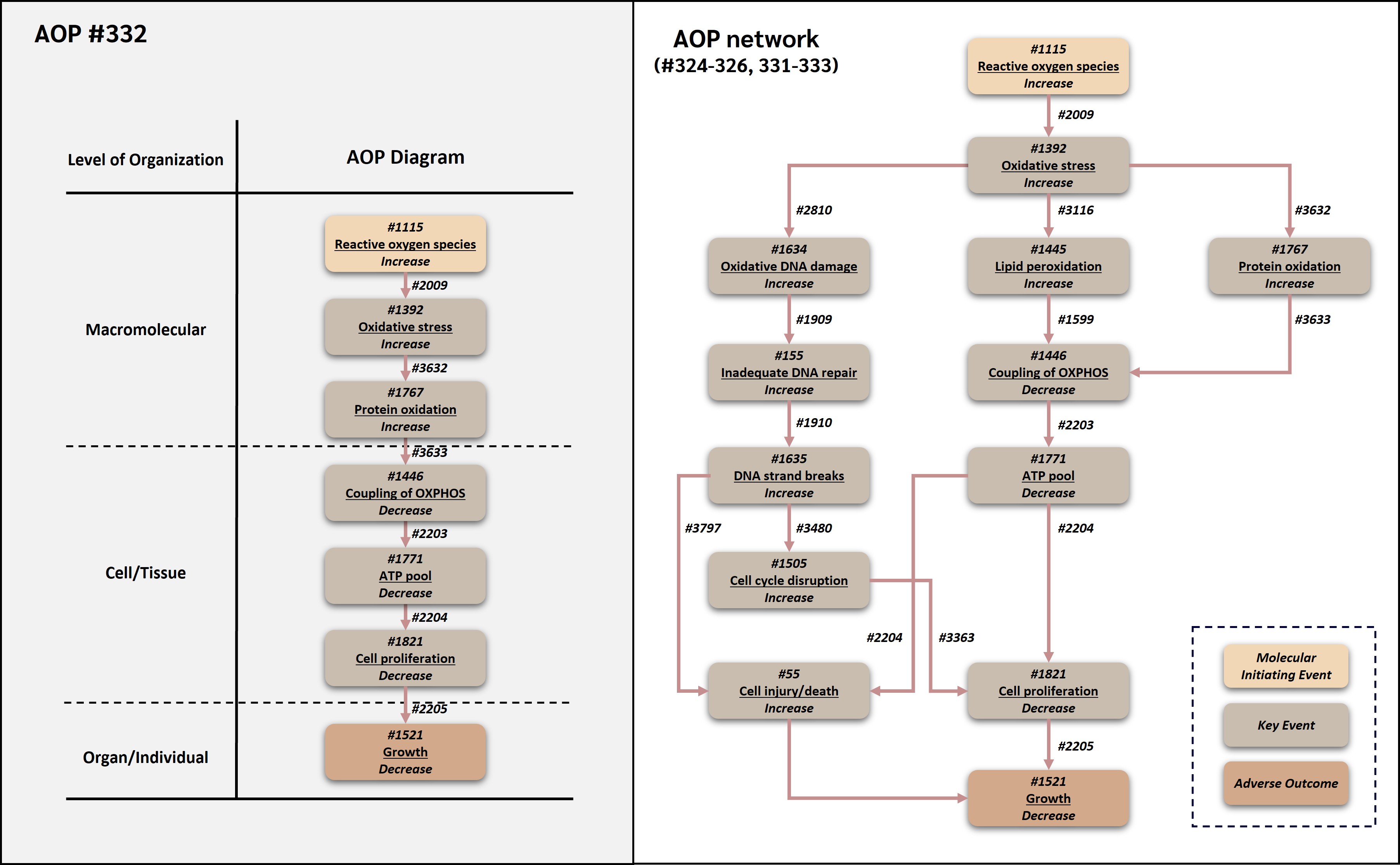
This adverse outcome pathway (AOP 332) describes a linear route by which increased reactive oxygen species (ROS) can lead to decreased organismal growth through oxidative stress-mediated protein oxidation and subsequent impairment of mitochondrial bioenergetics. Increased ROS is treated operationally as the molecular initiating event because it represents the earliest common measurable redox perturbation shared by diverse chemical and non-chemical stressors in the broader ROS-growth AOP network. Increased ROS leads to oxidative stress, which promotes oxidative modification of proteins. Oxidized proteins may lose catalytic, structural, transport, or regulatory function; they may also misfold, aggregate, or become targeted for degradation. When proteins required for mitochondrial electron transport, ATP synthase function, metabolite transport, or maintenance of mitochondrial membrane potential are oxidatively modified, coupling of oxidative phosphorylation (OXPHOS) can decrease. Reduced OXPHOS coupling decreases the cellular ATP pool, impairs ATP-dependent cell proliferation, and ultimately reduces growth.
AOP 332 reuses and connects established AOP-Wiki components from two major AOP contexts. The upstream ROS/oxidative stress module is associated with AOP 478, in which deposition of energy leads to oxidative stress through free radical generation and oxidative molecular damage (AOP-Wiki, 2026a). AOP 478 provides an AOP-Wiki precedent for oxidative stress as a conserved KE downstream of radical-generating stressors and for protein damage as an oxidative stress consequence. The downstream bioenergetic and growth module is directly associated with AOP 263, which causally links decreased coupling of OXPHOS to growth inhibition through ATP depletion and decreased cell proliferation (AOP-Wiki, 2026b; OECD, 2022; Song and Villeneuve, 2021). Thus, AOP 332 links an oxidative protein damage module to an OECD-endorsed OXPHOS-to-growth module. The AOP is relevant to environmental and human health contexts because ROS generation, protein oxidation, mitochondrial ATP production, cell proliferation, and organismal growth are broadly conserved in aerobic eukaryotes. Empirical support comes from studies in algae, fish, mollusks, mammalian systems, and human cells exposed to metals, hydrogen peroxide, hypoxia-reoxygenation, salinity or temperature stress, endogenous aging, and other oxidative stressors. This AOP can support mechanistic interpretation of oxidative stress-mediated growth impairment, assay selection, chemical prioritization, IATA development, and future quantitative AOP approaches for mitochondrial and oxidative stress-related toxicity.
Acknowledgement
This project was funded by the Research Council of Norway (RCN), grant no. RCN-315929 “EXPECT: In silico and experimental screening platform for characterizing environmental impact of industry development in the Arctic” (https://www.niva.no/en/projects/expect), the European Partnership for the Assessment of Risks from Chemicals (PARC) through European Union’s Horizon Europe research and innovation programme (Grant Agreement No 101057014, and supported by the NIVA Computational Toxicology Program, NCTP (https://www.niva.no/en/featured-pages/nctp, grant. No. RCN-342628).
AI disclosure
Artificial intelligence (AI) tools were used to support literature prioritization, review and AOP-Wiki page preparation in this work. AOP-helpFinder was used for automated literature mining, and ChatGPT (OpenAI) was used as an auxiliary tool for title and abstract screening, extraction of study metadata, and identification of potential weight-of-evidence indicators. AI-assisted outputs were used only to organize and prioritize information and were verified against the original sources by the authors before inclusion. Additional AI assistance was used for formatting, copy-editing, citation cross-checking, and harmonization of the AOP-Wiki pages. All scientific interpretations, weight-of-evidence judgments, final wording, and conclusions were determined and approved by the authors, who take full responsibility for the content and integrity of the work.
AOP Development Strategy
Context
ROS are continuously formed during aerobic metabolism and can also be generated in response to environmental stressors. At controlled levels, ROS participate in redox signaling, whereas excessive ROS can disturb redox homeostasis and initiate oxidative stress (Schieber and Chandel, 2014; Sies et al., 2017). Proteins are major targets of oxidative attack because many amino acid side chains and prosthetic groups are susceptible to oxidation, carbonylation, thiol oxidation, nitration, or other oxidative modifications. Protein oxidation can alter enzyme activity, protein-protein interactions, protein folding, stability, and degradation, and it is widely used as a marker of severe oxidative damage and cellular dysfunction (Dalle-Donne et al., 2006).
AOP 332 was developed to represent the protein oxidation-driven linear route within the broader ROS-growth AOP network. This route was selected because oxidative stress can directly modify proteins, including mitochondrial proteins involved in electron transport, OXPHOS coupling, ATP synthesis, metabolite transport, and maintenance of mitochondrial integrity. Oxidation of mitochondrial proteins can impair the efficiency of electron transfer and proton motive force utilization, thereby providing a mechanistically coherent bridge from oxidative stress to decreased coupling of OXPHOS (Murphy, 2009; Nicholls and Ferguson, 2013; Sokolov et al., 2019). The downstream sequence from decreased coupling of OXPHOS to decreased ATP pool, decreased cell proliferation, and decreased growth is represented in AOP 263 and provides the growth-relevant terminal module for AOP 332 (AOP-Wiki, 2026b; OECD, 2022; Song and Villeneuve, 2021).
Strategy
AOP 332 was developed using the principles described in OECD AOP guidance, including modular description of KEs and KERs, reuse of existing AOP-Wiki content where appropriate, evidence evaluation using biological plausibility, empirical support, essentiality, and quantitative understanding, and clear description of the biological domain of applicability (OECD, 2018, 2021). The objective was to assemble a focused linear pathway from reusable AOP-Wiki elements rather than to create an isolated de novo pathway. This is important because AOP 332 is one branch of the broader ROS-growth AOP network and because its downstream KEs and KERs overlap with the well-developed mitochondrial bioenergetics AOP 263.
Reuse of existing AOP-Wiki content was considered at the beginning of development. AOP 478 was reviewed because it provides an AOP-Wiki context for oxidative stress downstream of free radical generation and because it recognizes oxidative molecular damage, including protein damage, as a relevant consequence of oxidative stress. The KE increase in oxidative stress (Event 1392) and the conceptual linkage between radical generation, oxidative stress, and protein modification were therefore aligned with the AOP 478 context. AOP 263 was used as the primary source for the downstream module beginning with decreased coupling of OXPHOS (Event 1446), followed by decreased ATP pool (Event 1771), decreased cell proliferation (Event 1821), and decreased growth (Event 1521). The KERs decreased coupling of OXPHOS leading to decreased ATP pool, decreased ATP pool leading to decreased cell proliferation, and decreased cell proliferation leading to decreased growth were retained in the same causal order as AOP 263, supporting modular reuse and consistency with an OECD-endorsed pathway.
The literature review and evidence assembly process followed an AI-human hybrid workflow. First, event-specific search terms were developed for the KEs in AOP 332, including reactive oxygen species, oxidative stress, protein oxidation, protein carbonylation, oxidized proteins, mitochondrial protein oxidation, OXPHOS coupling, mitochondrial respiration, mitochondrial membrane potential, ATP depletion, cell proliferation, and growth. Synonyms, assay terms, representative stressors, taxa, and species names were also included. These terms were used in AOP-helpFinder to search PubMed for co-occurrence patterns between key events and mechanistic concepts, following text-mining approaches developed for AOP literature support (Carvaillo et al., 2019; Jornod et al., 2022). Exported records containing PMIDs, titles, abstracts, and matched terms were subjected to overlap analysis to remove redundant records and filter weakly relevant taxa- or endpoint-unrelated literature.
In the second phase, titles and abstracts from AOP-helpFinder and targeted manual searches were pre-screened using ChatGPT (GPT-4, OpenAI, San Francisco, CA, USA) as an auxiliary prioritization tool. The LLM was used to extract study metadata, including stressor, species, biological system, dose or concentration, and exposure duration; to identify evidence type, including biological plausibility, empirical support, and essentiality; and to flag weight-of-evidence indicators such as dose-response concordance, temporal concordance, incidence concordance, and intervention or rescue evidence. Studies were classified as high, medium, or low relevance. High-relevance studies were retrieved for full-text review, medium-relevance studies were reserved as supporting evidence, and low-relevance studies were documented as low priority or excluded. For full texts, LLM-assisted review was used only to organize candidate information; all outputs were verified against the original articles by human experts.
The final phase consisted of manual expert curation and weight-of-evidence evaluation. Experts verified relevance and interpretation of selected studies, extracted data into KER evidence tables, and evaluated biological plausibility, empirical support, essentiality, quantitative understanding, inconsistencies, and modulating factors. Targeted searches were also performed to fill evidence gaps for protein oxidation and mitochondrial dysfunction. Studies were prioritized when they measured two or more KEs in the same system, reported exposure duration and dose or concentration, or provided evidence relevant to concordance across the pathway. Mechanistic reviews and OECD reports were used to support well-established biological plausibility, whereas primary experimental studies were prioritized for empirical support (Dalle-Donne et al., 2006; Canesi et al., 2010; Almaida-Pagán et al., 2014; Sokolov et al., 2019; Song and Villeneuve, 2021; OECD, 2022).
Summary of the AOP
Events
Molecular Initiating Events (MIE), Key Events (KE), Adverse Outcomes (AO)
| Sequence | Type | Event ID | Title | Short name |
|---|---|---|---|---|
| MIE | 1115 | Increase, Reactive oxygen species | Increase, ROS | |
| KE | 1392 | Increase, Oxidative Stress | Increase, Oxidative Stress | |
| KE | 1767 | Increase, Protein oxidation | Increase, Protein oxidation | |
| KE | 1446 | Decrease, Coupling of oxidative phosphorylation | Decrease, Coupling of OXPHOS | |
| KE | 1771 | Decrease, Adenosine triphosphate pool | Decrease, ATP pool | |
| KE | 1821 | Decrease, Cell proliferation | Decrease, Cell proliferation | |
| AO | 1521 | Decrease, Growth | Decrease, Growth |
Key Event Relationships
| Upstream Event | Relationship Type | Downstream Event | Evidence | Quantitative Understanding |
|---|---|---|---|---|
| Increase, Reactive oxygen species | adjacent | Increase, Oxidative Stress | High | Moderate |
| Increase, Oxidative Stress | adjacent | Increase, Protein oxidation | High | Moderate |
| Increase, Protein oxidation | adjacent | Decrease, Coupling of oxidative phosphorylation | Moderate | Low |
| Decrease, Coupling of oxidative phosphorylation | adjacent | Decrease, Adenosine triphosphate pool | High | High |
| Decrease, Adenosine triphosphate pool | adjacent | Decrease, Cell proliferation | High | Moderate |
| Decrease, Cell proliferation | adjacent | Decrease, Growth | High | Moderate |
Stressors
| Name | Evidence |
|---|---|
| Hydrogen peroxide | |
| Heavy metals (cadmium, lead, copper, iron, nickel) | |
| Ionizing Radiation | |
| Ultraviolet B radiation |
Overall Assessment of the AOP
The overall weight of evidence supporting AOP 332 is considered moderate. Biological plausibility is high for all six KERs in the pathway. The chemical reactivity of ROS with proteins is well established, and the functional consequences of oxidative modification of mitochondrial respiratory proteins for OXPHOS coupling are mechanistically well supported. The downstream module from decreased OXPHOS coupling through ATP depletion, reduced cell proliferation, and decreased growth is directly reused from OECD-endorsed AOP 263, contributing high biological plausibility and established quantitative relationships for this segment (OECD, 2022; Song and Villeneuve, 2021). Empirical support is high for the upstream ROS-to-oxidative-stress and oxidative-stress-to-protein-oxidation relationships, where multiple stressors across algae, fish, mollusks, and mammalian systems demonstrate concordant increases in oxidative stress markers and protein carbonylation or oxidized protein products. Empirical support for the protein oxidation-to-OXPHOS coupling transition (KER 3633) is rated moderate, as evidence from aging, hypoxia-reoxygenation, and metal exposure studies links oxidative mitochondrial proteome changes to altered bioenergetics, but controlled intervention experiments specifically targeting protein oxidation without confounding other oxidative damage processes are limited. Essentiality is rated moderate to high overall, with the strongest direct support for the AOP 263 OXPHOS and ATP segment. Quantitative understanding is highest for the AOP 263-derived downstream module and low to moderate for the protein oxidation-to-OXPHOS transition, where generalizable cross-taxa quantitative models are lacking. The main uncertainties are the causal versus correlational nature of the protein oxidation-OXPHOS relationship, given that lipid peroxidation and other oxidative processes often co-occur, and the variable capacity of proteasomal and chaperone systems to mitigate protein oxidation-related mitochondrial dysfunction. AOP 332 is most suitable for mechanistic interpretation and chemical prioritisation for oxidative stress-related growth impairment (OECD, 2018; Becker et al., 2015).
Domain of Applicability
Life Stage Applicability| Life Stage | Evidence |
|---|---|
| All life stages | Moderate |
| Term | Scientific Term | Evidence | Links |
|---|---|---|---|
| humans | Homo sapiens | Moderate | NCBI |
| mammals | mammals | Moderate | NCBI |
| fish | fish | Moderate | NCBI |
| crustaceans | Daphnia magna | Moderate | NCBI |
| green algae | Ulva compressa | Moderate | NCBI |
| Sex | Evidence |
|---|---|
| Unspecific | Moderate |
The domain of applicability for AOP 332 is broad across aerobic eukaryotic organisms in which ROS generation, oxidative stress responses, protein oxidation, mitochondrial oxidative phosphorylation, ATP-dependent cell proliferation, and growth are biologically relevant. The AOP is most directly applicable to organisms or life stages in which growth depends substantially on mitochondrial ATP supply and cell proliferation. It is also applicable to cellular systems used to evaluate oxidative stress, mitochondrial dysfunction, ATP depletion, and proliferation outcomes.
The stressor domain includes direct oxidants, redox-cycling chemicals, metals, radiation, hypoxia-reoxygenation, temperature or salinity stress, and endogenous oxidative stress. Because the MIE is defined operationally as increased ROS rather than as a chemical-specific interaction, AOP 332 should be applied when evidence demonstrates increased ROS or oxidative stress and when downstream evidence supports protein oxidation and mitochondrial bioenergetic impairment. Important modifiers include antioxidant capacity, protein repair and degradation capacity, mitochondrial reserve capacity, temperature, oxygen availability, nutrient status, species metabolic rate, and growth stage.
Essentiality of the Key Events
Essentiality is evaluated for the overall AOP based on whether preventing or modifying upstream KEs changes downstream KEs or the AO. The strongest direct essentiality evidence is available for the downstream AOP 263 module, where restoring mitochondrial coupling or ATP production can recover downstream bioenergetic and proliferative functions. Essentiality for protein oxidation is mechanistically plausible and supported by intervention and association evidence, but direct experiments showing that selective prevention of protein oxidation blocks all downstream events remain limited.
|
Key event |
Essentiality |
Rationale |
Experimental manipulation evidence (KE knock-out / inhibition / rescue) |
Uncertainties |
|
Event 1115: Reactive oxygen species, increased |
Moderate |
ROS scavenging and antioxidant interventions often attenuate oxidative stress and protein oxidation in oxidative stress models (Schieber and Chandel, 2014; Sies et al., 2017). |
Indirect (stop/attenuation): antioxidant and ROS-scavenger pre-treatment reduces oxidative stress and downstream damage across oxidative-stress models (Schieber and Chandel, 2014; Sies et al., 2017). No selective single-source ROS knock-out is available. |
ROS also serve physiological signaling roles; increased ROS may not progress if antioxidant systems compensate. |
|
Event 1392: Oxidative stress, increased |
Moderate to high |
Oxidative stress is required for widespread oxidative protein modification when oxidant production exceeds antioxidant and repair capacity. AOP 478 supports oxidative stress as a central KE downstream of radical generation (AOP-Wiki, 2026a). |
Indirect: modulation of antioxidant capacity alters progression to oxidative macromolecular damage; oxidative stress is the curated hub KE in endorsed AOP 478 (AOP-Wiki, 2026a; Carrothers et al., 2025). |
Different oxidative stress biomarkers may capture different aspects of redox imbalance. |
|
Event 1767: Protein oxidation, increased |
Moderate |
Protein carbonylation and related oxidative modifications can impair enzyme activity, protein folding, degradation, and mitochondrial function (Dalle-Donne et al., 2006). Cadmium-induced protein carbonylation and actin glutathionylation in mussel hemocytes were reduced by oxidase/NOS inhibitors, supporting causal involvement of oxidative signaling (Canesi et al., 2010). |
Direct (partial): cadmium-induced protein carbonylation and actin glutathionylation reduced by oxidase/NOS inhibitors in mussel hemocytes (Canesi et al., 2010); GSTA4 silencing raised mitochondrial protein carbonylation and target knockdown reduced respiration (Curtis et al., 2012). |
Protein oxidation can be cause, consequence, or marker of cellular stress; selective intervention evidence is limited. |
|
Event 1446: Coupling of OXPHOS, decreased |
High |
This KE is reused from AOP 263. Evidence from AOP 263 supports essentiality because uncoupler removal or restoration of mitochondrial coupling can recover mitochondrial membrane potential and ATP levels (AOP-Wiki, 2026b; OECD, 2022; Song and Villeneuve, 2021). |
Direct (rescue): removal of uncouplers or restoration of coupling recovers mitochondrial membrane potential and ATP in the endorsed AOP 263 module (AOP-Wiki, 2026b; OECD, 2022; Song and Villeneuve, 2021). |
Mild uncoupling can be adaptive and may reduce ROS generation depending on context. |
|
Event 1771: ATP pool, decreased |
Moderate |
ATP depletion is associated with reduced proliferation and cytotoxicity in multiple systems and is a central KE in AOP 263 (AOP-Wiki, 2026b; OECD, 2022). |
Indirect: ATP-restoration experiments reduce downstream injury/proliferation deficits; central KE in endorsed AOP 263 (Leist et al., 1997; Nicotera et al., 1998; OECD, 2022). |
Cells may compensate via glycolysis or altered energy allocation. |
|
Event 1821: Cell proliferation, decreased |
Moderate |
Growth is dependent on cell number and biomass accumulation; AOP 263 supports decreased cell proliferation as a direct link between bioenergetic impairment and growth reduction (AOP-Wiki, 2026b; Conlon and Raff, 1999; OECD, 2022). |
Indirect: proliferation deficit links bioenergetic/genotoxic upstream to growth; reused from endorsed AOP 263 with KER 2205 (AOP-Wiki, 2026d; Conlon and Raff, 1999; OECD, 2022; Song and Villeneuve, 2021). |
Growth can also be influenced by cell size, nutrient status, development, and cell death. |
|
Event 1521: Growth, decreased (AO) |
Not applicable (AO) |
Growth is the adverse outcome and a regulatory-relevant endpoint across several OECD and ISO test systems; AOP 263 provides precedent for decreased growth as an AO downstream of mitochondrial bioenergetic impairment (OECD, 2022; Song and Villeneuve, 2021). |
As the adverse outcome, essentiality is assessed for upstream KEs; AOP 263 provides precedent for decreased growth as an AO downstream of these modules (OECD, 2022; Song and Villeneuve, 2021). |
Growth is integrative and can arise through multiple mechanisms. |
Weight of Evidence Summary
Evidence assessment is organized by KER. Calls follow OECD weight-of-evidence considerations for biological plausibility, empirical support, and quantitative understanding (OECD, 2018, 2021).
Biological plausibility of KERs
|
KER |
Biological plausibility call |
Rationale |
|
Relationship 2009: ROS increase leads to oxidative stress increase |
High |
Oxidative stress reflects an imbalance between oxidants and antioxidant defenses; ROS are major cellular oxidants and primary drivers of redox imbalance (Schieber and Chandel, 2014; Sies et al., 2017). AOP 478 provides a curated context for oxidative stress downstream of radical generation (AOP-Wiki, 2026a). |
|
Relationship 3632: oxidative stress increase leads to protein oxidation increase |
High |
ROS and related oxidants can modify protein side chains, thiols, metal centers, and prosthetic groups, producing carbonylated, glutathionylated, misfolded, or aggregated proteins (Dalle-Donne et al., 2006; Sies et al., 2017). |
|
Relationship 3633: protein oxidation increase leads to decreased coupling of OXPHOS |
Moderate to high |
Mitochondrial OXPHOS depends on the integrity of electron transport complexes, ATP synthase, carrier proteins, and membrane-associated protein assemblies. Oxidative modification of these proteins can impair electron transfer, proton pumping, membrane potential, and ATP synthesis efficiency (Murphy, 2009; Nicholls and Ferguson, 2013; Sokolov et al., 2019). |
|
Relationship 2203: decreased coupling of OXPHOS leads to decreased ATP pool |
High |
This relationship is reused from AOP 263. OXPHOS coupling is a major determinant of ATP production in aerobic eukaryotic cells; reduced coupling lowers ATP synthesis efficiency (AOP-Wiki, 2026b; OECD, 2022; Song and Villeneuve, 2021). |
|
Relationship 2204: decreased ATP pool leads to decreased cell proliferation |
High |
This relationship is reused from AOP 263. Cell proliferation requires ATP for DNA replication, mitosis, biosynthesis, and maintenance of cellular processes; ATP depletion therefore plausibly reduces proliferation (AOP-Wiki, 2026b; Bonora et al., 2012; OECD, 2022). |
|
Relationship 2205: decreased cell proliferation leads to decreased growth |
High |
This relationship is reused from AOP 263. Organismal, tissue, and population growth require accumulation of cells and biomass; sustained reduction in proliferation is therefore expected to reduce growth (AOP-Wiki, 2026b; Conlon and Raff, 1999; OECD, 2022). |
Empirical support for KERs
|
KER |
Empirical support call |
Rationale |
Inconsistencies or evidence gaps |
|
Relationship 2009: ROS increase leads to oxidative stress increase |
High |
Multiple studies demonstrate concordance between ROS-producing stressors and oxidative stress biomarkers. Paraquat increased ROS and antioxidant enzyme responses in Chlorella vulgaris (Qian et al., 2009). In fish, infection-induced ROS coincided with antioxidant and inflammatory responses (Gao et al., 2022). |
ROS is often transient and indirectly measured; oxidative stress endpoints differ across studies. |
|
Relationship 3632: oxidative stress increase leads to protein oxidation increase |
High |
Oxidative stressors increase protein carbonyls or related protein oxidation endpoints. Cadmium and hydrogen peroxide increased protein carbonylation and redox modification in Chlamydomonas systems (Zaffagnini et al., 2012). Cadmium induced protein carbonylation and actin glutathionylation in mussel hemocytes (Canesi et al., 2010). Thermal stress in zebrafish increased protein carbonyls with antioxidant responses (Tseng et al., 2011). |
Protein oxidation endpoints are heterogeneous; some studies measure total carbonyls whereas others identify specific oxidized proteins. |
|
Relationship 3633: protein oxidation increase leads to decreased coupling of OXPHOS |
Moderate |
Evidence links oxidative protein damage or mitochondrial proteome modification with altered mitochondrial function. Age-associated oxidative changes in zebrafish were associated with changes in mitochondrial oxidative status and aconitase activity (Almaida-Pagán et al., 2014). Hypoxia-reoxygenation altered mitochondrial proteome and bioenergetics in Crassostrea gigas (Sokolov et al., 2019). |
Many studies measure correlation rather than direct causation; protein oxidation may occur alongside lipid peroxidation or other mitochondrial damage. |
|
Relationship 2203: decreased coupling of OXPHOS leads to decreased ATP pool |
High |
This relationship is supported by AOP 263 and by multiple studies of mitochondrial uncouplers and mitochondrial toxicants showing ATP depletion following reduced OXPHOS efficiency (AOP-Wiki, 2026b; OECD, 2022; Song and Villeneuve, 2021). |
Cells may transiently compensate through glycolysis or substrate switching. |
|
Relationship 2204: decreased ATP pool leads to decreased cell proliferation |
Moderate to high |
AOP 263 reports concordance between ATP depletion and decreased cell proliferation across biological systems. ATP content is widely used as a quantitative indicator of cell viability and proliferative capacity (AOP-Wiki, 2026b; Bonora et al., 2012; OECD, 2022). |
ATP depletion may lead to either proliferation arrest or cell death depending on severity and duration. |
|
Relationship 2205: decreased cell proliferation leads to decreased growth |
Moderate |
AOP 263 provides empirical support for this relationship and identifies growth as a biologically and regulatory relevant endpoint downstream of reduced cell proliferation (AOP-Wiki, 2026b; OECD, 2022; Song and Villeneuve, 2021). |
Growth integrates many processes, and direct measurement of proliferation and organismal growth in the same study is less common. |
Inconsistencies and uncertainties
The main uncertainty in AOP 332 is the causal versus correlational nature of the protein oxidation to decreased OXPHOS coupling relationship (KER 3633), because lipid peroxidation and other oxidative processes frequently co-occur and can independently impair mitochondrial function. Controlled intervention experiments that target protein oxidation without confounding other oxidative damage are limited, and the capacity of proteasomal and chaperone systems to mitigate protein-oxidation-related mitochondrial dysfunction varies across taxa and exposure conditions. As with the other AOPs in this network, ROS-mediated growth inhibition can also proceed through genotoxic, lipid peroxidation, and cell death branches, so the protein oxidation branch represented here captures only one mechanistic route. Finally, growth is a multifactorial apical endpoint, which limits quantitative prediction of organismal growth from upstream protein oxidation and bioenergetic events.
Quantitative Consideration
Quantitative understanding of AOP 332 is strongest for the downstream AOP 263 module and more limited for the protein oxidation to OXPHOS transition. Protein oxidation is measurable using protein carbonyl assays, redox proteomics, and targeted detection of oxidized mitochondrial proteins, but translation from the extent of protein oxidation to a quantitative decrement in OXPHOS coupling remains context-dependent.
|
KER |
Quantitative understanding call |
Rationale |
|
Relationship 2009: ROS increase leads to oxidative stress increase |
Low to moderate |
ROS and oxidative stress biomarkers can be quantified, but ROS are short-lived and measurement is assay-dependent (Sies et al., 2017). |
|
Relationship 3632: oxidative stress increase leads to protein oxidation increase |
Moderate |
Protein carbonyls and redox proteomics provide quantitative measures of protein oxidation, but response-response models linking oxidative stress magnitude to protein oxidation are not broadly generalizable (Dalle-Donne et al., 2006). |
|
Relationship 3633: protein oxidation increase leads to decreased coupling of OXPHOS |
Low to moderate |
Specific oxidation of mitochondrial proteins can be linked to altered mitochondrial function in some systems, but predictive quantitative models are not yet established across taxa or stressors (Sokolov et al., 2019). |
|
Relationship 2203: decreased coupling of OXPHOS leads to decreased ATP pool |
High |
AOP 263 includes quantitative understanding for OXPHOS coupling and ATP depletion, supported by bioenergetic theory and experimental response-response relationships (AOP-Wiki, 2026b; OECD, 2022; Song and Villeneuve, 2021). |
|
Relationship 2204: decreased ATP pool leads to decreased cell proliferation |
Moderate |
ATP is often used as a quantitative indicator of cell status and proliferation, but thresholds vary by cell type and stress duration (Bonora et al., 2012; OECD, 2022). |
|
Relationship 2205: decreased cell proliferation leads to decreased growth |
Moderate |
Quantitative relationships between proliferation and growth exist in developmental and tissue growth biology, but stressor-specific models for this AOP remain limited (Conlon and Raff, 1999; OECD, 2022). |
BMD/POD-anchored concordance
The following benchmark-dose/point-of-departure (BMD/POD) concordance table anchors AOP 332 to quantitative cross-KE ordering, in line with Handbook section 4C. The multiomics point-of-departure (moPOD) dataset for gamma-irradiated Daphnia magna (Song et al., 2023) provides POD magnitudes for increased ROS, decreased ATP, decreased OXPHOS coupling, and cell death, demonstrating the expected upstream-to-downstream POD ordering (more sensitive PODs upstream). The moPOD is presented as POD magnitude evidence, not as a causal re-ordering of KEs. The Lemna minor EDR50 range provides a whole-pathway apical anchor in an aquatic primary producer.
|
Key event (functional category) |
POD metric |
POD value (mGy/h) |
POD ordering |
Source |
|
KE 1115: ROS, increased (mROS) |
moPOD (multiomics POD) |
0.4 |
1 (most sensitive) |
Song et al., 2023 |
|
KE 1771: ATP pool, decreased |
moPOD |
2.5 |
2 |
Song et al., 2023 |
|
KE 1446: OXPHOS coupling, decreased (UPS/OXPHOS module) |
moPOD |
42.3 |
3 |
Song et al., 2023 |
|
KE 55: Cell injury/death (apoptosis) |
moPOD |
42.3 |
3 (least sensitive) |
Song et al., 2023 |
|
Upstream KE chain → growth (Lemna minor, gamma) |
EDR50 (growth) |
31.5–54.8 (mGy/h) |
whole-pathway apical |
Xie et al., 2018, 2019, 2022 |
Considerations for Potential Applications of the AOP (optional)
AOP 332 can support mechanistic interpretation of growth impairment caused by oxidative stressors that produce protein oxidation and mitochondrial bioenergetic dysfunction. It is particularly useful for organizing evidence from assays measuring protein carbonyls, redox proteomics, mitochondrial respiration, mitochondrial membrane potential, ATP content, cell proliferation, and organismal growth. Because the downstream module is shared with AOP 263, the AOP can contribute to IATA and NAM-based screening strategies that use mitochondrial function, ATP status, and proliferation as early warning indicators for growth effects.
The AOP may also support chemical prioritization and grouping for stressors that induce ROS generation or oxidative stress and that show evidence for protein oxidation or mitochondrial impairment. Potential stressor classes include metals, redox-active organic chemicals, radiation, hypoxia-reoxygenation, and other environmental conditions that increase oxidative protein damage. The AOP should not be used as a stand-alone quantitative predictor of growth inhibition without additional empirical support, because the protein oxidation to OXPHOS transition remains less quantitatively resolved than the downstream AOP 263 module.
References
Almaida-Pagán, P.F., Lucas-Sánchez, A., & Tocher, D.R. (2014). Changes in mitochondrial membrane composition and oxidative status during rapid growth, maturation and aging in zebrafish, Danio rerio. Biochimica et Biophysica Acta - Molecular and Cell Biology of Lipids, 1841(7), 1003-1011. https://doi.org/10.1016/j.bbalip.2014.04.004
AOP-Wiki. (2026a). AOP 478: Deposition of energy leading to occurrence of cataracts. Adverse Outcome Pathway Wiki. https://aopwiki.org/aops/478
AOP-Wiki. (2026b). AOP 263: Uncoupling of oxidative phosphorylation leading to growth inhibition via decreased cell proliferation. Adverse Outcome Pathway Wiki. https://aopwiki.org/aops/263
Ayala, A., Munoz, M.F., & Arguelles, S. (2014). Lipid peroxidation: Production, metabolism, and signaling mechanisms of malondialdehyde and 4-hydroxy-2-nonenal. Oxidative Medicine and Cellular Longevity, 2014, 360438. https://doi.org/10.1155/2014/360438
Barata, C., Varo, I., Navarro, J.C., Arun, S., & Porte, C. (2005). Antioxidant enzyme activities and lipid peroxidation in the freshwater cladoceran Daphnia magna exposed to redox cycling compounds. Comparative Biochemistry and Physiology Part C: Toxicology & Pharmacology, 140(2), 175-186. https://doi.org/10.1016/j.cca.2005.01.013
Bonora, M., Patergnani, S., Rimessi, A., De Marchi, E., Suski, J.M., Bononi, A., Giorgi, C., Marchi, S., Missiroli, S., Poletti, F., Wieckowski, M.R., & Pinton, P. (2012). ATP synthesis and storage. Purinergic Signaling, 8(3), 343-357. https://doi.org/10.1007/s11302-012-9305-8
Canesi, L., Ciacci, C., Betti, M., Lorusso, L.C., Marchi, B., Burattini, S., Falcieri, E., & Gallo, G. (2010). The role of signaling molecules on actin glutathionylation and protein carbonylation induced by cadmium in hemocytes of mussel Mytilus galloprovincialis. Journal of Experimental Biology, 213(3), 361-372. https://doi.org/10.1242/jeb.035550
Carvaillo, J.C., Barouki, R., Coumoul, X., & Audouze, K. (2019). Linking bisphenol S to adverse outcome pathways using a combined text mining and systems biology approach. Environmental Health Perspectives, 127(4), 047005. https://doi.org/10.1289/EHP4200
Conlon, I., & Raff, M. (1999). Size control in animal development. Cell, 96(2), 235-244. https://doi.org/10.1016/S0092-8674(00)80563-2
Curtis, J. M., Hahn, W. S., Stone, M. D., Inda, J. J., Droullard, D. J., Kuzmicic, J. P., Donoghue, M. A., Long, E. K., Armien, A. G., Lavandero, S., Arriaga, E., Griffin, T. J., & Bernlohr, D. A. (2012). Protein carbonylation and adipocyte mitochondrial function. Journal of Biological Chemistry, 287(39), 32967-32980. https://doi.org/10.1074/jbc.M112.400663
Dalle-Donne, I., Aldini, G., Carini, M., Colombo, R., Rossi, R., & Milzani, A. (2006). Protein carbonylation, cellular dysfunction, and disease progression. Journal of Cellular and Molecular Medicine, 10(2), 389-406. https://doi.org/10.1111/j.1582-4934.2006.tb00407.x
Gao, J., Liu, M., Guo, H., Zhu, K., Liu, B., Liu, B., & Zhang, D. (2022). ROS induced by Streptococcus agalactiae activate inflammatory responses via the TNF-alpha/NF-kappaB signaling pathway in golden pompano Trachinotus ovatus (Linnaeus, 1758). Antioxidants, 11(9), 1809. https://doi.org/10.3390/antiox11091809
Jornod, F., Jaylet, T., Blaha, L., Sarigiannis, D., Tamisier, L., & Audouze, K. (2022). AOP-helpFinder webserver: A tool for comprehensive analysis of the literature to support adverse outcome pathways development. Bioinformatics, 38(4), 1173-1175. https://doi.org/10.1093/bioinformatics/btab750
Murphy, M.P. (2009). How mitochondria produce reactive oxygen species. Biochemical Journal, 417(1), 1-13. https://doi.org/10.1042/BJ20081386
Nicholls, D.G., & Ferguson, S.J. (2013). Bioenergetics 4. Academic Press.
Nicotera, P., Leist, M., & Ferrando-May, E. (1998). Intracellular ATP, a switch in the decision between apoptosis and necrosis. Toxicology Letters, 102-103, 139-142. https://doi.org/10.1016/S0378-4274(98)00298-7
OECD. (2018). Users' handbook supplement to the guidance document for developing and assessing adverse outcome pathways. OECD Series on Adverse Outcome Pathways No. 1. OECD Publishing.
OECD. (2021). Guidance document for the scientific review of adverse outcome pathways. OECD Series on Testing and Assessment No. 344. OECD Publishing.
OECD. (2022). Uncoupling of oxidative phosphorylation leading to growth inhibition via decreased cell proliferation. OECD Series on Adverse Outcome Pathways No. 28. OECD Publishing. https://doi.org/10.1787/f20867c1-en
Qian, H., Chen, W., Sun, L., Jin, Y., Liu, W., & Fu, Z. (2009). Inhibitory effects of paraquat on photosynthesis and the response to oxidative stress in Chlorella vulgaris. Ecotoxicology, 18(5), 537-543. https://doi.org/10.1007/s10646-009-0311-8
Schieber, M., & Chandel, N.S. (2014). ROS function in redox signaling and oxidative stress. Current Biology, 24(10), R453-R462. https://doi.org/10.1016/j.cub.2014.03.034
Sies, H., Berndt, C., & Jones, D.P. (2017). Oxidative stress. Annual Review of Biochemistry, 86, 715-748. https://doi.org/10.1146/annurev-biochem-061516-045037
Sokolov, E.P., Markert, S., Hinzke, T., Hirschfeld, C., Becher, D., Ponsuksili, S., & Sokolova, I.M. (2019). Effects of hypoxia-reoxygenation stress on mitochondrial proteome and bioenergetics of the hypoxia-tolerant marine bivalve Crassostrea gigas. Journal of Proteomics, 194, 99-111. https://doi.org/10.1016/j.jprot.2018.12.009
Song, Y., & Villeneuve, D.L. (2021). AOP report: Uncoupling of oxidative phosphorylation leading to growth inhibition via decreased cell proliferation. Environmental Toxicology and Chemistry, 40(11), 2951-2963. https://doi.org/10.1002/etc.5197
Tseng, Y.C., Chen, R.D., Lucassen, M., Schmidt, M.M., Dringen, R., Abele, D., & Hwang, P.P. (2011). Exploring uncoupling proteins and antioxidant mechanisms under acute cold exposure in brains of fish. PLoS ONE, 6(3), e18180. https://doi.org/10.1371/journal.pone.0018180
Zaffagnini, M., Bedhomme, M., Groni, H., Marchand, C.H., Puppo, C., Gontero, B., Cassier-Chauvat, C., Decottignies, P., & Lemaire, S.D. (2012). Glutathionylation in the photosynthetic model organism Chlamydomonas reinhardtii: A proteomic survey. Molecular & Cellular Proteomics, 11(8), M111.014142. https://doi.org/10.1074/mcp.M111.014142
Appendix 1
List of MIEs in this AOP
Event: 1115: Increase, Reactive oxygen species
Short Name: Increase, ROS
Event Component
| Process | Object | Action |
|---|---|---|
| reactive oxygen species biosynthetic process | reactive oxygen species | increased |
AOPs Including This Key Event
Biological Context
| Level of Biological Organization |
|---|
| Cellular |
Cell term
| Cell term |
|---|
| cell |
Organ term
| Organ term |
|---|
| organ |
Domain of Applicability
Taxonomic Applicability| Term | Scientific Term | Evidence | Links |
|---|---|---|---|
| Vertebrates | Vertebrates | High | NCBI |
| human | Homo sapiens | Moderate | NCBI |
| human and other cells in culture | human and other cells in culture | Moderate | NCBI |
| mouse | Mus musculus | Moderate | NCBI |
| crustaceans | Daphnia magna | High | NCBI |
| Lemna minor | Lemna minor | High | NCBI |
| zebrafish | Danio rerio | High | NCBI |
| Life Stage | Evidence |
|---|---|
| All life stages | High |
| Sex | Evidence |
|---|---|
| Unspecific | High |
| Mixed | High |
ROS is a normal constituent found in all organisms, lifestages, and sexes.
Key Event Description
Biological State: increased reactive oxygen species (ROS)
Biological compartment: an entire cell -- may be cytosolic, may also enter organelles.
Reactive oxygen species (ROS) are O2- derived molecules that can be both free radicals (e.g. superoxide, hydroxyl, peroxyl, alcoxyl) and non-radicals (hypochlorous acid, ozone and singlet oxygen) (Bedard and Krause 2007; Ozcan and Ogun 2015). ROS production occurs naturally in all kinds of tissues inside various cellular compartments, such as mitochondria and peroxisomes (Drew and Leeuwenburgh 2002; Ozcan and Ogun 2015). Furthermore, these molecules have an important function in the regulation of several biological processes – they might act as antimicrobial agents or triggers of animal gamete activation and capacitation (Goud et al. 2008; Parrish 2010; Bisht et al. 2017).
However, in environmental stress situations (exposure to radiation, chemicals, high temperatures) these molecules have its levels drastically increased, and overly interact with macromolecules, namely nucleic acids, proteins, carbohydrates and lipids, causing cell and tissue damage (Brieger et al. 2012; Ozcan and Ogun 2015).
Reactive oxygen species (ROS) refers to the chemical species superoxide, hydrogen peroxide, and their secondary reactive products. In the biological context, ROS are signaling molecules with important roles in cell energy metabolism, cell proliferation, and fate. Therefore, balancing ROS levels at the cellular and tissue level is an important part of many biological processes. Disbalance, mainly an increase in ROS levels, can cause cell dysfunction and irreversible cell damage.
ROS are produced from both exogenous stressors and normal endogenous cellular processes, such as the mitochondrial electron transport chain (ETC). Inhibition of the ETC can result in the accumulation of ROS. Exposure to chemicals, heavy metal ions, or ionizing radiation can also result in increased production of ROS. Chemicals and heavy metal ions can deplete cellular antioxidants reducing the cell’s ability to control cellular ROS and resulting in the accumulation of ROS. Cellular antioxidants include glutathione (GSH), protein sulfhydryl groups, superoxide dismutase (SOD).
ROS are radicals, ions, or molecules that have a single unpaired electron in their outermost shell of electrons, which can be categorized into two groups: free oxygen radicals and non-radical ROS [Liou et al., 2010].
<Free oxygen radicals>
|
superoxide |
O2·- |
|
hydroxyl radical |
·OH |
|
nitric oxide |
NO· |
|
organic radicals |
R· |
|
peroxyl radicals |
ROO· |
|
alkoxyl radicals |
RO· |
|
thiyl radicals |
RS· |
|
sulfonyl radicals |
ROS· |
|
thiyl peroxyl radicals |
RSOO· |
|
disulfides |
RSSR |
<Non-radical ROS>
|
hydrogen peroxide |
H2O2 |
|
singlet oxygen |
1O2 |
|
ozone/trioxygen |
O3 |
|
organic hydroperoxides |
ROOH |
|
hypochlorite |
ClO- |
|
peroxynitrite |
ONOO- |
|
nitrosoperoxycarbonate anion |
O=NOOCO2- |
|
nitrocarbonate anion |
O2NOCO2- |
|
dinitrogen dioxide |
N2O2 |
|
nitronium |
NO2+ |
|
highly reactive lipid- or carbohydrate-derived carbonyl compounds |
|
Potential sources of ROS include NADPH oxidase, xanthine oxidase, mitochondria, nitric oxide synthase, cytochrome P450, lipoxygenase/cyclooxygenase, and monoamine oxidase [Granger et al., 2015]. ROS are generated through NADPH oxidases consisting of p47phox and p67phox. ROS are generated through xanthine oxidase activation in sepsis [Ramos et al., 2018]. Arsenic produces ROS [Zhang et al., 2011]. Mitochondria-targeted paraquat and metformin mediate ROS production [Chowdhury et al., 2020]. ROS are generated by bleomycin [Lu et al., 2010]. Radiation induces dose-dependent ROS production [Ji et al., 2019].
ROS are generated in the course of cellular respiration, metabolism, cell signaling, and inflammation [Dickinson and Chang 2011; Egea et al. 2017]. Hydrogen peroxide is also made by the endoplasmic reticulum in the course of protein folding. Nitric oxide (NO) is produced at the highest levels by nitric oxide synthase in endothelial cells and phagocytes. NO production is one of the main mechanisms by which phagocytes kill bacteria [Wang et al., 2017]. The other species are produced by reactions with superoxide or peroxide, or by other free radicals or enzymes.
ROS activity is principally local. Most ROS have short half-lives, ranging from nano- to milliseconds, so diffusion is limited, while reactive nitrogen species (RNS) nitric oxide or peroxynitrite can survive long enough to diffuse across membranes [Calcerrada et al. 2011]. Consequently, local concentrations of ROS are much higher than average cellular concentrations, and signaling is typically controlled by colocalization with redox buffers [Dickinson and Chang 2011; Egea et al. 2017].
Although their existence is limited temporally and spatially, ROS interact with other ROS or with other nearby molecules to produce more ROS and participate in a feedback loop to amplify the ROS signal, which can increase RNS. Both ROS and RNS also move into neighboring cells, and ROS can increase intracellular ROS signaling in neighboring cells [Egea et al. 2017].
In the primary event, photoreactive chemicals are excited by the absorption of photon energy. The energy of the photoactivated chemicals transfer to oxygen and then generates the reactive oxygen species (ROS), including superoxide (O2−) via type I reaction and singlet oxygen (1O2) via type II reaction, as principal intermediate species in phototoxic reaction (Foote, 1991, Onoue et al. , 2009).
How it is Measured or Detected
Photocolorimetric assays (Sharma et al. 2017; Griendling et al. 2016) or through commercial kits purchased from specialized companies.
Yuan, Yan, et al., (2013) described ROS monitoring by using H2-DCF-DA, a redox-sensitive fluorescent dye. Briefly, the harvested cells were incubated with H2-DCF-DA (50 µmol/L final concentration) for 30 min in the dark at 37°C. After treatment, cells were immediately washed twice, re-suspended in PBS, and analyzed on a BD-FACS Aria flow cytometry. ROS generation was based on fluorescent intensity which was recorded by excitation at 504 nm and emission at 529 nm.
Lipid peroxidation (LPO) can be measured as an indicator of oxidative stress damage Yen, Cheng Chien, et al., (2013).
Chattopadhyay, Sukumar, et al. (2002) assayed the generation of free radicals within the cells and their extracellular release in the medium by addition of yellow NBT salt solution (Park et al., 1968). Extracellular release of ROS converted NBT to a purple colored formazan. The cells were incubated with 100 ml of 1 mg/ml NBT solution for 1 h at 37 °C and the product formed was assayed at 550 nm in an Anthos 2001 plate reader. The observations of the ‘cell-free system’ were confirmed by cytological examination of parallel set of explants stained with chromogenic reactions for NO and ROS.
On the basis of the pathogenesis of drug-induced phototoxicity, a reactive oxygen species (ROS) assay was proposed to evaluate the phototoxic risk of chemicals. The ROS assay can monitor generation of ROS, such as singlet oxygen and superoxide, from photoirradiated chemicals, and the ROS data can be used to evaluate the photoreactivity of chemicals (Onoue et al. , 2014, Onoue et al. , 2013, Onoue and Tsuda, 2006). The ROS assay is a recommended approach by guidelines to evaluate the phototoxic risk of chemicals (ICH, 2014, PCPC, 2014).
<Direct detection>
Many fluorescent compounds can be used to detect ROS, some of which are specific, and others are less specific.
・ROS can be detected by fluorescent probes such as p-methoxy-phenol derivative [Ashoka et al., 2020].
・Chemiluminescence analysis can detect the superoxide, where some probes have a wider range for detecting hydroxyl radical, hydrogen peroxide, and peroxynitrite [Fuloria et al., 2021].
・ROS in the blood can be detected using superparamagnetic iron oxide nanoparticles (SPION)-based biosensor [Lee et al., 2020].
・Hydrogen peroxide (H2O2) can be detected with a colorimetric probe, which reacts with H2O2 in a 1:1 stoichiometry to produce a bright pink colored product, followed by the detection with a standard colorimetric microplate reader with a filter in the 540-570 nm range.
・The levels of ROS can be quantified using multiple-step amperometry using a stainless steel counter electrode and non-leak Ag|AgCl reference node [Flaherty et al., 2017].
・Singlet oxygen can be measured by monitoring the bleaching of p-nitrosodimethylaniline at 440 nm using a spectrophotometer with imidazole as a selective acceptor of singlet oxygen [Onoue et al., 2014].
<Indirect Detection>
Alternative methods involve the detection of redox-dependent changes to cellular constituents such as proteins, DNA, lipids, or glutathione [Dickinson and Chang 2011; Wang et al. 2013; Griendling et al. 2016]. However, these methods cannot generally distinguish between the oxidative species behind the changes and cannot provide good resolution for the kinetics of oxidative activity.
References
Akai, K., et al. (2004). "Ability of ferric nitrilotriacetate complex with three pH-dependent conformations to induce lipid peroxidation." Free Radic Res. Sep;38(9):951-62. doi: 10.1080/1071576042000261945
Ashoka, A. H., et al. (2020). "Recent Advances in Fluorescent Probes for Detection of HOCl and HNO." ACS omega, 5(4), 1730-1742. doi:10.1021/acsomega.9b03420
B.H. Park, S.M. Fikrig, E.M. Smithwick Infection and nitroblue tetrazolium reduction by neutrophils: a diagnostic aid Lancet, 2 (1968), pp. 532-534
Bedard, Karen, and Karl-Heinz Krause. 2007. “The NOX Family of ROS-Generating NADPH Oxidases: Physiology and Pathophysiology.” Physiological Reviews 87 (1): 245–313.
Bisht, Shilpa, Muneeb Faiq, Madhuri Tolahunase, and Rima Dada. 2017. “Oxidative Stress and Male Infertility.” Nature Reviews. Urology 14 (8): 470–85.
Brieger, K., S. Schiavone, F. J. Miller Jr, and K-H Krause. 2012. “Reactive Oxygen Species: From Health to Disease.” Swiss Medical Weekly 142 (August): w13659.
Calcerrada, P., et al. (2011). "Nitric oxide-derived oxidants with a focus on peroxynitrite: molecular targets, cellular responses and therapeutic implications." Curr Pharm Des 17(35): 3905-3932.
Chattopadhyay, Sukumar, et al. "Apoptosis and necrosis in developing brain cells due to arsenic toxicity and protection with antioxidants." Toxicology letters 136.1 (2002): 65-76.
Chowdhury, A. R., et al. (2020). "Mitochondria-targeted paraquat and metformin mediate ROS production to induce multiple pathways of retrograde signaling: A dose-dependent phenomenon." Redox Biol. doi: 10.1016/j.redox.2020.101606. PMID: 32604037; PMCID: PMC7327929.
Dickinson, B. C. and Chang C. J. (2011). "Chemistry and biology of reactive oxygen species in signaling or stress responses." Nature chemical biology 7(8): 504-511.
Drew, Barry, and Christiaan Leeuwenburgh. 2002. “Aging and the Role of Reactive Nitrogen Species.” Annals of the New York Academy of Sciences 959 (April): 66–81.
Egea, J., et al. (2017). "European contribution to the study of ROS: A summary of the findings and prospects for the future from the COST action BM1203 (EU-ROS)." Redox biology 13: 94-162.
Flaherty, R. L., et al. (2017). "Glucocorticoids induce production of reactive oxygen species/reactive nitrogen species and DNA damage through an iNOS mediated pathway in breast cancer." Breast Cancer Research, 19(1), 1–13. https://doi.org/10.1186/s13058-017-0823-8
Foote CS. Definition of type I and type II photosensitized oxidation. Photochem Photobiol. 1991;54:659.
Fuloria, S., et al. (2021). "Comprehensive Review of Methodology to Detect Reactive Oxygen Species (ROS) in Mammalian Species and Establish Its Relationship with Antioxidants and Cancer." Antioxidants (Basel, Switzerland) 10(1) 128. doi:10.3390/antiox10010128
Go, Y. M. and Jones, D. P. (2013). "The redox proteome." J Biol Chem 288(37): 26512-26520.
Goud, Anuradha P., Pravin T. Goud, Michael P. Diamond, Bernard Gonik, and Husam M. Abu-Soud. 2008. “Reactive Oxygen Species and Oocyte Aging: Role of Superoxide, Hydrogen Peroxide, and Hypochlorous Acid.” Free Radical Biology & Medicine 44 (7): 1295–1304.
Granger, D. N. and Kvietys, P. R. (2015). "Reperfusion injury and reactive oxygen species: The evolution of a concept" Redox Biol. doi: 10.1016/j.redox.2015.08.020. PMID: 26484802; PMCID: PMC4625011.
Griendling, K. K., et al. (2016). "Measurement of Reactive Oxygen Species, Reactive Nitrogen Species, and Redox-Dependent Signaling in the Cardiovascular System: A Scientific Statement From the American Heart Association." Circulation research 119(5): e39-75.
Griendling, Kathy K., Rhian M. Touyz, Jay L. Zweier, Sergey Dikalov, William Chilian, Yeong-Renn Chen, David G. Harrison, Aruni Bhatnagar, and American Heart Association Council on Basic Cardiovascular Sciences. 2016. “Measurement of Reactive Oxygen Species, Reactive Nitrogen Species, and Redox-Dependent Signaling in the Cardiovascular System: A Scientific Statement From the American Heart Association.” Circulation Research 119 (5): e39–75.
ICH. ICH Guideline S10 Guidance on Photosafety Evaluation of Pharmaceuticals.: International Council on Harmonisation of Technical Requirements for Registration of Pharmaceuticals for Human Use; 2014.
Itziou, A., et al. (2011). "In vivo and in vitro effects of metals in reactive oxygen species production, protein carbonylation, and DNA damage in land snails Eobania vermiculata." Archives of Environmental Contamination and Toxicology, 60(4), 697–707. https://doi.org/10.1007/s00244-010-9583-5
Ji, W. O., et al. "Quantitation of the ROS production in plasma and radiation treatments of biotargets." Sci Rep. 2019 Dec 27;9(1):19837. doi: 10.1038/s41598-019-56160-0. PMID: 31882663; PMCID: PMC6934759.
Kruk, J. and Aboul-Enein, H. Y. (2017). "Reactive Oxygen and Nitrogen Species in Carcinogenesis: Implications of Oxidative Stress on the Progression and Development of Several Cancer Types." Mini-Reviews in Medicinal Chemistry, 17:11. doi:10.2174/1389557517666170228115324
Lee, D. Y., et al. (2020). "PEGylated Bilirubin-coated Iron Oxide Nanoparticles as a Biosensor for Magnetic Relaxation Switching-based ROS Detection in Whole Blood." Theranostics, 10(5), 1997-2007. doi:10.7150/thno.39662
Li, Z., et al. (2020). "Inhibition of MiR-25 attenuates doxorubicin-induced apoptosis, reactive oxygen species production and DNA damage by targeting pten." International Journal of Medical Sciences, 17(10), 1415–1427. https://doi.org/10.7150/ijms.41980
Liou, G. Y. and Storz, P. "Reactive oxygen species in cancer." Free Radic Res. 2010 May;44(5):479-96. doi:10.3109/10715761003667554. PMID: 20370557; PMCID: PMC3880197.
Lu, Y., et al. (2010). "Phosphatidylinositol-3-kinase/akt regulates bleomycin-induced fibroblast proliferation and collagen production." American journal of respiratory cell and molecular biology, 42(4), 432–441. https://doi.org/10.1165/rcmb.2009-0002OC
Onoue, S., et al. (2013). "Establishment and intra-/inter-laboratory validation of a standard protocol of reactive oxygen species assay for chemical photosafety evaluation." J Appl Toxicol. 33(11):1241-50. doi: 10.1002/jat.2776. Epub 2012 Jun 13. PMID: 22696462.
Onoue S, Hosoi K, Toda T, Takagi H, Osaki N, Matsumoto Y, et al. Intra-/inter-laboratory validation study on reactive oxygen species assay for chemical photosafety evaluation using two different solar simulators. Toxicology in vitro : an international journal published in association with BIBRA. 2014;28:515-23.
Onoue S, Hosoi K, Wakuri S, Iwase Y, Yamamoto T, Matsuoka N, et al. Establishment and intra-/inter-laboratory validation of a standard protocol of reactive oxygen species assay for chemical photosafety evaluation. Journal of applied toxicology : JAT. 2013;33:1241-50.
Onoue S, Kawamura K, Igarashi N, Zhou Y, Fujikawa M, Yamada H, et al. Reactive oxygen species assay-based risk assessment of drug-induced phototoxicity: classification criteria and application to drug candidates. J Pharm Biomed Anal. 2008;47:967-72.
Onoue S, Seto Y, Gandy G, Yamada S. Drug-induced phototoxicity; an early in vitro identification of phototoxic potential of new drug entities in drug discovery and development. Current drug safety. 2009;4:123-36.
Onoue S, Tsuda Y. Analytical studies on the prediction of photosensitive/phototoxic potential of pharmaceutical substances. Pharmaceutical research. 2006;23:156-64.
Ozcan, Ayla, and Metin Ogun. 2015. “Biochemistry of Reactive Oxygen and Nitrogen Species.” In Basic Principles and Clinical Significance of Oxidative Stress, edited by Sivakumar Joghi Thatha Gowder. Rijeka: IntechOpen.
Parrish, A. R. 2010. “2.27 - Hypoxia/Ischemia Signaling.” In Comprehensive Toxicology (Second Edition), edited by Charlene A. McQueen, 529–42. Oxford: Elsevier.
PCPC. PCPC 2014 safety evaluation guidelines; Chapter 7: Evaluation of Photoirritation and Photoallergy potential. Personal Care Products Council; 2014.
Ramos, M. F. P., et al. (2018). "Xanthine oxidase inhibitors and sepsis." Int J Immunopathol Pharmacol. 32:2058738418772210. doi:10.1177/2058738418772210
Ravanat, J. L., et al. (2014). "Radiation-mediated formation of complex damage to DNA: a chemical aspect overview." Br J Radiol 87(1035): 20130715.
Schutzendubel, A. and Polle, A. (2002). "Plant responses to abiotic stresses: heavy metal-induced oxidative stress and protection by mycorrhization." Journal of Experimental Botany, 53(372), 1351–1365. https://doi.org/10.1093/jexbot/53.372.1351
Seto Y, Kato M, Yamada S, Onoue S. Development of micellar reactive oxygen species assay for photosafety evaluation of poorly water-soluble chemicals. Toxicology in vitro : an international journal published in association with BIBRA. 2013;27:1838-46.
Sharma, Gunjan, Nishant Kumar Rana, Priya Singh, Pradeep Dubey, Daya Shankar Pandey, and Biplob Koch. 2017. “p53 Dependent Apoptosis and Cell Cycle Delay Induced by Heteroleptic Complexes in Human Cervical Cancer Cells.” Biomedicine & Pharmacotherapy = Biomedecine & Pharmacotherapie 88 (April): 218–31.
Silva, R., et al. (2019). "Light exposure during growth increases riboflavin production, reactive oxygen species accumulation and DNA damage in Ashbya gossypii riboflavin-overproducing strains." FEMS Yeast Research, 19(1), 1–7. https://doi.org/10.1093/femsyr/foy114
Tsuchiya K, et al. (2005). "Oxygen radicals photo-induced by ferric nitrilotriacetate complex." Biochim Biophys Acta. 1725(1):111-9. doi:10.1016/j.bbagen.2005.05.001
Wang, J., et al. (2017). "Glucocorticoids Suppress Antimicrobial Autophagy and Nitric Oxide Production and Facilitate Mycobacterial Survival in Macrophages." Scientific reports, 7(1), 982. https://doi.org/10.1038/s41598-017-01174-9
Wang, X., et al. (2013). "Imaging ROS signaling in cells and animals." Journal of molecular medicine 91(8): 917-927.
Yen, Cheng Chien, et al. "Inorganic arsenic causes cell apoptosis in mouse cerebrum through an oxidative stress-regulated signaling pathway." Archives of toxicology 85 (2011): 565-575.
Yuan, Yan, et al. "Cadmium-induced apoptosis in primary rat cerebral cortical neurons culture is mediated by a calcium signaling pathway." PloS one 8.5 (2013): e64330.
Zhang, Z., et al. (2011). "Reactive oxygen species mediate arsenic induced cell transformation and tumorigenesis through Wnt/β-catenin pathway in human colorectal adenocarcinoma DLD1 cells. " Toxicology and Applied Pharmacology, 256(2), 114-121. doi:10.1016/j.taap.2011.07.016
List of Key Events in the AOP
Event: 1392: Increase, Oxidative Stress
Short Name: Increase, Oxidative Stress
Event Component
| Process | Object | Action |
|---|---|---|
| oxidative stress | increased |
AOPs Including This Key Event
Stressors
| Name |
|---|
| Acetaminophen |
| Chloroform |
| furan |
| Platinum |
| Aluminum |
| Cadmium |
| Mercury |
| Uranium |
| Arsenic |
| Silver |
| Manganese |
| Nickel |
| Zinc |
| nanoparticles |
Biological Context
| Level of Biological Organization |
|---|
| Molecular |
Domain of Applicability
Taxonomic Applicability Life Stage Applicability| Life Stage | Evidence |
|---|---|
| All life stages | High |
| Sex | Evidence |
|---|---|
| Mixed | High |
Taxonomic applicability: Occurrence of oxidative stress is not species specific.
Life stage applicability: Occurrence of oxidative stress is not life stage specific.
Sex applicability: Occurrence of oxidative stress is not sex specific.
Evidence for perturbation by prototypic stressor: There is evidence of the increase of oxidative stress following perturbation from a variety of stressors including exposure to ionizing radiation and altered gravity (Bai et al., 2020; Ungvari et al., 2013; Zhang et al., 2009).
Key Event Description
Oxidative stress is defined as an imbalance in the production of reactive oxygen species (ROS) and antioxidant defenses. High levels of oxidizing free radicals can be very damaging to cells and molecules within the cell. As a result, the cell has important defense mechanisms to protect itself from ROS. For example, Nrf2 is a transcription factor and master regulator of the oxidative stress response. During periods of oxidative stress, Nrf2-dependent changes in gene expression are important in regaining cellular homeostasis (Nguyen, et al., 2009) and can be used as indicators of the presence of oxidative stress in the cell.
In addition to the directly damaging actions of ROS, cellular oxidative stress also changes cellular activities on a molecular level. Redox sensitive proteins have altered physiology in the presence and absence of ROS, which is caused by the oxidation of sulfhydryls to disulfides on neighboring amino acids (Antelmann & Helmann 2011). Importantly Keap1, the negative regulator of Nrf2, is regulated in this manner (Itoh, et al. 2010).
ROS also undermine the mitochondrial defense system from oxidative damage. The antioxidant systems consist of superoxide dismutase, catalase, glutathione peroxidase and glutathione reductase, as well as antioxidants such as α-tocopherol and ubiquinol, or antioxidant vitamins and minerals including vitamin E, C, carotene, lutein, zeaxanthin, selenium, and zinc (Fletcher, 2010). The enzymes, vitamins and minerals catalyze the conversion of ROS to non-toxic molecules such as water and O2. However, these antioxidant systems are not perfect and endogenous metabolic processes and/or exogenous oxidative influences can trigger cumulative oxidative injuries to the mitochondria, causing a decline in their functionality and efficiency, which further promotes cellular oxidative stress (Balasubramanian, 2000; Ganea & Harding, 2006; Guo et al., 2013; Karimi et al., 2017).
However, an emerging viewpoint suggests that ROS-induced modifications may not be as detrimental as previously thought, but rather contribute to signaling processes (Foyer et al., 2017).
Sources of ROS Production
Direct Sources: Direct sources involve the deposition of energy onto water molecules, breaking them into active radical species. When ionizing radiation hits water, it breaks it into hydrogen (H*) and hydroxyl (OH*) radicals by destroying its bonds. The hydrogen will create hydroxyperoxyl free radicals (HO2*) if oxygen is available, which can then react with another of itself to form hydrogen peroxide (H2O2) and more O2 (Elgazzar and Kazem, 2015). Antioxidant mechanisms are also affected by radiation, with catalase (CAT) and peroxidase (POD) levels rising as a result of exposure (Seen et al. 2018; Ahmad et al. 2021).
Indirect Sources: An indirect source of ROS is the mitochondria, which is one of the primary producers in eukaryotic cells (Powers et al., 2008). As much as 2% of the electrons that should be going through the electron transport chain in the mitochondria escape, allowing them an opportunity to interact with surrounding structures. Electron-oxygen reactions result in free radical production, including the formation of hydrogen peroxide (H2O2) (Zhao et al., 2019). The electron transport chain, which also creates ROS, is activated by free adenosine diphosphate (ADP), O2, and inorganic phosphate (Pi) (Hargreaves et al. 2020; Raimondi et al. 2020; Vargas-Mendoza et al. 2021). The first and third complexes of the transport chain are the most relevant to mammalian ROS production (Raimondi et al., 2020). The mitochondria has its own set of DNA and it is a prime target of oxidative damage (Guo et al., 2013). ROS is also produced through nicotinamide adenine dinucleotide phosphate oxidase (Nox) stimulation, an event commenced by angiotensin II, a product/effector of the renin-angiotensin system (Nguyen Dinh Cat et al. 2013; Forrester et al. 2018). Other ROS producers include xanthine oxidase, immune cells (macrophage, neutrophils, monocytes, and eosinophils), phospholipase A2 (PLA2), monoamine oxidase (MAO), and carbon-based nanomaterials (Powers et al. 2008; Jacobsen et al. 2008; Vargas-Mendoza et al. 2021).
How it is Measured or Detected
Oxidative Stress: Direct measurement of ROS is difficult because ROS are unstable. The presence of ROS can be assayed indirectly by measurement of cellular antioxidants, or by ROS-dependent cellular damage. Listed below are common methods for detecting the KE, however there may be other comparable methods that are not listed
- Detection of ROS by chemiluminescence (https://www.sciencedirect.com/science/article/abs/pii/S0165993606001683)
- Detection of ROS by chemiluminescence is also described in OECD TG 495 to assess phototoxic potential.
- Glutathione (GSH) depletion. GSH can be measured by assaying the ratio of reduced to oxidized glutathione (GSH:GSSG) using a commercially available kit (e.g., http://www.abcam.com/gshgssg-ratio-detection-assay-kit-fluorometric-green- ab138881.html).
- TBARS. Oxidative damage to lipids can be measured by assaying for lipid peroxidation using TBARS (thiobarbituric acid reactive substances) using a commercially available kit.
- 8-oxo-dG. Oxidative damage to nucleic acids can be assayed by measuring 8-oxo-dG adducts (for which there are a number of ELISA based commercially available kits),or HPLC, described in Chepelev et al. (Chepelev, et al. 2015).
Molecular Biology: Nrf2. Nrf2’s transcriptional activity is controlled post-translationally by oxidation of Keap1. Assay for Nrf2 activity include:
- Immunohistochemistry for increases in Nrf2 protein levels and translocation into the nucleus Western blot for increased Nrf2 protein levels
- Western blot of cytoplasmic and nuclear fractions to observe translocation of Nrf2 protein from the cytoplasm to the nucleus qPCR of Nrf2 target genes (e.g., Nqo1, Hmox-1, Gcl, Gst, Prx, TrxR, Srxn), or by commercially available pathway-based qPCR array (e.g., oxidative stress array from SABiosciences)
- Whole transcriptome profiling by microarray or RNA-seq followed by pathway analysis (in IPA, DAVID, metacore, etc.) for enrichment of the Nrf2 oxidative stress response pathway (e.g., Jackson et al. 2014)
- OECD TG422D describes an ARE-Nrf2 Luciferase test method
In general, there are a variety of commercially available colorimetric or fluorescent kits for detecting Nrf2 activation.
|
Assay Type & Measured Content |
Description |
Dose Range Studied |
Assay Characteristics (Length/Ease of use/Accuracy) |
|
ROS Formation in the Mitochondria assay (Shaki et al., 2012) |
“The mitochondrial ROS measurement was performed flow cytometry using DCFH-DA. Briefly, isolated kidney mitochondria were incubated with UA (0, 50, 100 and 200 µM) in respiration buffer containing (0.32 mM sucrose, 10mM Tris, 20 mM Mops, 50 µM EGTA, 0.5 mM MgCl2, 0.1 mM KH2PO4 and 5 mM sodium succinate) [32]. In the interval times of 5, 30 and 60 min following the UA addition, a sample was taken and DCFH-DA was added (final concentration, 10 µM) to mitochondria and was then incubated for 10 min.Uranyl acetate-induced ROS generation in isolated kidney mitochondria were determined through the flow cytometry (Partec, Deutschland) equipped with a 488-nm argon ion laser and supplied with the Flomax software and the signals were obtained using a 530-nm bandpass filter (FL-1 channel). Each determination is based on the mean fluorescence intensity of 15,000 counts.”
|
0, 50,100 and 200 µM of Uranyl Acetate
|
Long/ Easy High accuracy
|
|
Mitochondrial Antioxidant Content Assay Measuring GSH content (Shaki et al., 2012)
|
“GSH content was determined using DTNB as the indicator and spectrophotometer method for the isolated mitochondria. The mitochondrial fractions (0.5 mg protein/ml) were incubated with various concentrations of uranyl acetate for 1 h at 30 °C and then 0.1 ml of mitochondrial fractions was added into 0.1 mol/l of phosphate buffers and 0.04% DTNB in a total volume of 3.0 ml (pH 7.4). The developed yellow color was read at 412 nm on a spectrophotometer (UV-1601 PC, Shimadzu, Japan). GSH content was expressed as µg/mg protein.” |
0, 50, 100, or 200 µM Uranyl Acetate |
|
|
H2O2 Production Assay Measuring H2O2 Production in isolated mitochondria (Heyno et al., 2008)
|
“Effect of CdCl2 and antimycin A (AA) on H2O2 production in isolated mitochondria from potato. H2O2 production was measured as scopoletin oxidation. Mitochondria were incubated for 30 min in the measuring buffer (see the Materials and Methods) containing 0.5 mM succinate as an electron donor and 0.2 µM mesoxalonitrile 3‐chlorophenylhydrazone (CCCP) as an uncoupler, 10 U horseradish peroxidase and 5 µM scopoletin.” |
0, 10, 30 µM Cd2+
2 µM antimycin A |
|
|
Flow Cytometry ROS & Cell Viability (Kruiderig et al., 1997)
|
“For determination of ROS, samples taken at the indicated time points were directly transferred to FACScan tubes. Dih123 (10 mM, final concentration) was added and cells were incubated at 37°C in a humidified atmosphere (95% air/5% CO2) for 10 min. At t 5 9, propidium iodide (10 mM, final concentration) was added, and cells were analyzed by flow cytometry at 60 ml/min. Nonfluorescent Dih123 is cleaved by ROS to fluorescent R123 and detected by the FL1 detector as described above for Dc (Van de Water 1995)”“For determination of ROS, samples taken at the indicated time points were directly transferred to FACScan tubes. Dih123 (10 mM, final concentration) was added and cells were incubated at 37°C in a humidified atmosphere (95% air/5% CO2) for 10 min. At t 5 9, propidium iodide (10 mM, final concentration) was added, and cells were analyzed by flow cytometry at 60 ml/min. Nonfluorescent Dih123 is cleaved by ROS to fluorescent R123 and detected by the FL1 detector as described above for Dc (Van de Water 1995)” |
|
Strong/easy medium |
|
DCFH-DA Assay Detection of hydrogen peroxide production (Yuan et al., 2016) |
Intracellular ROS production was measured using DCFH-DA as a probe. Hydrogen peroxide oxidizes DCFH to DCF. The probe is hydrolyzed intracellularly to DCFH carboxylate anion. No direct reaction with H2O2 to form fluorescent production.
|
0-400 µM |
Long/ Easy High accuracy |
|
H2-DCF-DAAssay Detection of superoxide production (Thiebault etal., 2007)
|
This dye is a stable nonpolar compound which diffuses readily into the cells and yields H2-DCF. Intracellular OH or ONOO- react with H2-DCF when cells contain peroxides, to form the highly fluorescent compound DCF, which effluxes the cell. Fluorescence intensity of DCF is measured using a fluorescence spectrophotometer. |
0–600 µM |
Long/ Easy High accuracy |
|
CM-H2DCFDA Assay (Eruslanov & Kusmartsev, 2009) |
The dye (CM-H2DCFDA) diffuses into the cell and is cleaved by esterases, the thiol reactive chlormethyl group reacts with intracellular glutathione which can be detected using flow cytometry. |
|
Long/Easy/ High Accuracy |
|
Method of Measurement |
References |
Description |
OECD-Approved Assay |
|
|
Chemiluminescence |
(Lu, C. et al., 2006; Griendling, K. K., et al., 2016) |
ROS can induce electron transitions in molecules, leading to electronically excited products. When the electrons transition back to ground state, chemiluminescence is emitted and can be measured. Reagents such as luminol and lucigenin are commonly used to amplify the signal. |
No
|
|
|
Spectrophotometry |
(Griendling, K. K., et al., 2016) |
NO has a short half-life. However, if it has been reduced to nitrite (NO2-), stable azocompounds can be formed via the Griess Reaction, and further measured by spectrophotometry. |
No |
|
|
Direct or Spin Trapping-Based electron paramagnetic resonance (EPR) Spectroscopy |
(Griendling, K. K., et al., 2016) |
The unpaired electrons (free radicals) found in ROS can be detected with EPR and is known as electron paramagnetic resonance. A variety of spin traps can be used. |
No |
|
|
Nitroblue Tetrazolium Assay |
(Griendling, K. K., et al., 2016) |
The Nitroblue Tetrazolium assay is used to measure O2.− levels. O2.− reduces nitroblue tetrazolium (a yellow dye) to formazan (a blue dye), and can be measured at 620 nm. |
No |
|
|
Fluorescence analysis of dihydroethidium (DHE) or Hydrocyans |
(Griendling, K. K., et al., 2016) |
Fluorescence analysis of DHE is used to measure O2.− levels. O2.− is reduced to O2 as DHE is oxidized to 2-hydroxyethidium, and this reaction can be measured by fluorescence. Similarly, hydrocyans can be oxidized by any ROS, and measured via fluorescence. |
No |
|
|
Amplex Red Assay |
(Griendling, K. K., et al., 2016) |
Fluorescence analysis to measure extramitochondrial or extracellular H2O2 levels. In the presence of horseradish peroxidase and H2O2, Amplex Red is oxidized to resorufin, a fluorescent molecule measurable by plate reader. |
No |
|
|
Dichlorodihydrofluorescein Diacetate (DCFH-DA) |
(Griendling, K. K., et al., 2016) |
An indirect fluorescence analysis to measure intracellular H2O2 levels. H2O2 interacts with peroxidase or heme proteins, which further react with DCFH, oxidizing it to dichlorofluorescein (DCF), a fluorescent product. |
No |
|
|
HyPer Probe |
(Griendling, K. K., et al., 2016) |
Fluorescent measurement of intracellular H2O2 levels. HyPer is a genetically encoded fluorescent sensor that can be used for in vivo and in situ imaging. |
No |
|
|
Cytochrome c Reduction Assay |
(Griendling, K. K., et al., 2016) |
The cytochrome c reduction assay is used to measure O2.− levels. O O2.− is reduced to O2 as ferricytochrome c is oxidized to ferrocytochrome c, and this reaction can be measured by an absorbance increase at 550 nm. |
No |
|
|
Proton-electron double-resonance imaging (PEDRI) |
(Griendling, K. K., et al., 2016) |
The redox state of tissue is detected through nuclear magnetic resonance/magnetic resonance imaging, with the use of a nitroxide spin probe or biradical molecule. |
No
|
|
|
Glutathione (GSH) depletion |
(Biesemann, N. et al., 2018) |
A downstream target of the Nrf2 pathway is involved in GSH synthesis. As an indication of oxidation status, GSH can be measured by assaying the ratio of reduced to oxidized glutathione (GSH:GSSG) using a commercially available kit (e.g., http://www.abcam.com/gshgssg-ratio-detection-assay-kit-fluorometric-green-ab138881.html). |
No |
|
|
Thiobarbituric acid reactive substances (TBARS) |
(Griendling, K. K., et al., 2016) |
Oxidative damage to lipids can be measured by assaying for lipid peroxidation with TBARS using a commercially available kit. |
No |
|
|
Protein oxidation (carbonylation) |
(Azimzadeh et al., 2017; Azimzadeh et al., 2015; Ping et al., 2020) |
Can be determined with ELISA or a commercial assay kit. Protein oxidation can indicate the level of oxidative stress. |
No |
|
|
Seahorse XFp Analyzer |
Leung et al. 2018 |
The Seahorse XFp Analyzer provides information on mitochondrial function, oxidative stress, and metabolic dysfunction of viable cells by measuring respiration (oxygen consumption rate; OCR) and extracellular pH (extracellular acidification rate; ECAR). |
No |
|
Molecular Biology: Nrf2. Nrf2’s transcriptional activity is controlled post-translationally by oxidation of Keap1. Assays for Nrf2 activity include:
|
Method of Measurement |
References |
Description |
OECD-Approved Assay |
|
Immunohistochemistry |
(Amsen, D., de Visser, K. E., and Town, T., 2009) |
Immunohistochemistry for increases in Nrf2 protein levels and translocation into the nucleus |
No |
|
qPCR |
(Forlenza et al., 2012) |
qPCR of Nrf2 target genes (e.g., Nqo1, Hmox-1, Gcl, Gst, Prx, TrxR, Srxn), or by commercially available pathway-based qPCR array (e.g., oxidative stress array from SABiosciences) |
No |
|
Whole transcriptome profiling via microarray or via RNA-seq followed by a pathway analysis |
(Jackson, A. F. et al., 2014) |
Whole transcriptome profiling by microarray or RNA-seq followed by pathway analysis (in IPA, DAVID, metacore, etc.) for enrichment of the Nrf2 oxidative stress response pathway |
No |
References
Ahmad, S. et al. (2021), “60Co-γ Radiation Alters Developmental Stages of Zeugodacus cucurbitae (Diptera: Tephritidae) Through Apoptosis Pathways Gene Expression”, Journal Insect Science, Vol. 21/5, Oxford University Press, Oxford, https://doi.org/10.1093/jisesa/ieab080
Antelmann, H. and J. D. Helmann (2011), “Thiol-based redox switches and gene regulation.”, Antioxidants & Redox Signaling, Vol. 14/6, Mary Ann Leibert Inc., Larchmont, https://doi.org/10.1089/ars.2010.3400
Amsen, D., de Visser, K. E., and Town, T. (2009), “Approaches to determine expression of inflammatory cytokines”, in Inflammation and Cancer, Humana Press, Totowa, https://doi.org/10.1007/978-1-59745-447-6_5
Azimzadeh, O. et al. (2015), “Integrative Proteomics and Targeted Transcriptomics Analyses in Cardiac Endothelial Cells Unravel Mechanisms of Long-Term Radiation-Induced Vascular Dysfunction”, Journal of Proteome Research, Vol. 14/2, American Chemical Society, Washington, https://doi.org/10.1021/pr501141b
Azimzadeh, O. et al. (2017), “Proteome analysis of irradiated endothelial cells reveals persistent alteration in protein degradation and the RhoGDI and NO signalling pathways”, International Journal of Radiation Biology, Vol. 93/9, Informa, London, https://doi.org/10.1080/09553002.2017.1339332
Azzam, E. I. et al. (2012), “Ionizing radiation-induced metabolic oxidative stress and prolonged cell injury”, Cancer Letters, Vol. 327/1-2, Elsevier, Ireland, https://doi.org/10.1016/j.canlet.2011.12.012
Bai, J. et al. (2020), “Irradiation-induced senescence of bone marrow mesenchymal stem cells aggravates osteogenic differentiation dysfunction via paracrine signaling”, American Journal of Physiology - Cell Physiology, Vol. 318/5, American Physiological Society, Rockville, https://doi.org/10.1152/ajpcell.00520.2019.
Balasubramanian, D (2000), “Ultraviolet radiation and cataract”, Journal of ocular pharmacology and therapeutics, Vol. 16/3, Mary Ann Liebert Inc., Larchmont, https://doi.org/10.1089/jop.2000.16.285.
Biesemann, N. et al., (2018), “High Throughput Screening of Mitochondrial Bioenergetics in Human Differentiated Myotubes Identifies Novel Enhancers of Muscle Performance in Aged Mice”, Scientific Reports, Vol. 8/1, Nature Portfolio, London, https://doi.org/10.1038/s41598-018-27614-8.
Elgazzar, A. and N. Kazem. (2015), “Chapter 23: Biological effects of ionizing radiation” in The Pathophysiologic Basis of Nuclear Medicine, Springer, New York, pp. 540-548
Eruslanov, E., & Kusmartsev, S. (2010). Identification of ROS using oxidized DCFDA and flow-cytometry. Methods in molecular biology ,N.J., Vol. 594, https://doi.org/10.1007/978-1-60761-411-1_4
Fletcher, A. E (2010), “Free radicals, antioxidants and eye diseases: evidence from epidemiological studies on cataract and age-related macular degeneration”, Ophthalmic Research, Vol. 44, Karger International, Basel, https://doi.org/10.1159/000316476.
Forlenza, M. et al. (2012), “The use of real-time quantitative PCR for the analysis of cytokine mRNA levels” in Cytokine Protocols, Springer, New York, https://doi.org/10.1007/978-1-61779-439-1_2
Forrester, S.J. et al. (2018), “Angiotensin II Signal Transduction: An Update on Mechanisms of Physiology and Pathophysiology”, Physiological Reviews, Vol. 98/3, American Physiological Society, Rockville, https://doi.org/10.1152/physrev.00038.201
Foyer, C. H., A. V. Ruban, and G. Noctor (2017), “Viewing oxidative stress through the lens of oxidative signalling rather than damage”, Biochemical Journal, Vol. 474/6, Portland Press, England, https://doi.org/10.1042/BCJ20160814
Ganea, E. and J. J. Harding (2006), “Glutathione-related enzymes and the eye”, Current eye research, Vol. 31/1, Informa, London, https://doi.org/10.1080/02713680500477347.
Griendling, K. K. et al. (2016), “Measurement of reactive oxygen species, reactive nitrogen species, and redox-dependent signaling in the cardiovascular system: a scientific statement from the American Heart Association”, Circulation research, Vol. 119/5, Lippincott Williams & Wilkins, Philadelphia, https://doi.org/10.1161/RES.0000000000000110
Guo, C. et al. (2013), “Oxidative stress, mitochondrial damage and neurodegenerative diseases”, Neural regeneration research, Vol. 8/21, Publishing House of Neural Regeneration Research, China, https://doi.org/10.3969/j.issn.1673-5374.2013.21.009
Hargreaves, M., and L. L. Spriet (2020), “Skeletal muscle energy metabolism during exercise.”, Nature Metabolism, Vol. 2, Nature Portfolio, London, https://doi.org/10.1038/s42255-020-0251-4
Hladik, D. and S. Tapio (2016), “Effects of ionizing radiation on the mammalian brain”, Mutation Research/Reviews in Mutation Research, Vol. 770, Elsevier, Amsterdam, https://doi.org/10.1016/j.mrrev.2016.08.003
Itoh, K., J. Mimura and M. Yamamoto (2010), “Discovery of the negative regulator of Nrf2, Keap1: a historical overview”, Antioxidants & Redox Signaling, Vol. 13/11, Mary Ann Leibert Inc., Larchmont, https://doi.org/10.1089/ars.2010.3222
Jackson, A.F. et al. (2014), “Case study on the utility of hepatic global gene expression profiling in the risk assessment of the carcinogen furan.”, Toxicology and Applied Pharmacology, Vol. 274/11, Elsevier, Amsterdam, https://doi.org/10.1016/j.taap.2013.10.019
Jacobsen, N.R. et al. (2008), “Genotoxicity, cytotoxicity, and reactive oxygen species induced by single-walled carbon nanotubes and C60 fullerenes in the FE1-MutaTM Mouse lung epithelial cells”, Environmental and Molecular Mutagenesis, Vol. 49/6, John Wiley & Sons, Inc., Hoboken, https://doi.org/10.1002/em.20406
Karimi, N. et al. (2017), “Radioprotective effect of hesperidin on reducing oxidative stress in the lens tissue of rats”, International Journal of Pharmaceutical Investigation, Vol. 7/3, Phcog Net, Bengaluru, https://doi.org/10.4103/jphi.JPHI_60_17.
Leung, D.T.H., and Chu, S. (2018), “Measurement of Oxidative Stress: Mitochondrial Function Using the Seahorse System” In: Murthi, P., Vaillancourt, C. (eds) Preeclampsia. Methods in Molecular Biology, vol 1710. Humana Press, New York, NY. https://doi.org/10.1007/978-1-4939-7498-6_22
Lu, C., G. Song, and J. Lin (2006), “Reactive oxygen species and their chemiluminescence-detection methods”, TrAC Trends in Analytical Chemistry, Vol. 25/10, Elsevier, Amsterdam, https://doi.org/10.1016/j.trac.2006.07.007
Nguyen Dinh Cat, A. et al. (2013), “Angiotensin II, NADPH oxidase, and redox signaling in the vasculature”, Antioxidants & redox signaling, Vol. 19/10, Mary Ann Liebert, Larchmont, https://doi.org/10.1089/ars.2012.4641
Ping, Z. et al. (2020), “Oxidative Stress in Radiation-Induced Cardiotoxicity”, Oxidative Medicine and Cellular Longevity, Vol. 2020, Hindawi, https://doi.org/10.1155/2020/3579143
Powers, S.K. and M.J. Jackson. (2008), “Exercise-Induced Oxidative Stress: Cellular Mechanisms and Impact on Muscle Force Production”, Physiological Reviews, Vol. 88/4, American Physiological Society, Rockville, https://doi.org/10.1152/physrev.00031.2007
Raimondi, V., F. Ciccarese and V. Ciminale. (2020), “Oncogenic pathways and the electron transport chain: a dangeROS liason”, British Journal of Cancer, Vol. 122/2, Nature Portfolio, London, https://doi.org/10.1038/s41416-019-0651-y
Seen, S. and L. Tong. (2018), “Dry eye disease and oxidative stress”, Acta Ophthalmologica, Vol. 96/4, John Wiley & Sons, Inc., Hoboken, https://doi.org/10.1111/aos.13526
Ungvari, Z. et al. (2013), “Ionizing Radiation Promotes the Acquisition of a Senescence-Associated Secretory Phenotype and Impairs Angiogenic Capacity in Cerebromicrovascular Endothelial Cells: Role of Increased DNA Damage and Decreased DNA Repair Capacity in Microvascular Radiosensitivity”, The Journals of Gerontology Series A: Biological Sciences and Medical Sciences, Vol. 68/12, Oxford University Press, Oxford, https://doi.org/10.1093/gerona/glt057.
Vargas-Mendoza, N. et al. (2021), “Oxidative Stress, Mitochondrial Function and Adaptation to Exercise: New Perspectives in Nutrition”, Life, Vol. 11/11, Multidisciplinary Digital Publishing Institute, Basel, https://doi.org/10.3390/life11111269
Wang, H. et al. (2019), “Radiation-induced heart disease: a review of classification, mechanism and prevention”, International Journal of Biological Sciences, Vol. 15/10, Ivyspring International Publisher, Sydney, https://doi.org/10.7150/ijbs.35460
Zhang, R. et al. (2009), “Blockade of AT1 receptor partially restores vasoreactivity, NOS expression, and superoxide levels in cerebral and carotid arteries of hindlimb unweighting rats”, Journal of applied physiology, Vol. 106/1, American Physiological Society, Rockville, https://doi.org/10.1152/japplphysiol.01278.2007.
Zhao, R. Z. et al. (2019), “Mitochondrial electron transport chain, ROS generation and uncoupling”, International journal of molecular medicine, Vol. 44/1, Spandidos Publishing Ltd., Athens, https://doi.org/10.3892/ijmm.2019.4188
Event: 1767: Increase, Protein oxidation
Short Name: Increase, Protein oxidation
Event Component
| Process | Object | Action |
|---|---|---|
| protein oxidation | increased |
AOPs Including This Key Event
Biological Context
| Level of Biological Organization |
|---|
| Molecular |
Cell term
| Cell term |
|---|
| cell |
Organ term
| Organ term |
|---|
| organ |
Domain of Applicability
Taxonomic Applicability| Term | Scientific Term | Evidence | Links |
|---|---|---|---|
| humans | Homo sapiens | High | NCBI |
| mammals | mammals | High | NCBI |
| fish | fish | High | NCBI |
| crustaceans | Daphnia magna | Moderate | NCBI |
| green algae | Ulva compressa | Moderate | NCBI |
| Life Stage | Evidence |
|---|---|
| All life stages | Moderate |
| Sex | Evidence |
|---|---|
| Unspecific | Moderate |
The biological domain of applicability for this KE is broad because proteins are universal biological macromolecules and many amino-acid residues are susceptible to oxidative modification. The KE is applicable wherever proteins are exposed to oxidants and where oxidative modification can be measured. It is therefore relevant across unicellular algae, invertebrates, fish, mammals, plants and human-derived cell systems. The evidence base is strongest in mammalian toxicology and biomedical studies, but ecotoxicological evidence supports relevance in algae, fish, mollusks and crustaceans.
The KE is not intrinsically limited by life stage or sex. However, the magnitude and toxicological importance of protein oxidation may be modified by antioxidant capacity, proteasomal and lysosomal degradation capacity, protein turnover, metal ion availability, oxygen availability, temperature, inflammatory status, nutritional status, mitochondrial activity, and exposure duration. Tissues or cells with high metabolic demand, high mitochondrial density, high inflammatory activity, or low proteostatic reserve may be especially susceptible.
Within the ROS-growth AOP network, this KE functions as a molecular damage event linking oxidative stress to downstream impairment of mitochondrial function and cellular injury. Nevertheless, the KE should remain modular. It may be reused in any AOP in which increased oxidative modification of proteins is measured or inferred as a discrete biological state, regardless of whether the downstream effect is impaired oxidative phosphorylation, cell death, altered signaling, immune dysfunction, neurotoxicity, growth inhibition or another adverse outcome.
Key Event Description
Protein oxidation refers to an increase in oxidative modification of proteins relative to an appropriate control state. Proteins are abundant and chemically diverse macromolecules that contain amino-acid side chains and peptide backbones susceptible to attack by ROS and related oxidants. Oxidation can lead to formation of protein carbonyls, oxidation of sulfur-containing amino acids such as cysteine and methionine, nitration or hydroxylation of aromatic residues, disulfide formation, S-glutathionylation, fragmentation, cross-linking, aggregation, altered folding and changes in enzymatic or structural function (Stadtman and Levine, 2003; Dalle-Donne et al., 2006; Fedorova et al., 2014).
The KE is defined by the observed or measured increase in oxidatively modified proteins rather than by a particular upstream stressor or downstream consequence. Protein oxidation can be reversible or irreversible depending on the chemical modification. Reversible thiol oxidation, disulfide formation, S-glutathionylation and methionine oxidation may participate in redox signaling and adaptive regulation, whereas irreversible carbonylation, backbone cleavage and protein aggregation are more commonly associated with protein dysfunction, proteostatic burden and cellular injury (Stadtman and Levine, 2003; Dalle-Donne et al., 2006; Reichmann et al., 2018).
Within oxidative stress AOPs, protein oxidation is an important molecular damage KE because it links redox imbalance to functional impairment of enzymes, structural proteins, signaling proteins and organelle proteins. In the ROS-growth AOP network, oxidation of mitochondrial respiratory proteins, cytoskeletal proteins or metabolic enzymes may contribute to decreased coupling of oxidative phosphorylation, impaired ATP production, altered cell cycle regulation, increased cell injury/death and reduced growth. However, these downstream consequences should be described on separate KER and AOP pages so that KE 1767 remains modular and reusable.
How it is Measured or Detected
Protein oxidation can be measured using biochemical, immunochemical, fluorescence-based and proteomic approaches. No single method captures all forms of protein oxidation. Protein carbonylation is one of the most widely used and relatively stable indicators of oxidative protein damage, but other modifications such as methionine sulfoxide, cysteine oxidation, nitrotyrosine, S-glutathionylation and protein cross-linking may be more appropriate in particular biological contexts. Confidence is highest when the method directly detects a defined oxidized protein modification or oxidized peptide, and lower when broad assays are used without complementary specificity checks.
|
Measurement approach |
Endpoint measured |
Representative method names |
Scientific confidence and limitations |
|
Protein carbonyl assays |
Protein carbonyl groups formed by direct oxidation or by adduction of reactive carbonyl species |
DNPH derivatization with spectrophotometry, ELISA, immunoblotting or OxyBlot; hydrazide-based probes |
Widely used, relatively stable and broadly accepted as a marker of protein oxidation. DNPH methods are sensitive but do not identify individual proteins unless combined with immunoblotting or proteomics. Carbonyls may arise from direct oxidation or from secondary reactions with lipid peroxidation products (Levine et al., 1990; Dalle-Donne et al., 2006; Fedorova et al., 2014). |
|
Redox proteomics |
Oxidized proteins or oxidized amino-acid residues at protein or peptide level |
2D gel electrophoresis plus anti-DNP immunoblotting; LC-MS/MS redox proteomics; carbonyl-reactive enrichment workflows |
High mechanistic value because it can identify protein targets and modification sites. Requires careful sample handling, derivatization or enrichment, and appropriate bioinformatic analysis (McDonagh et al., 2005; Fedorova et al., 2014; Butterfield and Dalle-Donne, 2014). |
|
Thiol oxidation assays |
Oxidation state of protein thiols and disulfides |
Biotin-switch methods; maleimide labeling; redox Western blot; differential alkylation; thiol redox proteomics |
Useful for reversible cysteine oxidation and redox signaling. Interpretation depends on preservation of redox state during sampling and on whether reversible signaling events or irreversible damage are being assessed (Dalle-Donne et al., 2006; Reichmann et al., 2018). |
|
S-glutathionylation assays |
Protein S-glutathionylation as a reversible thiol redox modification |
Anti-glutathione immunoblotting; redox proteomics; mass spectrometry |
Mechanistically informative for redox regulation and oxidative stress responses. It may represent adaptive regulation rather than irreversible damage and should be interpreted in biological context (Dailianis et al., 2009; Zaffagnini et al., 2012). |
|
Nitrotyrosine and other specific oxidized residue assays |
Specific oxidized or nitrated amino-acid residues |
Anti-nitrotyrosine immunoassays; LC-MS/MS; targeted proteomics |
Provides higher chemical specificity for particular oxidant pathways, such as peroxynitrite-associated nitration, but does not capture all protein oxidation. Best used when the expected chemistry is known. |
|
Advanced oxidation protein products and aggregate assays |
Bulk oxidized protein products, cross-linked proteins or protein aggregates |
AOPP assays; aggregate detection; protein insolubility assays |
Useful for broad screening of oxidative protein burden but less specific than defined chemical or proteomic measurements. Should be interpreted as supportive evidence, especially when combined with carbonyl or mass-spectrometric endpoints. |
References
AOP-Wiki. 2026. Key Event 1767: Increase, Protein oxidation. AOP-Wiki. Available at: https://aopwiki.org/events/1767. Accessed 14 May 2026.
Almaida-Pagán PF, Lucas-Sánchez A, Tocher DR. 2014. Changes in mitochondrial membrane composition and oxidative status during rapid growth, maturation and aging in zebrafish, Danio rerio. Biochimica et Biophysica Acta - Molecular and Cell Biology of Lipids 1841(7):1003-1011. https://doi.org/10.1016/j.bbalip.2014.04.004.
Butterfield DA, Dalle-Donne I. 2014. Redox proteomics: from protein modifications to cellular dysfunction and diseases. Mass Spectrometry Reviews 33(1):1-6. https://doi.org/10.1002/mas.21382.
Dailianis S, Patetsini E, Kaloyianni M. 2009. The role of signaling molecules on actin glutathionylation and protein carbonylation induced by cadmium in hemocytes of mussel Mytilus galloprovincialis. Journal of Experimental Biology 212(22):3612-3620. https://doi.org/10.1242/jeb.031211.
Dalle-Donne I, Aldini G, Carini M, Colombo R, Rossi R, Milzani A. 2006. Protein carbonylation, cellular dysfunction, and disease progression. Journal of Cellular and Molecular Medicine 10(2):389-406. https://doi.org/10.1111/j.1582-4934.2006.tb00407.x.
Fedorova M, Bollineni RC, Hoffmann R. 2014. Protein carbonylation as a major hallmark of oxidative damage: update of analytical strategies. Mass Spectrometry Reviews 33(2):79-97. https://doi.org/10.1002/mas.21381.
Levine RL, Garland D, Oliver CN, Amici A, Climent I, Lenz AG, Ahn BW, Shaltiel S, Stadtman ER. 1990. Determination of carbonyl content in oxidatively modified proteins. Methods in Enzymology 186:464-478. https://doi.org/10.1016/0076-6879(90)86141-H.
Martínez M, Rodríguez-Graña L, Santos L, Denicola A, Calliari D. 2020. Long-term exposure to salinity variations induces protein carbonylation in the copepod Acartia tonsa. Journal of Experimental Marine Biology and Ecology 526:151337. https://doi.org/10.1016/j.jembe.2020.151337.
McDonagh B, Tyther R, Sheehan D. 2005. Carbonylation and glutathionylation of proteins in the blue mussel Mytilus edulis detected by proteomic analysis and Western blotting: actin as a target for oxidative stress. Aquatic Toxicology 73(3):315-326. https://doi.org/10.1016/j.aquatox.2005.03.020.
Mukherjee K, Chio TI, Sackett DL, Bane SL. 2015. Detection of oxidative stress-induced carbonylation in live mammalian cells using a hydrazine-based fluorescent probe. Free Radical Biology and Medicine 84:11-21. https://doi.org/10.1016/j.freeradbiomed.2015.03.011.
Parvez S, Raisuddin S. 2005. Protein carbonyls: novel biomarkers of exposure to oxidative stress-inducing pesticides in freshwater fish Channa punctata (Bloch). Environmental Toxicology and Pharmacology 20(1):112-117. https://doi.org/10.1016/j.etap.2004.11.002.
Reichmann D, Voth W, Jakob U. 2018. Maintaining a healthy proteome during oxidative stress. Molecular Cell 69(2):203-213. https://doi.org/10.1016/j.molcel.2017.12.021.
Sokolov EP, Markert S, Hinzke T, Hirschfeld C, Becher D, Ponsuksili S, Sokolova IM. 2019. Effects of hypoxia-reoxygenation stress on mitochondrial proteome and bioenergetics of the hypoxia-tolerant marine bivalve Crassostrea gigas. Journal of Proteomics 194:99-111. https://doi.org/10.1016/j.jprot.2018.12.009.
Stadtman ER, Levine RL. 2003. Free radical-mediated oxidation of free amino acids and amino acid residues in proteins. Amino Acids 25(3-4):207-218. https://doi.org/10.1007/s00726-003-0011-2.
Tseng YC, Chen RD, Lucassen M, Schmidt MM, Dringen R, Abele D, Hwang PP. 2011. Exploring uncoupling proteins and antioxidant mechanisms under acute cold exposure in brains of fish. PLoS ONE 6(3):e18180. https://doi.org/10.1371/journal.pone.0018180.
Zaffagnini M, Bedhomme M, Groni H, Marchand CH, Puppo C, Gontero B, Cassier-Chauvat C, Decottignies P, Lemaire SD. 2012. Glutathionylation in the photosynthetic model organism Chlamydomonas reinhardtii: a proteomic survey. Molecular & Cellular Proteomics 11(2):M111.014142. https://doi.org/10.1074/mcp.M111.014142.
Event: 1446: Decrease, Coupling of oxidative phosphorylation
Short Name: Decrease, Coupling of OXPHOS
Event Component
| Process | Object | Action |
|---|---|---|
| proton binding | mitochondrion | increased |
| oxidative phosphorylation uncoupler activity | mitochondrion | increased |
| regulation of mitochondrial membrane potential | mitochondrion | decreased |
AOPs Including This Key Event
Stressors
| Name |
|---|
| 2,4-Dinitrophenol |
| Carbonyl cyanide-p-trifluoromethoxyphenylhydrazone |
| Carbonyl cyanide m-chlorophenyl hydrazone |
| Pentachlorophenol |
| Triclosan |
| Emodin |
| Malonoben |
Biological Context
| Level of Biological Organization |
|---|
| Cellular |
Cell term
| Cell term |
|---|
| cell |
Domain of Applicability
Taxonomic Applicability| Term | Scientific Term | Evidence | Links |
|---|---|---|---|
| zebrafish | Danio rerio | High | NCBI |
| human | Homo sapiens | High | NCBI |
| mouse | Mus musculus | High | NCBI |
| rat | Rattus norvegicus | High | NCBI |
| Lemna minor | Lemna minor | High | NCBI |
| Life Stage | Evidence |
|---|---|
| Embryo | High |
| Juvenile | High |
| Adult, reproductively mature | Moderate |
| Sex | Evidence |
|---|---|
| Unspecific | High |
Taxonomic applicability domain
This key event is in general considered applicable to most eukaryotes, as the mitochondrion and oxidative phosphorylation are highly conserved (Roger 2017).
Life stage applicability domain
This key event is considered applicable to all life stages, as ATP synthesis by oxidative phosphorylation is an essential biological process for most living organisms.
Sex applicability domain
This key event is considered sex-unspecific, as both males and females use oxidative phosphorylation as a main process to generate ATP.
Key Event Description
Decreased coupling of oxidative phosphorylation (OXPHOS), or uncoupling of OXPHOS, describes dissipation of protonmotive force (PMF) across the inner mitochondrial membrane (IMM) by environmental stressors. In eukaryotes, the mitochondrial electron transport chain mediates a series of redox reactions to create a PMF across the IMM. The PMF is used as energy to drive adenosine triphosphate (ATP) synthesis through phosphorylation of adenosine diphosphate (ADP). These processes are coupled and referred to as OXPHOS. A number of chemicals can dissipate the PMF, leading to uncoupling of OXPHOS. This key event describes the main outcome of the interactions between an uncoupler and the transmembrane PMF. An uncoupler can bind to a proton in the mitochondrial inter membrane space, transport the proton to the matrix side of the IMM, release the proton and move back to the inter membrane space. These processes are repeated until the transmembrane PMF is dissipated. This KE is therefore a lumped term of these processes and represents the final consequence of the interactions.
How it is Measured or Detected
Uncoupling of oxidative phosphorylation can be indicated by reduced mitochondrial membrane potential, increased proton leak and/or increased oxygen consumption rate.
- Mitochondrial membrane potential can be determined using ToxCast high-throughput screening bioassays such as “APR_HepG2_MitoMembPot”, “APR_Hepat_MitoFxnI”, and “APR_Mitochondrial_membrane_potential”, and the Tox21 high-throughput screening assay “tox21-mitotox-p1”.
- Mitochondrial membrane potential can also be measured using commercially available fluorescent probes such as TMRM (tetramethylrhodamine, methyl ester, perchlorate), TMRE (tetramethylrhodamine, ethyl ester, perchlorate) and JC-1 (Perry 2011).
- Proton leak and oxygen consumption rate can be measured using a high-resolution respirometry (Affourtit 2018) or a Seahorse XF analyzer (Divakaruni 2014).
References
Affourtit C, Wong H-S, Brand MD. 2018. Measurement of proton leak in isolated mitochondria. In Palmeira CM, Moreno AJ, eds, Mitochondrial Bioenergetics: Methods and Protocols. Springer New York, New York, NY, pp 157-170.
Attene-Ramos MS, Huang R, Sakamuru S, Witt KL, Beeson GC, Shou L, Schnellmann RG, Beeson CC, Tice RR, Austin CP, Xia M. 2013. Systematic study of mitochondrial toxicity of environmental chemicals using quantitative high throughput screening. Chemical Research in Toxicology 26:1323-1332. DOI: 10.1021/tx4001754.
Attene-Ramos MS, Huang RL, Michael S, Witt KL, Richard A, Tice RR, Simeonov A, Austin CP, Xia MH. 2015. Profiling of the Tox21 chemical collection for mitochondrial function to identify compounds that acutely decrease mitochondrial membrane potential. Environ Health Persp 123:49-56. DOI: 10.1289/ehp.1408642.
Divakaruni AS, Paradyse A, Ferrick DA, Murphy AN, Jastroch M. 2014. Chapter Sixteen - Analysis and Interpretation of Microplate-Based Oxygen Consumption and pH Data. In Murphy AN, Chan DC, eds, Methods in Enzymology. Vol 547. Academic Press, pp 309-354.
Dreier DA, Denslow ND, Martyniuk CJ. 2019. Computational in vitro toxicology uncovers chemical structures impairing mitochondrial membrane potential. J Chem Inf Model 59:702-712. DOI: 10.1021/acs.jcim.8b00433.
Escher BI, Schwarzenbach RP. 2002. Mechanistic studies on baseline toxicity and uncoupling of organic compounds as a basis for modeling effective membrane concentrations in aquatic organisms. Aquatic Sciences 64:20-35. DOI: 10.1007/s00027-002-8052-2.
Legradi J, Dahlberg A-K, Cenijn P, Marsh G, Asplund L, Bergman Å, Legler J. 2014. Disruption of Oxidative Phosphorylation (OXPHOS) by Hydroxylated Polybrominated Diphenyl Ethers (OH-PBDEs) Present in the Marine Environment. Environmental Science & Technology 48:14703-14711. DOI: 10.1021/es5039744.
Naven RT, Swiss R, Klug-Mcleod J, Will Y, Greene N. 2012. The development of structure-activity relationships for mitochondrial dysfunction: Uncoupling of oxidative phosphorylation. Toxicol Sci 131:271-278. DOI: 10.1093/toxsci/kfs279.
Perry SW, Norman JP, Barbieri J, Brown EB, Gelbard HA. 2011. Mitochondrial membrane potential probes and the proton gradient: a practical usage guide. BioTechniques 50:98-115. DOI: 10.2144/000113610.
Roger AJ, Munoz-Gomez SA, Kamikawa R. 2017. The origin and diversification of mitochondria. Curr Biol 27:R1177-R1192. DOI: 10.1016/j.cub.2017.09.015.
Russom CL, Bradbury SP, Broderius SJ, Hammermeister DE, Drummond RA. 1997. Predicting modes of toxic action from chemical structure: Acute toxicity in the fathead minnow (Pimephales promelas). Environ Toxicol Chem 16:948-967. DOI: https://doi.org/10.1002/etc.5620160514.
Schultz TW, Cronin MTD. 1997. Quantitative structure-activity relationships for weak acid respiratory uncouplers to Vibrio fisheri. Environ Toxicol Chem 16:357-360. DOI: https://doi.org/10.1002/etc.5620160235.
Shim J, Weatherly LM, Luc RH, Dorman MT, Neilson A, Ng R, Kim CH, Millard PJ, Gosse JA. 2016. Triclosan is a mitochondrial uncoupler in live zebrafish. J Appl Toxicol 36:1662-1667. DOI: 10.1002/jat.3311.
Sugiyama Y, Shudo T, Hosokawa S, Watanabe A, Nakano M, Kakizuka A. 2019. Emodin, as a mitochondrial uncoupler, induces strong decreases in adenosine triphosphate (ATP) levels and proliferation of B16F10 cells, owing to their poor glycolytic reserve. Genes to Cells 24:569-584. DOI: https://doi.org/10.1111/gtc.12712.
Terada H. 1990. Uncouplers of oxidative phosphorylation. Environ Health Perspect 87:213-218. DOI: 10.1289/ehp.9087213.
Troger F, Delp J, Funke M, van der Stel W, Colas C, Leist M, van de Water B, Ecker GF. 2020. Identification of mitochondrial toxicants by combined in silico and in vitro studies – A structure-based view on the adverse outcome pathway. Computational Toxicology 14:100123. DOI: https://doi.org/10.1016/j.comtox.2020.100123.
Weatherly LM, Shim J, Hashmi HN, Kennedy RH, Hess ST, Gosse JA. 2016. Antimicrobial agent triclosan is a proton ionophore uncoupler of mitochondria in living rat and human mast cells and in primary human keratinocytes. Journal of Applied Toxicology 36:777-789. DOI: https://doi.org/10.1002/jat.3209.
Xia M, Huang R, Shi Q, Boyd WA, Zhao J, Sun N, Rice JR, Dunlap PE, Hackstadt AJ, Bridge MF, Smith MV, Dai S, Zheng W, Chu PH, Gerhold D, Witt KL, DeVito M, Freedman JH, Austin CP, Houck KA, Thomas RS, Paules RS, Tice RR, Simeonov A. 2018. Comprehensive analyses and prioritization of Tox21 10K chemicals affecting mitochondrial function by in-depth mechanistic studies. Environ Health Perspect 126:077010. DOI: 10.1289/EHP2589.
Event: 1771: Decrease, Adenosine triphosphate pool
Short Name: Decrease, ATP pool
Event Component
| Process | Object | Action |
|---|---|---|
| ATP biosynthetic process | ATP | decreased |
AOPs Including This Key Event
Stressors
| Name |
|---|
| Carbonyl cyanide-p-trifluoromethoxyphenylhydrazone |
| Carbonyl cyanide m-chlorophenyl hydrazone |
| 2,4-Dinitrophenol |
| Malonoben |
| Pentachlorophenol |
| Triclosan |
| Emodin |
Biological Context
| Level of Biological Organization |
|---|
| Cellular |
Cell term
| Cell term |
|---|
| cell |
Domain of Applicability
Taxonomic Applicability| Term | Scientific Term | Evidence | Links |
|---|---|---|---|
| zebrafish | Danio rerio | High | NCBI |
| human | Homo sapiens | High | NCBI |
| rat | Rattus norvegicus | High | NCBI |
| mouse | Mus musculus | High | NCBI |
| Lemna minor | Lemna minor | High | NCBI |
| Life Stage | Evidence |
|---|---|
| Embryo | High |
| Juvenile | High |
| Adult, reproductively mature | Moderate |
| Sex | Evidence |
|---|---|
| Unspecific | High |
Taxonomic applicability domain
This key event is in general considered applicable to all eukaryotes utilizing ATP as a direct source of energy and signaling molecule.
Life stage applicability domain
This key event is considered applicable to all life stages, as all developmental stages require energy supply to maintain necessary physiological processes.
Sex applicability domain
This key event is considered sex-unspecific, as both males and females use ATP as an essential energy molecule.
Key Event Description
Decreased adenosine triphosphate (ATP) pool describes the loss of balance between ATP synthesis and ATP consumption, leading to reduced total ATP. As a primary form of biological energy, ATP is used by many biological processes (Bonora 2012). Decrease in ATP level normally attributes to metabolic disorders in major ATP synthetic pathways, such as mitochondrial oxidative phosphorylation, fatty acid β-oxidation, glycolysis and plant photophosphorylation.
How it is Measured or Detected
-The ATP pool in cells or tissue can be quantified using a well-established ATP bioluminescent assay (Lemasters 1978; Wibom 1990). Assay principles: ATP can react with luciferase and luciferin from firefly and the luminescence emitted from the reaction is proportional to the ATP concentration:
ATP + D-Luciferin + O2 è Oxyluciferin + AMP + PPi + CO2 + Light
-ToxCast high-throughput screening bioassays, such as “NCCT_HEK293T_CellTiterGLO” and “NIS_HEK293T_CTG_Cytotoxicity” can be used to measure this KE.
References
Bonora M, Patergnani S, Rimessi A, De Marchi E, Suski JM, Bononi A, Giorgi C, Marchi S, Missiroli S, Poletti F, Wieckowski MR, Pinton P. 2012. ATP synthesis and storage. Purinergic Signalling 8:343-357. DOI: 10.1007/s11302-012-9305-8.
Lemasters JJ, Hackenbrock CR. 1978. [4] Firefly luciferase assay for ATP production by mitochondria. Methods in Enzymology. Vol 57. Academic Press, pp 36-50.
Wibom R, Lundin A, Hultman E. 1990. A sensitive method for measuring ATP-formation in rat muscle mitochondria. Scandinavian Journal of Clinical and Laboratory Investigation 50:143-152. DOI: 10.1080/00365519009089146.
Event: 1821: Decrease, Cell proliferation
Short Name: Decrease, Cell proliferation
Event Component
| Process | Object | Action |
|---|---|---|
| cell proliferation | cell | decreased |
AOPs Including This Key Event
Stressors
| Name |
|---|
| 2,4-Dinitrophenol |
| Carbonyl cyanide-p-trifluoromethoxyphenylhydrazone |
| Carbonyl cyanide m-chlorophenyl hydrazone |
| Pentachlorophenol |
| Triclosan |
| Emodin |
| Malonoben |
Biological Context
| Level of Biological Organization |
|---|
| Cellular |
Cell term
| Cell term |
|---|
| cell |
Domain of Applicability
Taxonomic Applicability| Term | Scientific Term | Evidence | Links |
|---|---|---|---|
| zebrafish | Danio rerio | High | NCBI |
| human | Homo sapiens | High | NCBI |
| rat | Rattus norvegicus | High | NCBI |
| mouse | Mus musculus | High | NCBI |
| Life Stage | Evidence |
|---|---|
| Embryo | High |
| Juvenile | High |
| Sex | Evidence |
|---|---|
| Unspecific | High |
Taxonomic applicability domain
This key event is in general applicable to all eukaryotes, as most organisms are known to use cell proliferation to achieve growth.
Life stage applicability domain
This key event is in general applicable to all life stages. As cell proliferation not only occurs in developing organisms, but also in adults.
Sex applicability domain
This key event is sex-unspecific, as both genders use the same cell proliferation mechanisms.
Key Event Description
Decreased cell proliferation describes the outcome of reduced cell division and cell growth. Cell proliferation is considered the main mechanism of tissue and organismal growth (Conlon 1999). Decreased cell proliferation has been associated with abnormal growth-factor signaling and cellular energy depletion (DeBerardinis 2008).
How it is Measured or Detected
Multiple types of in vitro bioassays can be used to measure this key event:
- ToxCast high-throughput screening bioassays such as “BSK_3C_Proliferation”, “BSK_CASM3C_Proliferation” and “BSK_SAg_Proliferation” can be used to measure cell proliferation status.
- Commercially available methods such as the well-established 5-bromo-2’-deoxyuridine (BrdU) (Raza 1985; Muir 1990) or 5-ethynyl-2’-deoxyuridine (EdU) assay. Both assays measure DNA synthesis in dividing cells to indicate proliferation status.
References
Conlon I, Raff M. 1999. Size control in animal development. Cell 96:235-244. DOI: 10.1016/s0092-8674(00)80563-2.
DeBerardinis RJ, Lum JJ, Hatzivassiliou G, Thompson CB. 2008. The biology of cancer: metabolic reprogramming fuels cell growth and proliferation. Cell Metabolism 7:11-20. DOI: https://doi.org/10.1016/j.cmet.2007.10.002.
Muir D, Varon S, Manthorpe M. 1990. An enzyme-linked immunosorbent assay for bromodeoxyuridine incorporation using fixed microcultures. Analytical Biochemistry 185:377-382. DOI: https://doi.org/10.1016/0003-2697(90)90310-6.
Raza A, Spiridonidis C, Ucar K, Mayers G, Bankert R, Preisler HD. 1985. Double labeling of S-phase murine cells with bromodeoxyuridine and a second DNA-specific probe. Cancer Research 45:2283-2287.
List of Adverse Outcomes in this AOP
Event: 1521: Decrease, Growth
Short Name: Decrease, Growth
Event Component
| Process | Object | Action |
|---|---|---|
| growth | multicellular organism | decreased |
AOPs Including This Key Event
Stressors
| Name |
|---|
| 2,4-Dinitrophenol |
| Carbonyl cyanide-p-trifluoromethoxyphenylhydrazone |
| Carbonyl cyanide m-chlorophenyl hydrazone |
| Pentachlorophenol |
| Triclosan |
| Emodin |
| Malonoben |
Biological Context
| Level of Biological Organization |
|---|
| Individual |
Domain of Applicability
Taxonomic Applicability| Term | Scientific Term | Evidence | Links |
|---|---|---|---|
| human | Homo sapiens | Moderate | NCBI |
| rat | Rattus norvegicus | Moderate | NCBI |
| mouse | Mus musculus | Moderate | NCBI |
| zebrafish | Danio rerio | High | NCBI |
| fathead minnow | Pimephales promelas | High | NCBI |
| Lemna minor | Lemna minor | High | NCBI |
| Daphnia magna | Daphnia magna | Moderate | NCBI |
| Life Stage | Evidence |
|---|---|
| Embryo | High |
| Juvenile | High |
| Sex | Evidence |
|---|---|
| Unspecific | High |
Taxonomic applicability domain
This key event is in general applicable to all eukaryotes.
Life stage applicability domain
This key event is applicable to early life stages such as embryo and juvenile.
Sex applicability domain
This key event is sex-unspecific.
Key Event Description
Decreased growth refers to a reduction in size and/or weight of a tissue, organ or individual organism. Growth is normally controlled by growth factors and mainly achieved through cell proliferation (Conlon 1999).
How it is Measured or Detected
Growth can be indicated by measuring weight, length, total volume, and/or total area of a tissue, organ or individual organism.
Regulatory Significance of the AO
Growth is a regulatory relevant chronic toxicity endpoint for almost all organisms. Multiple OECD test guidelines have included growth either as a main endpoint of concern, or as an additional endpoint to be considered in the toxicity assessments. Relevant test guidelines include, but not only limited to:
-Test No. 201: Freshwater Alga and Cyanobacteria, Growth Inhibition Test
-Test No. 208: Terrestrial Plant Test: Seedling Emergence and Seedling Growth Test
-Test No. 211: Daphnia magna Reproduction Test
-Test No. 212: Fish, Short-term Toxicity Test on Embryo and Sac-Fry Stages
-Test No. 215: Fish, Juvenile Growth Test
-Test No. 221: Lemna sp. Growth Inhibition Test
-Test No. 228: Determination of Developmental Toxicity to Dipteran Dung Flies (Scathophaga stercoraria L. (Scathophagidae), Musca autumnalis De Geer (Muscidae))
-Test No. 241: The Larval Amphibian Growth and Development Assay (LAGDA)
-Test No. 407: Repeated Dose 28-day Oral Toxicity Study in Rodents
-Test No. 408: Repeated Dose 90-Day Oral Toxicity Study in Rodents
-Test No. 416: Two-Generation Reproduction Toxicity
-Test No. 422: Combined Repeated Dose Toxicity Study with the Reproduction/Developmental Toxicity Screening Test
-Test No. 443: Extended One-Generation Reproductive Toxicity Study
-Test No. 453: Combined Chronic Toxicity/Carcinogenicity Studies
References
Conlon I, Raff M. 1999. Size control in animal development. Cell 96:235-244. DOI: 10.1016/s0092-8674(00)80563-2.
Appendix 2
List of Key Event Relationships in the AOP
List of Adjacent Key Event Relationships
Relationship: 2009: Increase, ROS leads to Increase, Oxidative Stress
AOPs Referencing Relationship
Evidence Supporting Applicability of this Relationship
| Life Stage | Evidence |
|---|---|
| All life stages | High |
| Sex | Evidence |
|---|---|
| Unspecific | High |
This KER is broadly applicable to aerobic eukaryotic systems in which ROS production and antioxidant buffering can be measured. The current AOP-Wiki relationship page identifies human, mouse and rat with high evidence, but the ROS-growth evidence base supports extension to algae, fish, crustaceans, mollusks and other organisms relevant to environmental toxicology (AOP-Wiki, 2026a). The relationship is expected to be conserved because it is based on redox chemistry and conserved antioxidant-defense systems rather than on a taxon-specific receptor or signaling pathway.
The applicability domain should nevertheless be bounded by biological context and measurement feasibility. This KER is most relevant when the upstream KE is a measurable increase in ROS and the downstream KE is a measurable redox imbalance or antioxidant-response state rather than a distal oxidative damage endpoint alone. In organisms or compartments where ROS cannot be measured directly, evidence may rely on antioxidant-response or oxidative damage biomarkers, but these should be interpreted as indirect support. Applicability is strongest when ROS and oxidative stress endpoints are measured in the same system under the same exposure conditions.
Key Event Relationship Description
This KER describes the causal and predictive relationship by which an increase in reactive oxygen species leads to oxidative stress. ROS include superoxide, hydrogen peroxide, hydroxyl radical and secondary oxygen-derived reactive products. At low or transient levels, ROS can participate in normal cell signaling. However, when ROS production, flux or local concentration exceeds the capacity of enzymatic and non-enzymatic antioxidant defenses, the redox balance of the biological system shifts toward an oxidizing state, producing oxidative stress (Schieber and Chandel, 2014; Sies et al., 2017).
The downstream KE, oxidative stress, is not identical to increased ROS. Rather, it represents a systems-level imbalance between pro-oxidant pressure and antioxidant or repair capacity. The KER therefore depends not only on the magnitude of ROS increase, but also on the duration, localization and chemical identity of the ROS, the capacity of scavenging systems such as glutathione, superoxide dismutase, catalase and glutathione peroxidases, and the ability of the cell or organism to activate adaptive redox responses such as NRF2 signaling (Halliwell and Gutteridge, 2015; Griendling et al., 2016; Sies et al., 2017).
Within the ROS-growth AOP network, Relationship 2009 functions as a shared upstream KER. It connects the early measurable perturbation of increased ROS to the central hub event of oxidative stress, from which downstream AOP branches proceed through oxidative DNA damage, lipid peroxidation, protein oxidation, mitochondrial dysfunction, ATP depletion, altered cell proliferation, cell injury/death and decreased growth. This KER should remain modular and stressor-agnostic; stressor-specific mechanisms of ROS generation should be described in MIE or stressor sections where appropriate.
Evidence Supporting this KER
Biological PlausibilityBiological plausibility of Relationship 2009 is high. ROS are produced endogenously by mitochondrial electron transport, oxidase enzymes, peroxisomal reactions, photosynthetic electron transport and immune-cell oxidant systems, and they may also be generated by redox-cycling chemicals, metals, radiation and other stressors (Bedard and Krause, 2007; Murphy, 2009; Halliwell and Gutteridge, 2015). Oxidative stress is defined as a disturbance in the balance between oxidants and antioxidants in favor of oxidants, leading to disruption of redox signaling and/or molecular damage (Sies et al., 2017). Therefore, a sufficient increase in ROS has a direct mechanistic basis for causing oxidative stress when antioxidant and repair capacity are exceeded.
This relationship is also strongly supported by the known biology of antioxidant defenses. Superoxide dismutases convert superoxide to hydrogen peroxide; catalase, glutathione peroxidases and peroxiredoxins reduce hydrogen peroxide and organic peroxides; and glutathione and thioredoxin systems maintain protein thiol redox balance. Increased ROS can consume these defenses, oxidize redox-sensitive proteins, activate NRF2-dependent antioxidant response pathways, and produce oxidative modification of lipids, proteins and nucleic acids (Schieber and Chandel, 2014; Griendling et al., 2016; Sies et al., 2017).
Empirical EvidenceEmpirical support for this KER is high. Numerous studies across taxa and stressor classes demonstrate concordant increases in ROS or ROS-generating conditions and oxidative stress endpoints. The strongest evidence comes from studies measuring both ROS and antioxidant-response or oxidative-stress biomarkers in the same biological system. Several examples from the ROS-growth concordance table are summarized below.
|
Biological system |
Stressor |
Exposure |
Evidence for KE1115 (ROS increase) |
Evidence for KE1392 (oxidative stress increase) |
Concordance interpretation |
Reference |
|
Chlorella vulgaris |
Paraquat |
24 h; 0-1.0 uM |
DCFH-DA fluorescence increased; LOEC for ROS approximately 0.5 uM paraquat. |
SOD, POD and CAT activities increased at similar concentrations; antioxidant enzymes were approximately 3-5-fold above control at 0.5 uM. |
Dose concordance supports ROS increase leading to oxidative stress in a photosynthetic eukaryote. |
Qian et al. (2009) |
|
Daphnia magna |
Paraquat |
48 h; 0.01-10 uM |
ROS induction threshold reported around 0.1 uM paraquat. |
SOD, CAT and GPx induction observed around 0.5 uM; TBARS increased around 1 uM. |
ROS occurs at lower or similar concentrations than antioxidant and damage markers, supporting dose concordance. |
Barata et al. (2005) |
|
Trachinotus ovatus |
Streptococcus agalactiae infection |
0-120 h; 2 x 10^7 CFU/fish |
ROS increased early, with maximum response around 6 h. |
Antioxidant enzyme activities and antioxidant gene expression changed following the ROS response. |
Temporal concordance supports ROS preceding redox-response activation during pathogen-induced oxidative stress. |
Gao et al. (2022) |
|
Mus musculus |
Copper sulfate |
42 days; 0-40 mg/kg bw |
ROS increased at the lowest tested dose by day 42. |
Antioxidant markers including SOD, GSH-related responses and oxidative stress/inflammatory indicators changed with exposure. |
Concordant ROS and antioxidant-response changes support the relationship in mammals. |
Jian et al. (2020) |
|
Marine bivalves |
Chlorothalonil |
96 h; 0.1-10 ug/L |
Stressor is thiol-reactive and associated with oxidative challenge; direct ROS was not the primary endpoint. |
SOD, CAT and GPx activity changes and MDA/TBARS increases occurred in gill tissues. |
Supports downstream oxidative stress following a stressor known to disturb redox balance; direct ROS evidence is weaker than in rows with ROS measurement. |
Haque et al. (2019) |
|
Mya arenaria |
Cyclic hypoxia/reoxygenation |
3 weeks; repeated low oxygen exposure |
Hypoxia/reoxygenation is a recognized ROS-generating condition in mitochondria. |
Mitochondrial proton leak and oxidative stress-related bioenergetic changes were elevated under cyclic hypoxia. |
Supports environmental modulation of ROS-associated oxidative stress and mitochondrial response. |
Ouillon et al. (2021) |
Uncertainties and Inconsistencies
The main uncertainties relate to measurement specificity and context dependence. ROS are chemically diverse and often short-lived, so different assays may detect different ROS species or generalized oxidant-dependent probe oxidation rather than a single ROS concentration. DCFH-DA and related probes are useful screening tools but can be influenced by peroxidases, metals, light, probe loading and cellular esterase activity (Wardman, 2007; Kalyanaraman et al., 2012). Consequently, apparent ROS increases must be interpreted with assay limitations in mind.
A second uncertainty is that ROS increases are not always adverse. Transient or localized ROS signals may activate adaptive stress responses and restore redox homeostasis without producing sustained oxidative stress. Conversely, oxidative stress may be inferred from antioxidant enzyme induction or oxidative damage biomarkers in studies where ROS were not directly measured. These cases support the KER less strongly than studies with direct, temporally resolved ROS measurements. Differences among taxa, life stages, tissues, exposure durations and antioxidant capacities may alter the threshold at which increased ROS becomes oxidative stress.
Quantitative Understanding of the Linkage
Quantitative understanding of this KER is low to moderate. The qualitative relationship is well established: oxidative stress occurs when ROS production or flux exceeds antioxidant and repair capacity. However, a universal quantitative threshold for ROS leading to oxidative stress cannot be defined because the relationship depends strongly on ROS species, subcellular localization, measurement method, antioxidant capacity, exposure duration, organism, cell type and co-stressors (Kalyanaraman et al., 2012; Griendling et al., 2016; Sies et al., 2017).
Response-response relationshipResponse-response information is available in specific systems. For example, in Chlorella vulgaris exposed to paraquat, ROS and antioxidant enzyme responses were observed at approximately 0.5 uM after 24 h, indicating local dose concordance between the upstream and downstream events (Qian et al., 2009). In Daphnia magna exposed to paraquat, ROS induction was reported at lower concentrations than antioxidant enzyme and TBARS responses, supporting an expected dose sequence in which ROS increases precede oxidative stress endpoints (Barata et al., 2005). These examples provide semi-quantitative support, but they cannot be generalized across all taxa or stressors.
Time-scaleThe time scale of the KER can range from minutes to hours for ROS-sensitive signaling and antioxidant pathway activation, and from hours to days for measurable changes in antioxidant enzyme activities, glutathione status or oxidative damage biomarkers. In pathogen-exposed golden pompano, ROS increased early, followed by antioxidant enzyme and gene expression responses over subsequent hours to days, supporting temporal concordance (Gao et al., 2022).
Known modulating factors|
Modulating factor |
Details |
Effect on the KER |
Supporting evidence |
|
Antioxidant capacity |
Levels and activities of GSH, SOD, CAT, GPx, peroxiredoxins, thioredoxin systems and antioxidant vitamins. |
Higher antioxidant capacity buffers ROS and raises the threshold for oxidative stress; depleted or impaired antioxidant systems lower the threshold. |
Halliwell and Gutteridge (2015); Sies et al. (2017). |
|
NRF2/ARE pathway activation |
Induction of antioxidant and detoxification genes through NRF2-dependent signaling. |
Adaptive NRF2 activation may reduce progression from increased ROS to sustained oxidative stress, but strong NRF2 activation can also serve as evidence that ROS has perturbed redox homeostasis. |
Schieber and Chandel (2014); Sies et al. (2017); AOP-Wiki (2026c). |
|
Subcellular localization of ROS |
Mitochondria, chloroplasts, peroxisomes, membranes, nuclei and phagosomes differ in ROS production and local antioxidant buffering. |
Localized ROS production can cause oxidative stress in a specific compartment even when whole-cell ROS measurements are modest. |
Murphy (2009); Griendling et al. (2016). |
|
Exposure duration and recovery time |
Acute pulses, chronic low-level exposure and repeated stress can produce different redox outcomes. |
Short pulses may be buffered or adaptive; sustained or repeated ROS elevations increase the probability of oxidative stress. |
Sies et al. (2017); Ouillon et al. (2021). |
|
Oxygen availability and hypoxia/reoxygenation |
Oxygen tension affects mitochondrial electron transport and ROS formation. |
Reoxygenation after hypoxia can increase mitochondrial ROS and enhance oxidative stress. |
Ouillon et al. (2021). |
|
Temperature and metabolic rate |
Temperature and metabolic demand alter oxygen flux, mitochondrial activity and antioxidant capacity. |
Higher metabolic activity or thermal stress can increase ROS formation and shift the balance toward oxidative stress. |
Tseng et al. (2011). |
|
Stressor chemistry |
Redox cycling, metal-catalyzed reactions, radiation and mitochondrial inhibition generate ROS by different mechanisms. |
Stressor type influences the ROS species, localization, time course and threshold for oxidative stress. |
Bedard and Krause (2007); Murphy (2009); Qian et al. (2009); Gao et al. (2022). |
Known feedback and feedforward mechanisms influence the linkage. NRF2-dependent antioxidant responses can reduce ROS and restore homeostasis, whereas mitochondrial dysfunction, lipid peroxidation, inflammation and redox-sensitive signaling can amplify ROS generation and sustain oxidative stress. These feedbacks make the KER dynamic and nonlinear, particularly under chronic exposure or repeated stress.
References
AOP-Wiki. 2026a. Relationship 2009: Increase, ROS leads to Increase, Oxidative stress. AOP-Wiki. Available at: https://aopwiki.org/relationships/2009. Accessed 14 May 2026.
AOP-Wiki. 2026b. Event 1115: Increase, Reactive oxygen species. AOP-Wiki. Available at: https://aopwiki.org/events/1115. Accessed 14 May 2026.
AOP-Wiki. 2026c. Event 1392: Increase, Oxidative stress. AOP-Wiki. Available at: https://aopwiki.org/events/1392. Accessed 14 May 2026.
Barata C, Varo I, Navarro JC, Arun S, Porte C. 2005. Antioxidant enzyme activities and lipid peroxidation in the freshwater cladoceran Daphnia magna exposed to redox cycling compounds. Comparative Biochemistry and Physiology Part C: Toxicology & Pharmacology 140(2):175-186. https://doi.org/10.1016/j.cca.2005.01.013.
Bedard K, Krause KH. 2007. The NOX family of ROS-generating NADPH oxidases: physiology and pathophysiology. Physiological Reviews 87(1):245-313. https://doi.org/10.1152/physrev.00044.2005.
Dickinson BC, Chang CJ. 2011. Chemistry and biology of reactive oxygen species in signaling or stress responses. Nature Chemical Biology 7(8):504-511. https://doi.org/10.1038/nchembio.607.
Esperanza M, Cid A, Herrero C, Rioboo C. 2015. Acute effects of a prooxidant herbicide on the microalga Chlamydomonas reinhardtii: screening cytotoxicity and genotoxicity endpoints. Aquatic Toxicology 165:210-221. https://doi.org/10.1016/j.aquatox.2015.06.004.
Gao J, Liu M, Guo H, Zhu K, Liu B, Liu B, Zhang N, Sun X, Jiang S, Zhang D. 2022. ROS induced by Streptococcus agalactiae activate inflammatory responses via the TNF-alpha/NF-kappaB signaling pathway in golden pompano Trachinotus ovatus (Linnaeus, 1758). Antioxidants 11(9):1809. https://doi.org/10.3390/antiox11091809.
Griendling KK, Touyz RM, Zweier JL, Dikalov S, Chilian W, Chen YR, Harrison DG, Bhatnagar A. 2016. Measurement of reactive oxygen species, reactive nitrogen species, and redox-dependent signaling in the cardiovascular system: a scientific statement from the American Heart Association. Circulation Research 119(5):e39-e75. https://doi.org/10.1161/RES.0000000000000110.
Halliwell B, Gutteridge JMC. 2015. Free Radicals in Biology and Medicine. 5th ed. Oxford: Oxford University Press.
Haque MN, Eom HJ, Nam SE, Shin YK, Rhee JS. 2019. Chlorothalonil induces oxidative stress and reduces enzymatic activities of Na+/K+-ATPase and acetylcholinesterase in gill tissues of marine bivalves. PLoS ONE 14(4):e0214236. https://doi.org/10.1371/journal.pone.0214236.
Jian Z, Guo H, Liu H, Cui H, Fang J, Zuo Z, Deng J, Li Y, Wang X, Zhao L. 2020. Oxidative stress, apoptosis and inflammatory responses involved in copper-induced pulmonary toxicity in mice. Aging 12(17):16867-16886. https://doi.org/10.18632/aging.103585.
Kalyanaraman B, Darley-Usmar V, Davies KJA, Dennery PA, Forman HJ, Grisham MB, Mann GE, Moore K, Roberts LJ II, Ischiropoulos H. 2012. Measuring reactive oxygen and nitrogen species with fluorescent probes: challenges and limitations. Free Radical Biology and Medicine 52(1):1-6. https://doi.org/10.1016/j.freeradbiomed.2011.09.030.
Murphy MP. 2009. How mitochondria produce reactive oxygen species. Biochemical Journal 417(1):1-13. https://doi.org/10.1042/BJ20081386.
Ouillon N, Sokolov EP, Otto S, Rehder G, Sokolova IM. 2021. Effects of variable oxygen regimes on mitochondrial bioenergetics and reactive oxygen species production in a marine bivalve, Mya arenaria. Journal of Experimental Biology 224(4):jeb237156. https://doi.org/10.1242/jeb.237156.
Pan YX, Luo Z, Zhuo MQ, Wei CC, Chen GH, Song YF. 2018. Oxidative stress and mitochondrial dysfunction mediated Cd-induced hepatic lipid accumulation in zebrafish Danio rerio. Aquatic Toxicology 199:12-20. https://doi.org/10.1016/j.aquatox.2018.03.017.
Qian H, Chen W, Sun L, Jin Y, Liu W, Fu Z. 2009. Inhibitory effects of paraquat on photosynthesis and the response to oxidative stress in Chlorella vulgaris. Ecotoxicology 18(5):537-543. https://doi.org/10.1007/s10646-009-0311-8.
Schieber M, Chandel NS. 2014. ROS function in redox signaling and oxidative stress. Current Biology 24(10):R453-R462. https://doi.org/10.1016/j.cub.2014.03.034.
Sies H, Berndt C, Jones DP. 2017. Oxidative stress. Annual Review of Biochemistry 86:715-748. https://doi.org/10.1146/annurev-biochem-061516-045037.
Tseng YC, Chen RD, Lucassen M, Schmidt MM, Dringen R, Abele D, Hwang PP. 2011. Exploring uncoupling proteins and antioxidant mechanisms under acute cold exposure in brains of fish. PLoS ONE 6(3):e18180. https://doi.org/10.1371/journal.pone.0018180.
Wardman P. 2007. Fluorescent and luminescent probes for measurement of oxidative and nitrosative species in cells and tissues: progress, pitfalls, and prospects. Free Radical Biology and Medicine 43(7):995-1022. https://doi.org/10.1016/j.freeradbiomed.2007.06.026.
Relationship: 3632: Increase, Oxidative Stress leads to Increase, Protein oxidation
AOPs Referencing Relationship
Evidence Supporting Applicability of this Relationship
| Life Stage | Evidence |
|---|---|
| All life stages | Moderate |
| Sex | Evidence |
|---|---|
| Unspecific | Moderate |
This KER is broadly applicable to aerobic biological systems in which oxidative stress and protein oxidation can be measured. It is particularly relevant to tissues and cellular compartments exposed to high oxidant flux, including mitochondria, chloroplasts, peroxisomes, inflammatory cells, gill and digestive tissues, nervous tissues and rapidly metabolizing cells. The relationship is expected to be conserved because it is based on fundamental redox chemistry and protein chemistry rather than on a taxon-specific receptor or signaling pathway.
The KER should be applied with greatest confidence when upstream oxidative stress is assessed using direct or mechanistically interpretable redox endpoints and downstream protein oxidation is measured using specific markers such as protein carbonyls, oxidized thiols, methionine oxidation, AOPP, or redox proteomics. Applicability is weaker when protein oxidation is inferred only from broad stress responses or when oxidative stress and protein oxidation are not measured in the same biological context. Species, life stage and sex should be considered mainly as modifiers of sensitivity rather than determinants of whether the relationship can occur.
Key Event Relationship Description
This KER describes the relationship by which an increase in oxidative stress leads to increased protein oxidation. Oxidative stress represents a state in which oxidant generation or antioxidant depletion shifts the biological system toward a pro-oxidant condition. Under these conditions, reactive oxygen and nitrogen species, lipid-derived reactive aldehydes, metal-catalyzed oxidants and oxidized thiol/disulfide systems can modify proteins directly or indirectly. Protein oxidation includes irreversible modifications such as protein carbonyl formation, oxidation of aromatic and sulfur-containing amino acids, backbone fragmentation, crosslinking and aggregation, as well as reversible or regulatory modifications such as disulfide formation, S-glutathionylation, S-nitrosylation and other redox post-translational modifications (Stadtman and Levine, 2003; Dalle-Donne et al., 2006; Davies, 2016).
The relationship is biologically plausible because proteins are abundant cellular targets and many amino acid side chains react with oxidants or with secondary products of oxidative stress. Increased oxidative stress raises the probability that susceptible proteins will undergo oxidation, particularly when antioxidant defenses, reductive repair systems, proteasomal degradation or protein turnover cannot maintain proteostasis. The downstream KE therefore reflects a measurable biochemical consequence of the upstream oxidative-stress state.
Evidence Supporting this KER
Biological PlausibilityBiological plausibility of this KER is high. Oxidative stress produces or reflects oxidizing conditions that can modify proteins through multiple well-established chemical mechanisms. Hydroxyl radicals, peroxyl radicals, singlet oxygen, hypochlorous acid, peroxynitrite and metal-catalyzed oxidants can oxidize amino acid side chains, while secondary products of lipid peroxidation, such as reactive aldehydes, can form protein adducts and carbonyl derivatives. These processes produce measurable protein carbonyls, oxidized methionine, oxidized cysteine residues, disulfides, protein hydroperoxides, crosslinks and fragmented or aggregated proteins (Stadtman and Levine, 2003; Dalle-Donne et al., 2006; Davies, 2016).
The structural and functional relationship between the two KEs is direct. The upstream KE increases the oxidizing chemical environment, and the downstream KE is the covalent modification of protein targets under that oxidizing environment. Because proteins are abundant and essential for enzyme activity, signaling, structural integrity and energy metabolism, protein oxidation is a broadly expected consequence of oxidative stress when protective and repair mechanisms are insufficient.
Empirical EvidenceEmpirical support for this KER is moderate to high. Multiple studies in diverse systems show that oxidative-stress conditions coincide with or precede increases in protein oxidation markers, especially protein carbonylation, oxidized thiols or glutathionylated proteins. The strongest evidence comes from experiments in which oxidative stress biomarkers and protein oxidation endpoints were measured in the same biological system and exposure context. However, the empirical evidence is not uniformly high because many studies measure protein oxidation alone as an oxidative damage endpoint, without direct upstream ROS or redox measurements in the same time course.
|
Biological system |
Stressor or condition |
Evidence relevant to KER |
Interpretation |
|
Chlamydomonas reinhardtii |
Cadmium or hydrogen peroxide / oxidative stress conditions |
Proteomic analyses identified protein carbonylation and redox modifications including glutathionylation of photosynthetic and metabolic proteins under oxidative stress conditions (Zaffagnini et al., 2012). |
Supports occurrence of protein oxidation under oxidative-stress conditions in photosynthetic eukaryotes. |
|
Zebrafish brain |
Acute cold exposure |
Protein carbonyls increased by 38% within 1 h after cold exposure, with increased antioxidant response markers over the same early time frame (Tseng et al., 2011). |
Supports temporal association between oxidative stress response and protein oxidation in fish. |
|
Freshwater fish Channa punctata |
Deltamethrin, endosulfan and paraquat |
Protein carbonyls were proposed and measured as biomarkers of exposure to oxidative-stress-inducing pesticides (Parvez and Raisuddin, 2005). |
Supports stressor-induced protein oxidation in fish exposed to pro-oxidant pesticides. |
|
Mytilus galloprovincialis hemocytes |
Cadmium or 17 beta-estradiol |
Redox parameters were altered by micromolar concentrations of stressors, consistent with oxidative stress and linked signaling processes in mussel hemocytes (Koutsogiannaki et al., 2014). |
Supports relevance of oxidative stress/protein-oxidation processes in molluscan immune cells. |
|
Mammalian / human cell systems |
Hydrogen peroxide and related oxidants |
Live-cell fluorescent detection approaches demonstrate oxidative stress-induced carbonylation of biomolecules, including proteins (Mukherjee et al., 2015). |
Supports direct oxidative stress-induced carbonylation in mammalian cell systems. |
Uncertainties and Inconsistencies
A major uncertainty is that protein oxidation comprises many different chemical modifications with different reversibility, biological consequences and measurement approaches. Protein carbonyls are widely used as relatively stable markers, but they represent only one subset of oxidative protein damage. Thiol oxidation, methionine oxidation and glutathionylation may be reversible or regulatory, while carbonylation and aggregation are often associated with irreversible damage. Therefore, different studies may use different operational definitions of protein oxidation, making quantitative comparison difficult (Dalle-Donne et al., 2006; Davies, 2016).
A second uncertainty is that oxidative stress is often inferred from antioxidant enzyme activity, glutathione status or damage endpoints rather than directly measured ROS flux. As a result, some empirical studies demonstrate co-occurrence of oxidative-stress markers and protein oxidation but cannot establish the exact sequence of events. Conversely, protein oxidation may arise secondarily from lipid peroxidation products, inflammation, metal-catalyzed reactions or impaired protein turnover, so the upstream oxidative-stress KE should be interpreted as a redox-state driver rather than a single chemical species. No strong contradictory evidence was identified for the general relationship that oxidative stress can increase protein oxidation.
Quantitative Understanding of the Linkage
Quantitative understanding of this KER is moderate. The qualitative biochemical relationship between oxidative stress and protein oxidation is well established, and response-response relationships exist in some experimental systems.
Response-response relationshipHowever, a general quantitative function predicting the magnitude of protein oxidation from a given oxidative-stress measurement has not been established across taxa, tissues, protein classes, stressors and assay methods. Quantitative prediction is complicated because the upstream KE can be measured by multiple endpoints, including ROS probes, glutathione status, antioxidant enzyme responses or pathway activation, while the downstream KE can be measured by protein carbonyls, oxidized thiols, methionine oxidation, glutathionylation, AOPP or redox proteomics.
Time-scaleThe time scale of the linkage can range from minutes to days. Oxidation of susceptible amino acid residues may occur rapidly during an acute oxidant pulse, whereas accumulation of stable carbonylated proteins, protein aggregates or proteomic changes may require longer exposure or exceed the capacity of repair and degradation systems. In zebrafish exposed to acute cold stress, protein carbonylation increased within 1 h, showing that the downstream KE can occur rapidly in vivo under oxidative-stress conditions (Tseng et al., 2011). In Chlamydomonas and mammalian cell systems, protein oxidation and carbonylation are also detectable under defined pro-oxidant exposure conditions (Zaffagnini et al., 2012; Mukherjee et al., 2015).
Known modulating factors|
Modulating factor |
Details |
Influence on KER |
Supporting evidence |
|
Antioxidant and reductive capacity |
Glutathione, thioredoxin, glutaredoxin, peroxiredoxins, catalase, superoxide dismutase and related systems. |
Higher antioxidant/reductive capacity decreases the probability or magnitude of protein oxidation for a given oxidative challenge; depletion increases sensitivity. |
Sies et al., 2017; Rouhier et al., 2015; Zaffagnini et al., 2012. |
|
Metal availability |
Iron, copper, cadmium and other redox-active or thiol-reactive metals. |
Transition metals and thiol-reactive metals can promote site-specific oxidation, protein carbonylation or altered thiol redox state. |
Stadtman and Levine, 2003; Parvez and Raisuddin, 2005; Koutsogiannaki et al., 2014. |
|
Protein composition and localization |
Proteins rich in cysteine, methionine, aromatic residues or metal-binding sites; mitochondrial, chloroplast and membrane-associated proteins. |
Susceptible proteins and proteins located near ROS sources are more likely to undergo oxidation. |
Davies, 2016; Dalle-Donne et al., 2006. |
|
Proteostasis and repair capacity |
Proteasome activity, autophagy, methionine sulfoxide reductases, thiol-disulfide exchange systems and protein turnover. |
Efficient repair and degradation can reduce accumulation of oxidized proteins even when oxidative stress occurs. |
Dalle-Donne et al., 2006; Davies, 2016. |
|
Exposure duration and intensity |
Acute versus chronic oxidative stress; pulse versus sustained oxidant generation. |
Longer or more intense oxidative stress increases accumulation of stable oxidative protein damage, especially carbonyls and aggregates. |
Mukherjee et al., 2015; Tseng et al., 2011. |
The linkage is expected to be nonlinear and threshold-dependent. Low or transient oxidative stress may lead to reversible redox signaling or repairable thiol modifications, whereas stronger or persistent oxidative stress is more likely to cause irreversible carbonylation, aggregation or loss of protein function. Quantitative evaluation is therefore strongest when upstream oxidative stress and downstream protein oxidation are measured in the same biological system across multiple exposure concentrations and time points.
References
AOP-Wiki. Relationship 3632: Increase, Oxidative Stress leads to Increase, Protein oxidation. https://aopwiki.org/relationships/3632. Accessed 14 May 2026.
Dalle-Donne I, Aldini G, Carini M, Colombo R, Rossi R, Milzani A. 2006. Protein carbonylation, cellular dysfunction, and disease progression. Journal of Cellular and Molecular Medicine 10(2):389-406. https://doi.org/10.1111/j.1582-4934.2006.tb00407.x.
Davies MJ. 2016. Protein oxidation and peroxidation. Biochemical Journal 473(7):805-825. https://doi.org/10.1042/BJ20151227.
Halliwell B, Gutteridge JMC. 2015. Free Radicals in Biology and Medicine. 5th ed. Oxford: Oxford University Press.
Koutsogiannaki S, Franzellitti S, Fabbri E, Kaloyianni M. 2014. Oxidative stress parameters induced by exposure to either cadmium or 17 beta-estradiol on Mytilus galloprovincialis hemocytes: the role of signaling molecules. Aquatic Toxicology 146:186-195. https://doi.org/10.1016/j.aquatox.2013.11.005.
Mukherjee K, Chio TI, Sackett DL, Bane SL. 2015. Detection of oxidative stress-induced carbonylation in live mammalian cells using a hydrazine-based fluorescent probe. Free Radical Biology and Medicine 84:11-21. https://doi.org/10.1016/j.freeradbiomed.2015.03.011.
Parvez S, Raisuddin S. 2005. Protein carbonyls: novel biomarkers of exposure to oxidative stress-inducing pesticides in freshwater fish Channa punctata (Bloch). Environmental Toxicology and Pharmacology 20(1):112-117. https://doi.org/10.1016/j.etap.2004.11.002.
Rouhier N, Cerveau D, Couturier J, Reichheld JP, Rey P. 2015. Involvement of thiol-based mechanisms in plant development. FEBS Letters 589(1):37-44. https://doi.org/10.1016/j.febslet.2014.11.021.
Schieber M, Chandel NS. 2014. ROS function in redox signaling and oxidative stress. Current Biology 24(10):R453-R462. https://doi.org/10.1016/j.cub.2014.03.034.
Sies H, Berndt C, Jones DP. 2017. Oxidative stress. Annual Review of Biochemistry 86:715-748. https://doi.org/10.1146/annurev-biochem-061516-045037.
Stadtman ER, Levine RL. 2003. Free radical-mediated oxidation of free amino acids and amino acid residues in proteins. Amino Acids 25(3-4):207-218. https://doi.org/10.1007/s00726-003-0011-2.
Tseng YC, Chen RD, Lucassen M, Schmidt MM, Dringen R, Abele D, Hwang PP. 2011. Exploring uncoupling proteins and antioxidant mechanisms under acute cold exposure in brains of fish. PLoS ONE 6(3):e18180. https://doi.org/10.1371/journal.pone.0018180.
Zaffagnini M, Bedhomme M, Groni H, Marchand CH, Puppo C, Gontero B, Cassier-Chauvat C, Decottignies P, Lemaire SD. 2012. Glutathionylation in the photosynthetic model organism Chlamydomonas reinhardtii: a proteomic survey. Molecular & Cellular Proteomics 11(2):M111.014142. https://doi.org/10.1074/mcp.M111.014142.
Relationship: 3633: Increase, Protein oxidation leads to Decrease, Coupling of OXPHOS
AOPs Referencing Relationship
| AOP Name | Adjacency | Weight of Evidence | Quantitative Understanding |
|---|---|---|---|
| Excessive reactive oxygen species leading to growth inhibition via protein oxidation and cell injury/death | adjacent | ||
| Reactive oxygen species leading to growth inhibition via protein oxidation and decreased cell proliferation | adjacent | Moderate | Low |
| Reactive oxygen species leading to growth inhibition via protein oxidation and cell death | adjacent | Moderate | Low |
Evidence Supporting Applicability of this Relationship
| Life Stage | Evidence |
|---|---|
| All life stages | Moderate |
| Sex | Evidence |
|---|---|
| Unspecific | Moderate |
This KER is most applicable to aerobic eukaryotic cells and tissues in which mitochondria are important for ATP production and in which protein oxidation affects proteins involved in mitochondrial respiration, membrane potential, substrate transport or ATP synthesis. It is applicable across a broad range of taxa because the underlying chemistry of protein oxidation and the core architecture of OXPHOS are conserved. Applicability is strongest when the upstream KE is measured using specific protein oxidation endpoints, such as protein carbonyls, oxidized thiols, nitrated proteins, methionine oxidation, glutathionylation or redox proteomics, and when the downstream KE is measured using mechanistically informative mitochondrial endpoints such as membrane potential, oxygen consumption rate, respiratory control ratio, proton leak, ATP-linked respiration, or complex activity.
Confidence is lower when protein oxidation is measured only as a nonspecific bulk endpoint, when mitochondrial dysfunction is measured only as general cytotoxicity, or when the two KEs are not measured in the same biological system. The KER should also be interpreted cautiously under conditions where direct chemical uncoupling, lipid peroxidation, mitochondrial DNA damage, or generalized cell injury may be the dominant cause of decreased OXPHOS coupling.
Key Event Relationship Description
This KER describes the relationship by which increased protein oxidation leads to decreased coupling of oxidative phosphorylation. Protein oxidation refers to oxidative modification of protein amino acid residues or protein-associated cofactors, including carbonylation, thiol oxidation, methionine oxidation, tyrosine nitration, protein-peroxide formation, glutathionylation, and adduction by reactive aldehydes generated during lipid peroxidation. When such modifications affect mitochondrial proteins involved in electron transport, proton pumping, substrate transport, ATP synthase function, or maintenance of the inner mitochondrial membrane potential, OXPHOS efficiency can decline.
The downstream KE, decreased coupling of OXPHOS, describes a reduction in the efficiency with which electron transport and protonmotive force are coupled to ATP synthesis. AOP-Wiki Event 1446 describes this KE as dissipation or impairment of the protonmotive force across the inner mitochondrial membrane, measurable through decreased mitochondrial membrane potential, increased proton leak, altered oxygen consumption, or reduced respiratory control (AOP-Wiki, 2026c). Protein oxidation can contribute to this KE by impairing respiratory chain complexes, phosphate or nucleotide transporters, ATP synthase, redox cofactors, or mitochondrial membrane-associated proteins. This KER therefore links molecular damage to proteins with a cellular bioenergetic consequence.
The relationship is not intended to imply that all protein oxidation is adverse or that all oxidized proteins impair OXPHOS. Many reversible thiol modifications participate in redox regulation. The KER is most applicable when protein oxidation is persistent, extensive, affects mitochondrial or bioenergetic proteins, or exceeds cellular repair, reduction, and proteolytic capacity.
Evidence Supporting this KER
Biological PlausibilityBiological plausibility of this KER is high. Proteins are major targets of oxidants because they are abundant, contain redox-active residues and cofactors, and often catalyze or participate in electron-transfer reactions. Reactive oxygen and nitrogen species, metal-catalyzed oxidants, lipid-derived aldehydes, and protein peroxides can modify cysteine, methionine, histidine, lysine, arginine, tyrosine and other residues, resulting in altered protein conformation, catalytic activity, complex assembly, stability, or degradation (Stadtman and Levine, 2003; Dalle-Donne et al., 2006; Davies, 2016).
Mitochondrial OXPHOS is particularly vulnerable to protein oxidation because it relies on multi-subunit protein complexes embedded in the inner mitochondrial membrane, iron-sulfur clusters, redox-active cofactors, substrate and nucleotide transporters, and maintenance of a protonmotive force. Oxidative modification of respiratory-chain subunits or transport proteins can reduce electron transfer, increase electron leak, impair proton pumping, alter substrate availability, or decrease membrane potential, thereby reducing coupling efficiency. Curtis et al. (2012) provided direct mechanistic evidence in 3T3-L1 adipocytes that increased carbonylation of mitochondrial proteins, including complex I-related proteins and transport proteins, was accompanied by decreased complex I activity, impaired respiration and reduced mitochondrial membrane potential. This provides a strong mechanistic bridge from the upstream KE to the downstream KE.
The relationship is also coherent with the broader OXPHOS AOP module. Decreased coupling of OXPHOS is a recognized measurable KE in AOP-Wiki and in the OECD-endorsed OXPHOS uncoupling leading to growth inhibition AOP. Although classical uncouplers act primarily through protonophoric mechanisms, oxidative damage to mitochondrial proteins provides an additional route to reduced coupling efficiency (AOP-Wiki, 2026c; OECD, 2022).
Empirical EvidenceEmpirical support for this KER is moderate. The strongest empirical evidence comes from studies in which increased mitochondrial protein carbonylation or oxidative protein damage is measured together with reduced mitochondrial membrane potential, impaired respiration, decreased complex activity, or reduced coupling efficiency. However, many studies report either protein oxidation or mitochondrial dysfunction without measuring both KEs in a manner that allows complete temporal, dose-response and incidence concordance assessment.
|
Evidence type |
Summary of evidence |
References |
|
Direct mechanistic evidence in mammalian cells |
GSTA4-silenced 3T3-L1 adipocytes displayed elevated carbonylation of mitochondrial proteins, including NADH dehydrogenase 1 alpha subcomplexes and phosphate carrier protein. Elevated protein carbonylation was accompanied by diminished complex I activity, impaired respiration, increased superoxide production and reduced mitochondrial membrane potential. Knockdown of selected carbonylation targets reduced basal and maximal respiration. |
Curtis et al., 2012 |
|
Association in invertebrate life-history and mitochondrial function |
Short-lived Daphnia pulex clones showed reduced complex I activity, increased oxidative damage and altered expression of ROS-scavenging enzymes. This supports an association between oxidative damage to cellular components and impaired mitochondrial respiratory function, although it does not isolate protein oxidation as the sole cause. |
Ukhueduan et al., 2022 |
|
Bivalve hypoxia-reoxygenation evidence |
Hypoxia-reoxygenation stress in the oyster Crassostrea gigas induced mitochondrial proteome and phosphoproteome shifts together with altered bioenergetic responses. This supports environmental relevance of oxidative/proteomic stress coupled to mitochondrial bioenergetic impairment, but direct protein oxidation-to-OXPHOS causality is not fully resolved. |
Sokolov et al., 2019 |
|
Fish oxidative stress and mitochondrial response |
Acute cold exposure in zebrafish brain induced oxidative stress responses and changes in uncoupling-protein/antioxidant mechanisms; protein carbonylation increased rapidly in the time course. This supports temporal feasibility of oxidative protein damage in relation to mitochondrial stress responses but does not provide a fully quantitative KER model. |
Tseng et al., 2011 |
|
Photosynthetic eukaryote evidence |
Large-scale redox proteomics in Chlamydomonas reinhardtii identified extensive protein glutathionylation under oxidative conditions, showing broad susceptibility of cellular proteins to redox modification. Evidence directly linking these modifications to decreased mitochondrial coupling in the same study is limited. |
Zaffagnini et al., 2012 |
Uncertainties and Inconsistencies
A key uncertainty is that protein oxidation is chemically diverse. Reversible thiol oxidation and glutathionylation can act as regulatory or protective modifications, whereas carbonylation, nitration, aggregation or irreversible oxidation are more likely to be associated with functional impairment. As a result, the biological consequence of the upstream KE depends strongly on the specific protein target, modification type, dose, duration and cellular context.
A second uncertainty is that decreased coupling of OXPHOS may result from multiple upstream mechanisms, including lipid peroxidation, direct chemical uncoupling, mitochondrial DNA damage, calcium dysregulation, permeability transition, complex inhibition, or changes in mitochondrial dynamics. Protein oxidation may be causal, contributory, or secondary to these other mechanisms. Empirical support is strongest when oxidative modification of mitochondrial proteins is measured together with respiratory endpoints, but such studies remain relatively limited across ecotoxicological species.
Temporal concordance may also be difficult to establish. Protein oxidation of susceptible residues can occur within minutes to hours, but detectable impairment of OXPHOS coupling may require accumulation of damage, modification of key targets, or failure of repair and proteolytic systems. Conversely, mitochondrial dysfunction can increase ROS production and promote further protein oxidation, creating a feedforward loop that complicates the assignment of a strictly unidirectional sequence.
Quantitative Understanding of the Linkage
Quantitative understanding of this KER is low to moderate. The qualitative linkage between oxidative modification of mitochondrial proteins and impaired mitochondrial coupling is well supported, and individual studies provide quantitative data on protein carbonylation, complex I activity, oxygen consumption and mitochondrial membrane potential. However, there is currently no generalizable mathematical model that predicts the magnitude of decreased OXPHOS coupling from a given amount of total protein oxidation across taxa, tissues, stressors and measurement platforms.
Response-response relationshipThe response-response relationship is expected to be nonlinear and target-dependent. Total protein carbonyls or other bulk oxidation markers may correlate poorly with OXPHOS impairment if oxidation occurs mainly in proteins unrelated to mitochondrial respiration. Conversely, relatively small amounts of oxidation affecting key respiratory-chain subunits, ATP synthase, inner membrane transporters, or proteins required for maintenance of mitochondrial membrane potential may have substantial bioenergetic consequences. Curtis et al. (2012) provide a strong example of response-response evidence because elevated carbonylation of specific mitochondrial proteins was accompanied by reduced complex I activity, altered oxygen consumption and reduced membrane potential.
Time-scaleThe time scale can range from minutes to days. Oxidation of susceptible mitochondrial protein residues may occur rapidly during an oxidant pulse, while measurable decreases in coupling efficiency may appear after sufficient oxidation of functionally important targets or after compensatory mechanisms are overwhelmed. In vivo studies of oxidative stress responses in fish show that protein oxidation can increase within hours under acute stress (Tseng et al., 2011), whereas environmentally relevant hypoxia-reoxygenation or chronic oxidative damage may alter mitochondrial proteome and function over longer time scales (Sokolov et al., 2019; Ukhueduan et al., 2022).
Known modulating factors|
Modulating factor |
Details |
Influence on KER |
Supporting evidence |
|
Protein target and modification type |
Carbonylation, nitration and irreversible oxidation of mitochondrial proteins are more likely to impair function than transient reversible thiol modifications. |
Alters magnitude and probability of downstream OXPHOS impairment; oxidation of respiratory-chain subunits or transporters has higher expected impact. |
Stadtman and Levine, 2003; Dalle-Donne et al., 2006; Davies, 2016; Curtis et al., 2012 |
|
Antioxidant and reductive repair capacity |
Glutathione, thioredoxin, peroxiredoxins, methionine sulfoxide reductases and related systems can reverse or limit some oxidative protein modifications. |
Higher antioxidant/repair capacity raises the threshold for downstream mitochondrial impairment. |
Sies et al., 2017; Davies, 2016 |
|
Proteostasis capacity |
Proteasomal and mitochondrial protein quality-control systems remove damaged proteins; reduced turnover permits accumulation. |
Impaired proteostasis increases persistence of oxidized mitochondrial proteins and may increase downstream effect size. |
Dalle-Donne et al., 2006; Davies, 2016 |
|
Mitochondrial abundance and energy demand |
Cells with high mitochondrial density or high ATP demand may show stronger consequences of oxidation of OXPHOS proteins. |
May increase sensitivity of downstream coupling endpoints to upstream protein oxidation. |
Murphy, 2009; Nicholls and Ferguson, 2013 |
|
Exposure duration and intensity |
Short transient oxidant pulses may cause reversible modification; persistent or high-intensity exposures can produce irreversible carbonylation and dysfunction. |
Determines whether protein oxidation remains adaptive/regulatory or becomes damaging and functionally linked to OXPHOS impairment. |
Davies, 2016; Curtis et al., 2012 |
|
Temperature, hypoxia-reoxygenation and oxygen availability |
Environmental oxygen fluctuations and temperature stress affect ROS production, mitochondrial function and protein oxidation. |
Can amplify oxidative modification and alter the timing and magnitude of downstream mitochondrial impairment. |
Tseng et al., 2011; Sokolov et al., 2019 |
A biologically important feedforward loop may occur because impairment of mitochondrial OXPHOS can increase electron leak and ROS production, which can further oxidize mitochondrial proteins. This loop can amplify the KER once mitochondrial protein oxidation begins to impair electron transport or membrane coupling. Negative feedback or adaptive responses may include activation of antioxidant pathways, increased protein turnover, mitophagy, mitochondrial biogenesis, and metabolic compensation through glycolysis. These feedback mechanisms are expected to influence the threshold and persistence of the downstream KE but are not yet sufficiently quantified for general application.
References
Almaida-Pagán PF, Lucas-Sánchez A, Tocher DR. 2014. Changes in mitochondrial membrane composition and oxidative status during rapid growth, maturation and aging in zebrafish, Danio rerio. Biochimica et Biophysica Acta - Molecular and Cell Biology of Lipids 1841(7):1003-1011. https://doi.org/10.1016/j.bbalip.2014.04.004.
AOP-Wiki. 2026a. Relationship 3633: Increase, Protein oxidation leads to Decrease, Coupling of OXPHOS. https://aopwiki.org/relationships/3633. Accessed 14 May 2026.
AOP-Wiki. 2026b. Event 1767: Increase, Protein oxidation. https://aopwiki.org/events/1767. Accessed 14 May 2026.
AOP-Wiki. 2026c. Event 1446: Decrease, Coupling of oxidative phosphorylation. https://aopwiki.org/events/1446. Accessed 14 May 2026.
Curtis JM, Hahn WS, Stone MD, Inda JJ, Droullard DJ, Kuzmicic JP, Donoghue MA, Long EK, Armien AG, Lavandero S, Arriaga E, Griffin TJ, Bernlohr DA. 2012. Protein carbonylation and adipocyte mitochondrial function. Journal of Biological Chemistry 287(39):32967-32980. https://doi.org/10.1074/jbc.M112.400663.
Dalle-Donne I, Aldini G, Carini M, Colombo R, Rossi R, Milzani A. 2006. Protein carbonylation, cellular dysfunction, and disease progression. Journal of Cellular and Molecular Medicine 10(2):389-406. https://doi.org/10.1111/j.1582-4934.2006.tb00407.x.
Davies MJ. 2016. Protein oxidation and peroxidation. Biochemical Journal 473(7):805-825. https://doi.org/10.1042/BJ20151227.
Murphy MP. 2009. How mitochondria produce reactive oxygen species. Biochemical Journal 417(1):1-13. https://doi.org/10.1042/BJ20081386.
Nicholls DG, Ferguson SJ. 2013. Bioenergetics 4. London: Academic Press.
OECD. 2022. Uncoupling of oxidative phosphorylation leading to growth inhibition via decreased cell proliferation. OECD Series on Adverse Outcome Pathways No. 28. Paris: OECD Publishing. https://doi.org/10.1787/f20867c1-en.
Perry SW, Norman JP, Barbieri J, Brown EB, Gelbard HA. 2011. Mitochondrial membrane potential probes and the proton gradient: a practical usage guide. BioTechniques 50(2):98-115. https://doi.org/10.2144/000113610.
Schieber M, Chandel NS. 2014. ROS function in redox signaling and oxidative stress. Current Biology 24(10):R453-R462. https://doi.org/10.1016/j.cub.2014.03.034.
Sies H, Berndt C, Jones DP. 2017. Oxidative stress. Annual Review of Biochemistry 86:715-748. https://doi.org/10.1146/annurev-biochem-061516-045037.
Sokolov EP, Markert S, Hinzke T, Hirschfeld C, Becher D, Ponsuksili S, Sokolova IM. 2019. Effects of hypoxia-reoxygenation stress on mitochondrial proteome and bioenergetics of the hypoxia-tolerant marine bivalve Crassostrea gigas. Journal of Proteomics 194:99-111. https://doi.org/10.1016/j.jprot.2018.12.009.
Stadtman ER, Levine RL. 2003. Free radical-mediated oxidation of free amino acids and amino acid residues in proteins. Amino Acids 25(3-4):207-218. https://doi.org/10.1007/s00726-003-0011-2.
Tseng YC, Chen RD, Lucassen M, Schmidt MM, Dringen R, Abele D, Hwang PP. 2011. Exploring uncoupling proteins and antioxidant mechanisms under acute cold exposure in brains of fish. PLoS ONE 6(3):e18180. https://doi.org/10.1371/journal.pone.0018180.
Ukhueduan B, Schumpert C, Kim E, Dudycha JL, Patel RC. 2022. Relationship between oxidative stress and lifespan in Daphnia pulex. Scientific Reports 12:2354. https://doi.org/10.1038/s41598-022-06279-4.
Zaffagnini M, Bedhomme M, Groni H, Marchand CH, Puppo C, Gontero B, Cassier-Chauvat C, Decottignies P, Lemaire SD. 2012. Glutathionylation in the photosynthetic model organism Chlamydomonas reinhardtii: a proteomic survey. Molecular & Cellular Proteomics 11(2):M111.014142. https://doi.org/10.1074/mcp.M111.014142.
Relationship: 2203: Decrease, Coupling of OXPHOS leads to Decrease, ATP pool
AOPs Referencing Relationship
Evidence Supporting Applicability of this Relationship
| Life Stage | Evidence |
|---|---|
| Embryo | High |
| Juvenile | High |
| Sex | Evidence |
|---|---|
| Unspecific | High |
Taxonomic applicability
Relationship 2203 is considered applicable to eukaryotes, as mitochondrial oxidative phosphorylation and ATP synthesis are highly conserved in these organisms. Uncoupling of oxidative phosphorylation leading to ATP depletion is a well-documented relationship in many taxa, such as human, rodents and fish.
Sex applicability
Relationship 2203 is considered applicable to all genders, as mitochondrial oxidative phosphorylation and ATP synthesis are fundamental biological processes and are not sex-pecific.
Life-stage applicability
Relationship 2203 is considered applicable to all life-stages, as mitochondrial oxidative phosphorylation and ATP synthesis are essential energy production processes for maintaining basic biological activities.
Key Event Relationship Description
This key event relationship describes the dissipation of protonmotive force across the inner mitochondrial membrane by uncouplers (uncoupling of oxidative phosphorylation), leading to reduced total adenosine triphosphate (ATP) pool in cells or organisms.
Evidence Supporting this KER
The overall evidence supporting Relationship 2203 is considered high.
Biological PlausibilityThe biological plausibility of Relationship 2203 is considered high.
Rationale: In eukaryotic cells, the major metabolic pathways responsible for ATP production are OXPHOS, citric acid (TCA) cycle, glycolysis and photosynthesis. Oxidative phosphorylation is much (theoretically 15-18 times) more efficient than the rest due to high energy derived from oxygen during aerobic respiration (Schmidt-Rohr 2020). As the ATP level is relatively balanced between production and consumption (Bonora 2012), ATP depletion is a plausible consequence of reduced ATP synthetic efficiency following uncoupling of OXPHOS.
Empirical EvidenceThe empirical support of Relationship 2203 is considered high.
Rationale: The majority of relevant studies show good incidence, temporal and/or dose concordance in different organisms and cell types after exposure to known uncouplers, with relatively few exceptions.
Evidence:
- Temporal concordance: Exposure of zebrafish embryos to 0.5 µM of the classical uncoupler 2,4-DNP led to significantly uncoupling of OXPHOS after 21h, whereas significant reduction in ATP was only observed after 45h (Bestman 2015).
- Dose concordance: The uncoupler triclosan induced significant uncoupling of OXPHOS in zebrafish embryos at 15 µM, whereas higher (30 µM) concentration was required to caused significant ATP depletion (Shim 2016).
- Dose concordance: Exposure to 1 µM of of the uncoupler CCCP led to 40% uncoupling of OXPHOS in rat RBL-2H3 cells, whereas the same magnitude of effect for ATP reduction required 1.6 µM of CCCP (Weatherly 2016).
- Dose concordance: Exposure to 10 µM of the uncoupler triclosan caused significant uncoupling of OXPHOS in rat RBL-2H3 cells, whereas significant reduction in ATP was observed at a higher concentration (30 µM) (Weatherly 2018).
- Dose concordance: Significant effect on uncoupling of OXPHOS required 2 µM FCCP, whereas a significant reduction in ATP required 20 µM FCCP in human RD cells (Kuruvilla 2003).
- Incidence concordance: In human colon cancer cells (SW480), exposure to 150 µM of the uncoupler flavanoid morin caused 60% reduction in MMP, whereas only around 35% decrease in ATP (Sithara 2017).
- Incidence concordance: Exposure of rat RBL-2H3 cells to 10 µM of the uncoupler triclosan led to 50% uncoupling of OXPHOS, whereas only 40% reduction in ATP (Weatherly 2016).
- Incidence concordance: Exposure to 5 µM of the uncoupler CCCP caused 71% uncoupling of OXPHOS, whereas only 64% reduction of ATP in human HL-60 cells (Sweet 1999).
- Incidence concordance: Exposure of human HeLa cells to 50 µM of the uncoupler CCCP for 1h led to 77% uncoupling of OXPHOS and 25% reduction in ATP (Koczor 2009).
- Incidence concordance: Exposure of the nematode Caenorhabditis elegans to 50 µM Arsenite for 1h led to approximately 45% uncoupling of OXPHOS and 20% reduction in ATP (Luz 2016).
- A significant decrease followed by a significant increase in total ATP was observed in human RD cells during a 48h exposure to the uncoupler FCCP (Kuruvilla 2003), possibly due to the enhancement of other ATP synthetic pathways (e.g., glycolysis) as a compensatory action to impaired OXPHOS (Jose 2011
Quantitative Understanding of the Linkage
The quantitative understanding of Relationship 2203 is high.
Rationale: Multiple mathematical models have been developed for describing the quantitative relationships between uncoupling of OXPHOS and ATP synthesis in vertebrates (Beard 2005; Schmitz 2011; Heiske 2017; Kubo 2020). These models, however, are highly complex metabolic or systems biological models and warrant further simplification to be used for this AOP.
Response-response relationshipA regression based quantitative response-response relationship between uncoupling of OXPHOS and ATP depletion was proposed for the crustacean Daphnia magna under UVB stress (Song 2020).
Known Feedforward/Feedback loops influencing this KER- It is known that mild uncoupling of oxidative phosphorylation can enhance the activity of the mitochondrial electron transport chain to produce more ATP, and/or activate other ATP synthetic pathways (e.g., glycolysis) as a compensatory action to impaired OXPHOS (Jose 2011).
References
Beard DA. 2005. A biophysical model of the mitochondrial respiratory system and oxidative phosphorylation. PLOS Computational Biology 1:e36. DOI: 10.1371/journal.pcbi.0010036.
Bestman JE, Stackley KD, Rahn JJ, Williamson TJ, Chan SS. 2015. The cellular and molecular progression of mitochondrial dysfunction induced by 2,4-dinitrophenol in developing zebrafish embryos. Differentiation 89:51-69. DOI: 10.1016/j.diff.2015.01.001.
Bonora M, Patergnani S, Rimessi A, De Marchi E, Suski JM, Bononi A, Giorgi C, Marchi S, Missiroli S, Poletti F, Wieckowski MR, Pinton P. 2012. ATP synthesis and storage. Purinergic Signalling 8:343-357. DOI: 10.1007/s11302-012-9305-8.
Heiske M, Letellier T, Klipp E. 2017. Comprehensive mathematical model of oxidative phosphorylation valid for physiological and pathological conditions. The FEBS Journal 284:2802-2828. DOI: https://doi.org/10.1111/febs.14151.
Jose C, Bellance N, Rossignol R. 2011. Choosing between glycolysis and oxidative phosphorylation: A tumor's dilemma? Biochimica et Biophysica Acta (BBA) - Bioenergetics 1807:552-561. DOI: https://doi.org/10.1016/j.bbabio.2010.10.012.
Koczor CA, Shokolenko IN, Boyd AK, Balk SP, Wilson GL, Ledoux SP. 2009. Mitochondrial DNA damage initiates a cell cycle arrest by a Chk2-associated mechanism in mammalian cells. J Biol Chem 284:36191-36201. DOI: 10.1074/jbc.M109.036020.
Kubo S, Niina T, Takada S. 2020. Molecular dynamics simulation of proton-transfer coupled rotations in ATP synthase FO motor. Scientific Reports 10:8225. DOI: 10.1038/s41598-020-65004-1.
Kuruvilla S, Qualls CW, Jr., Tyler RD, Witherspoon SM, Benavides GR, Yoon LW, Dold K, Brown RH, Sangiah S, Morgan KT. 2003. Effects of minimally toxic levels of carbonyl cyanide P-(trifluoromethoxy) phenylhydrazone (FCCP), elucidated through differential gene expression with biochemical and morphological correlations. Toxicol Sci 73:348-361. DOI: 10.1093/toxsci/kfg084.
Luz AT, Godebo TR, Bhatt DP, Ilkayeva OR, Maurer LL, Hirschey MD, Meyer JN. 2016. Arsenite Uncouples Mitochondrial Respiration and Induces a Warburg-Like Effect in Caenorhabditis elegans. Toxicol Sci 154:195-195. DOI: 10.1093/toxsci/kfw185.
Schmidt-Rohr K. 2020. Oxygen is the high-energy molecule powering complex multicellular life: fundamental corrections to traditional bioenergetics. ACS Omega 5:2221-2233. DOI: 10.1021/acsomega.9b03352.
Schmitz JPJ, Vanlier J, van Riel NAW, Jeneson JAL. 2011. Computational modeling of mitochondrial energy transduction. 39:363-377. DOI: 10.1615/CritRevBiomedEng.v39.i5.20.
Shim J, Weatherly LM, Luc RH, Dorman MT, Neilson A, Ng R, Kim CH, Millard PJ, Gosse JA. 2016. Triclosan is a mitochondrial uncoupler in live zebrafish. J Appl Toxicol 36:1662-1667. DOI: 10.1002/jat.3311.
Sithara T, Arun KB, Syama HP, Reshmitha TR, Nisha P. 2017. Morin inhibits proliferation of SW480 colorectal cancer cells by inducing apoptosis mediated by reactive oxygen species formation and uncoupling of Warburg effect. Frontiers in Pharmacology 8. DOI: 10.3389/fphar.2017.00640.
Song Y, Xie L, Lee Y, Tollefsen KE. 2020. De novo development of a quantitative adverse outcome pathway (qAOP) network for ultraviolet B (UVB) radiation using targeted laboratory tests and automated data mining. Environmental Science & Technology 54:13147-13156. DOI: 10.1021/acs.est.0c03794.
Sweet S, Singh G. 1999. Changes in mitochondrial mass, membrane potential, and cellular adenosine triphosphate content during the cell cycle of human leukemic (HL-60) cells. Journal of Cellular Physiology 180:91-96. DOI: https://doi.org/10.1002/(SICI)1097-4652(199907)180:1<91::AID-JCP10>3.0.CO;2-6.
Weatherly LM, Nelson AJ, Shim J, Riitano AM, Gerson ED, Hart AJ, de Juan-Sanz J, Ryan TA, Sher R, Hess ST, Gosse JA. 2018. Antimicrobial agent triclosan disrupts mitochondrial structure, revealed by super-resolution microscopy, and inhibits mast cell signaling via calcium modulation. Toxicol Appl Pharmacol 349:39-54. DOI: 10.1016/j.taap.2018.04.005.
Weatherly LM, Shim J, Hashmi HN, Kennedy RH, Hess ST, Gosse JA. 2016. Antimicrobial agent triclosan is a proton ionophore uncoupler of mitochondria in living rat and human mast cells and in primary human keratinocytes. Journal of Applied Toxicology 36:777-789. DOI: https://doi.org/10.1002/jat.3209.
Relationship: 2204: Decrease, ATP pool leads to Decrease, Cell proliferation
AOPs Referencing Relationship
| AOP Name | Adjacency | Weight of Evidence | Quantitative Understanding |
|---|---|---|---|
| Uncoupling of oxidative phosphorylation leading to growth inhibition via decreased cell proliferation | adjacent | Moderate | Moderate |
| Mitochondrial ATP synthase antagonism leading to growth inhibition (1) | adjacent | ||
| Mitochondrial complex III antagonism leading to growth inhibition (1) | adjacent | ||
| Excessive reactive oxygen species leading to growth inhibition via fatty acid oxidation and reduced cell proliferation | adjacent | ||
| Reactive oxygen species leading to growth inhibition via lipid peroxidation and decreased cell proliferation | adjacent | High | Moderate |
| Reactive oxygen species leading to growth inhibition via protein oxidation and decreased cell proliferation | adjacent | High | Moderate |
Evidence Supporting Applicability of this Relationship
| Life Stage | Evidence |
|---|---|
| Embryo | High |
| Sex | Evidence |
|---|---|
| Unspecific | High |
Taxonomic applicability
Relationship 2204 is considered applicable to all eukaryotes, as ATP and cell proliferation are known to be tightly coupled in animals, plants and some microorganisms.
Sex applicability
Relationship 2204 is considered applicable to all sexes, as ATP-dependent cell proliferation are used by both males and females in eukaryotes.
Life-stage applicability
Relationship 2204 is considered applicable to all life stages, as ATP-dependent cell proliferation is an essential process for an organism throughout the entire life.
Key Event Relationship Description
This key event relationship describes reduced adenosine triphosphate (ATP) supply leading to reduced cell proliferation (cell growth, division or a combination of these).
Evidence Supporting this KER
The overall evidence supporting Relationship 2204 is considered moderate.
Biological PlausibilityThe biological plausibility of Relationship 2204 is considered high.
Rationale: Cell proliferation is a well-known ATP-dependent process. Cell division processes, such as the mitotic cell cycle uses ATP for chromosome movements and DNA replication (Kingston 1999). The synthetic processes of major cellular components that are necessary for cell structure and growth, such as proteins and lipids, also require sufficient ATP supply (Bonora 2012). Depletion of ATP therefore has a negative impact on these processes.
Empirical EvidenceThe empirical support of Relationship 2204 is considered moderate.
Evidence:
- Incidence concordance: Exposure of human HeLa cells to 50 µM of the uncoupler CCCP for 1h led to 25% reduction in ATP, whereas a non-significant reduction in cell proliferation (Koczor 2009).
- Incidence concordance: Exposure of human RD cells to 20 µM of the uncoupler CCCP for 2h led to 20% ATP depletion, whereas a non-significant decrease in cell proliferation (Kuruvilla 2003).
- Incidence concordance: Exposure of human SE480 cells to 150 µM of the uncoupler flavanoid morin for 48h led to 35% ATP depletion and 35% reduction in cell proliferation (Sithara 2017).
There are currently no inconsistencies based on the supporting literature.
References
Ahmann FR, Garewal HS, Schifman R, Celniker A, Rodney S. 1987. Intracellular adenosine triphosphate as a measure of human tumor cell viability and drug modulated growth. In Vitro Cellular & Developmental Biology 23:474-480. DOI: 10.1007/BF02628417.
Bonora M, Patergnani S, Rimessi A, De Marchi E, Suski JM, Bononi A, Giorgi C, Marchi S, Missiroli S, Poletti F, Wieckowski MR, Pinton P. 2012. ATP synthesis and storage. Purinergic Signalling 8:343-357. DOI: 10.1007/s11302-012-9305-8.
Crouch SPM, Kozlowski R, Slater KJ, Fletcher J. 1993. The use of ATP bioluminescence as a measure of cell proliferation and cytotoxicity. Journal of Immunological Methods 160:81-88. DOI: https://doi.org/10.1016/0022-1759(93)90011-U.
Kingston RE, Narlikar GJ. 1999. ATP-dependent remodeling and acetylation as regulators of chromatin fluidity. Genes Dev 13:2339-2352. DOI: 10.1101/gad.13.18.2339.
Koczor CA, Shokolenko IN, Boyd AK, Balk SP, Wilson GL, Ledoux SP. 2009. Mitochondrial DNA damage initiates a cell cycle arrest by a Chk2-associated mechanism in mammalian cells. J Biol Chem 284:36191-36201. DOI: 10.1074/jbc.M109.036020.
Kuruvilla S, Qualls CW, Jr., Tyler RD, Witherspoon SM, Benavides GR, Yoon LW, Dold K, Brown RH, Sangiah S, Morgan KT. 2003. Effects of minimally toxic levels of carbonyl cyanide P-(trifluoromethoxy) phenylhydrazone (FCCP), elucidated through differential gene expression with biochemical and morphological correlations. Toxicol Sci 73:348-361. DOI: 10.1093/toxsci/kfg084.
Nieminen AL, Saylor AK, Herman B, Lemasters JJ. 1994. ATP depletion rather than mitochondrial depolarization mediates hepatocyte killing after metabolic inhibition. Am J Physiol 267:C67-74. DOI: 10.1152/ajpcell.1994.267.1.C67.
Sithara T, Arun KB, Syama HP, Reshmitha TR, Nisha P. 2017. Morin inhibits proliferation of SW480 colorectal cancer cells by inducing apoptosis mediated by reactive oxygen species formation and uncoupling of Warburg effect. Frontiers in Pharmacology 8. DOI: 10.3389/fphar.2017.00640.
Relationship: 2205: Decrease, Cell proliferation leads to Decrease, Growth
AOPs Referencing Relationship
Evidence Supporting Applicability of this Relationship
| Term | Scientific Term | Evidence | Links |
|---|---|---|---|
| zebrafish | Danio rerio | High | NCBI |
| Life Stage | Evidence |
|---|---|
| Embryo | High |
| Sex | Evidence |
|---|---|
| Unspecific | High |
Taxonomic applicability
Relationship 2205 is considered applicable to all eukaryotes (both unicellular and multicellular), as growth (or population growth of alga) is well known to be achieved through cell proliferation in animals, plants and some microorganisms.
Sex applicability
Relationship 2205 is considered applicable to both all sexes, as cell proliferation leading to growth is a fundamental process and not sex-specific.
Life-stage applicability
Relationship 2205 is considered applicable to all life stages, as cell proliferation leading to growth is essential for maintaining basic biological processes throughout an organism’s life.
Key Event Relationship Description
This key event relationship describes reduced cell proliferation (cell growth, division or a combination of these) leading to reduced tissue, organ or individual growth.
Evidence Supporting this KER
The overall evidence supporting Relationship 2205 is considered moderate.
Biological PlausibilityThe biological plausibility of Relationship 2205 is considered high.
Rationale: The biological structural and functional relationship between cell proliferation and growth is well established. It is commonly accepted that the size of an organism, organ or tissue is dependent on the total number and volume of the cells it contains, and the amount of extracellular matrix and fluids (Conlon 1999). Impairment to cell proliferation can logically affect tissue and organismal growth.
Empirical EvidenceThe empirical support of Relationship 2205 is considered low.
Rationale: Because cell proliferation is typically measured in vitro, while growth of an organism is measured in vivo, few studies have measured both in the same experiment. There is one zebrafish study reporting concordant relationship between reduced cell proliferation and embryo growth with some inconsistencies (Bestman 2015).
Uncertainties and Inconsistencies- In zebrafish embryos exposed to 2,4-DNP, significant growth inhibition (AO), as indicated by whole embryo length, caudal primary (CaP) motor neuron axons and otic vesicle length (OVL) ratio after 21h, somite width and eye diameter after 45h exposure was identified, after 21h, whereas a non- significant reduction in cell proliferation was observed (Bestman 2015).
References
Bestman JE, Stackley KD, Rahn JJ, Williamson TJ, Chan SS. 2015. The cellular and molecular progression of mitochondrial dysfunction induced by 2,4-dinitrophenol in developing zebrafish embryos. Differentiation 89:51-69. DOI: 10.1016/j.diff.2015.01.001.
Binder BJ, Landman KA, Simpson MJ, Mariani M, Newgreen DF. 2008. Modeling proliferative tissue growth: a general approach and an avian case study. Phys Rev E Stat Nonlin Soft Matter Phys 78:031912. DOI: 10.1103/PhysRevE.78.031912.
Conlon I, Raff M. 1999. Size control in animal development. Cell 96:235-244. DOI: 10.1016/s0092-8674(00)80563-2.
Jarrett AM, Lima EABF, Hormuth DA, McKenna MT, Feng X, Ekrut DA, Resende ACM, Brock A, Yankeelov TE. 2018. Mathematical models of tumor cell proliferation: A review of the literature. Expert Review of Anticancer Therapy 18:1271-1286. DOI: 10.1080/14737140.2018.1527689.
Mosca G, Adibi, M., Strauss, S., Runions, A., Sapala, A., Smith, R.S. 2018. Modeling Plant Tissue Growth and Cell Division. In Morris R., ed, Mathematical Modelling in Plant Biology. Springer, Cham.